
Filters
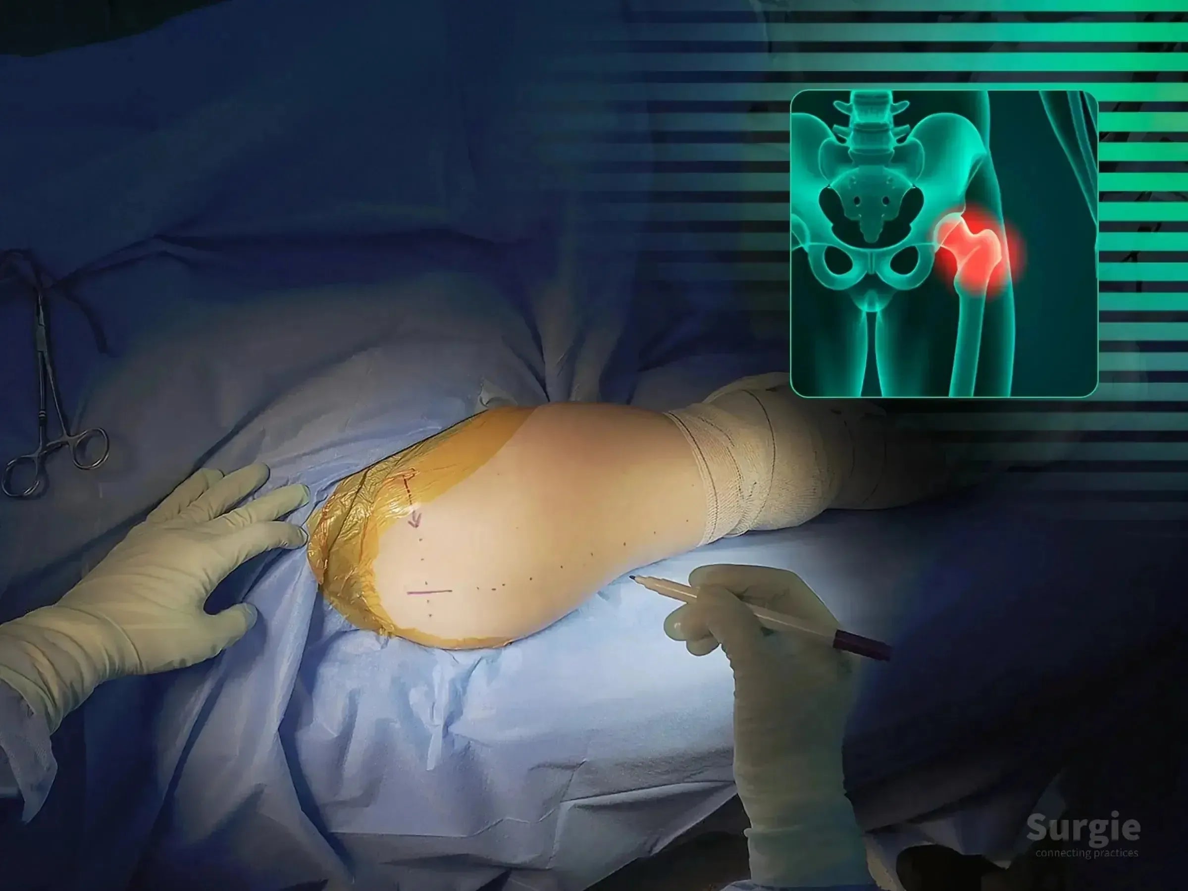
Rocker-assisted calcar reduction without a traction table
 डॉ. सावीओ चामीऑर्थोपेडिस्ट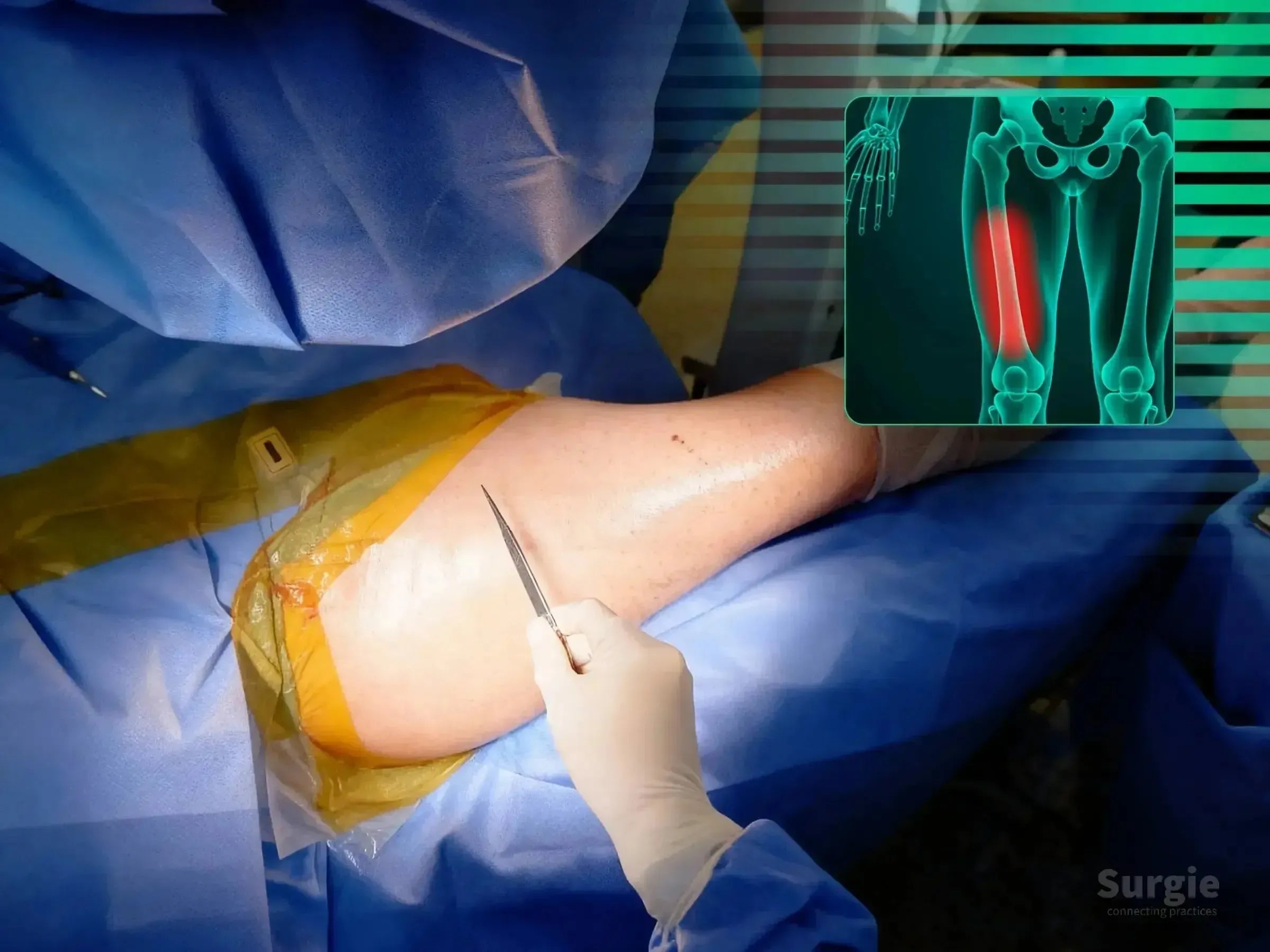
Double see-saw reduction for shaft and calcar realignment
डॉ. सावीओ चामीऑर्थोपेडिस्ट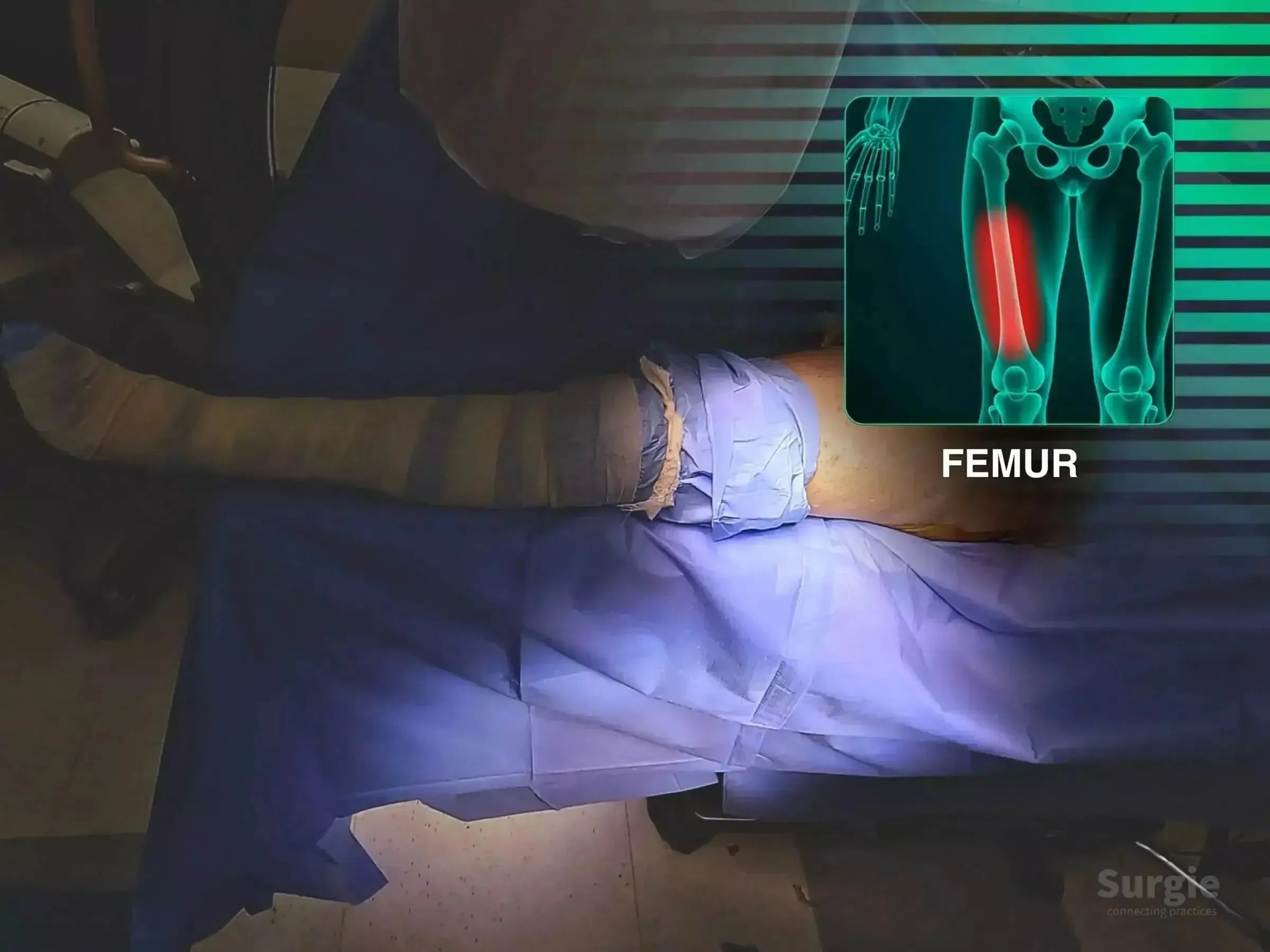
See-saw reduction of an unstable proximal femur fracture
डॉ. सावीओ चामीऑर्थोपेडिस्ट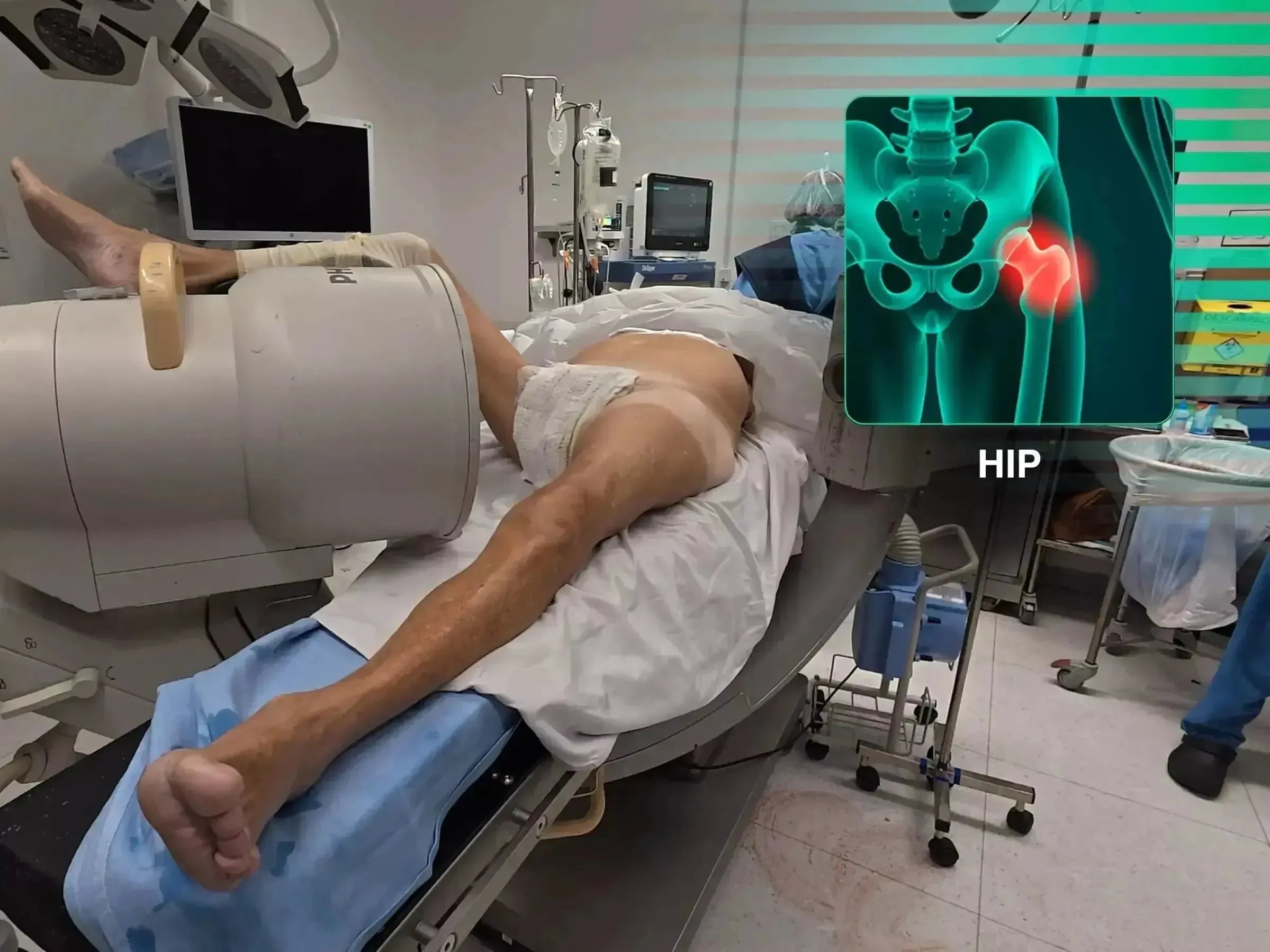
Free-fragment manipulation using a conventional operating table
डॉ. सावीओ चामीऑर्थोपेडिस्ट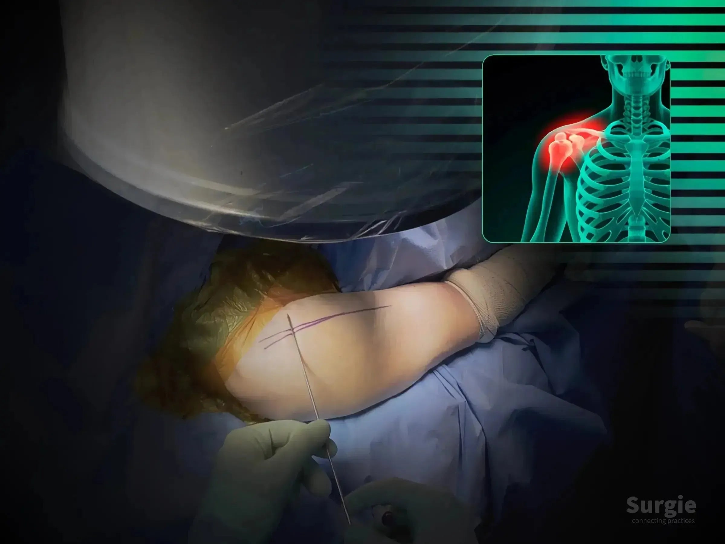
Joystick reduction and long-nail fixation with cuff preservation
डॉ. सावीओ चामीऑर्थोपेडिस्ट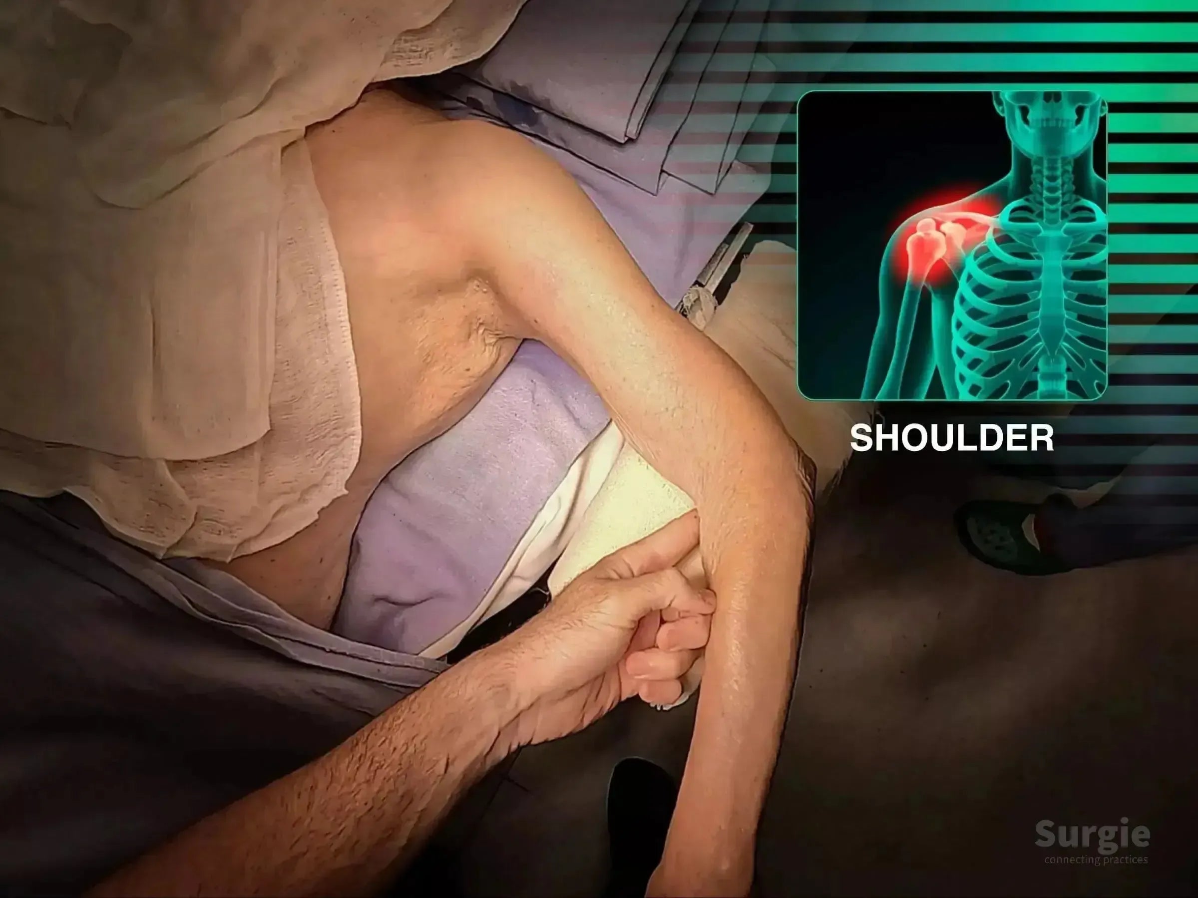
Deltopectoral exposure with cuff management and plate fixation
डॉ. सावीओ चामीऑर्थोपेडिस्ट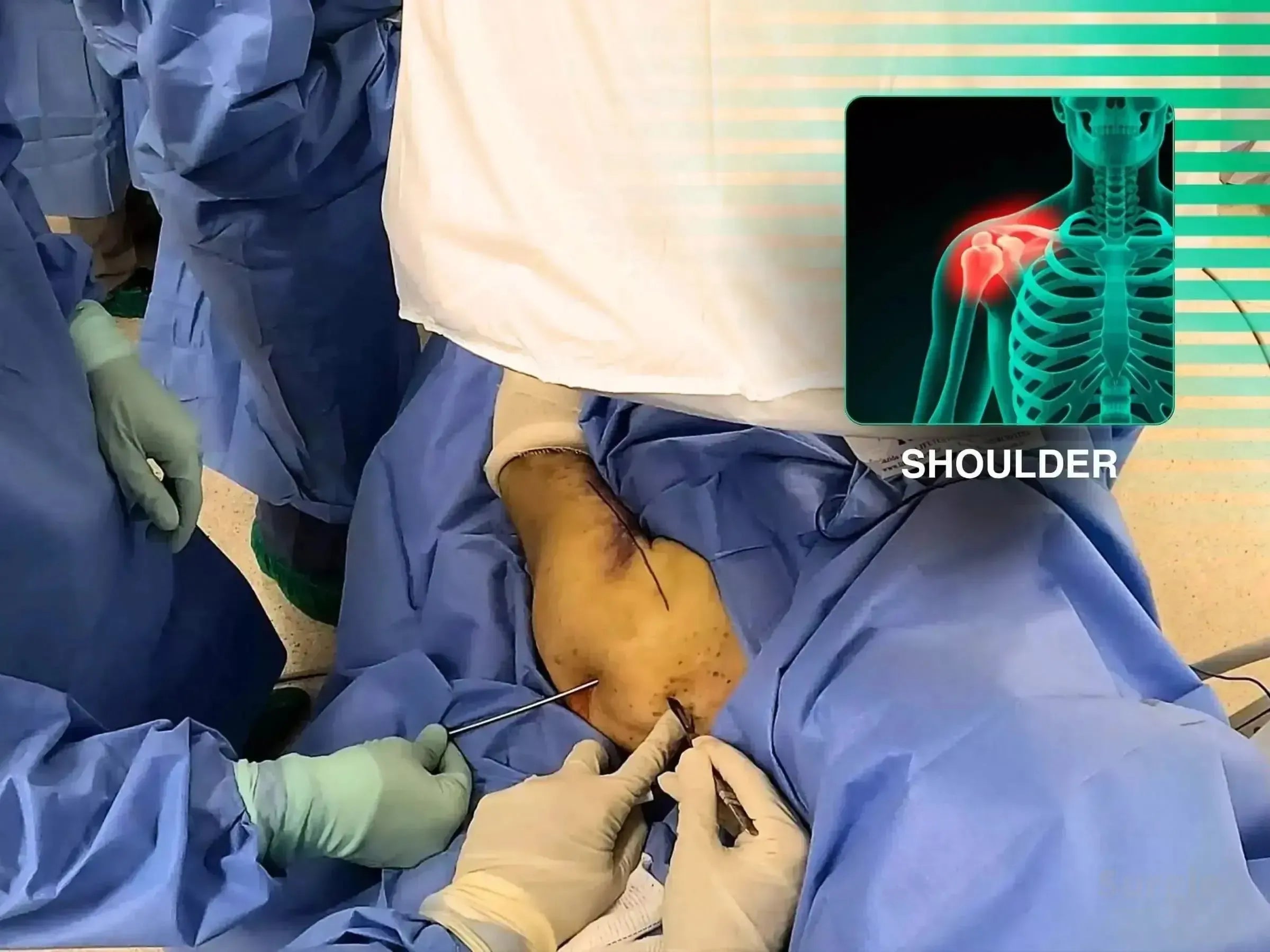
Joystick-guided reduction and intramedullary fixation
डॉ. सावीओ चामीऑर्थोपेडिस्ट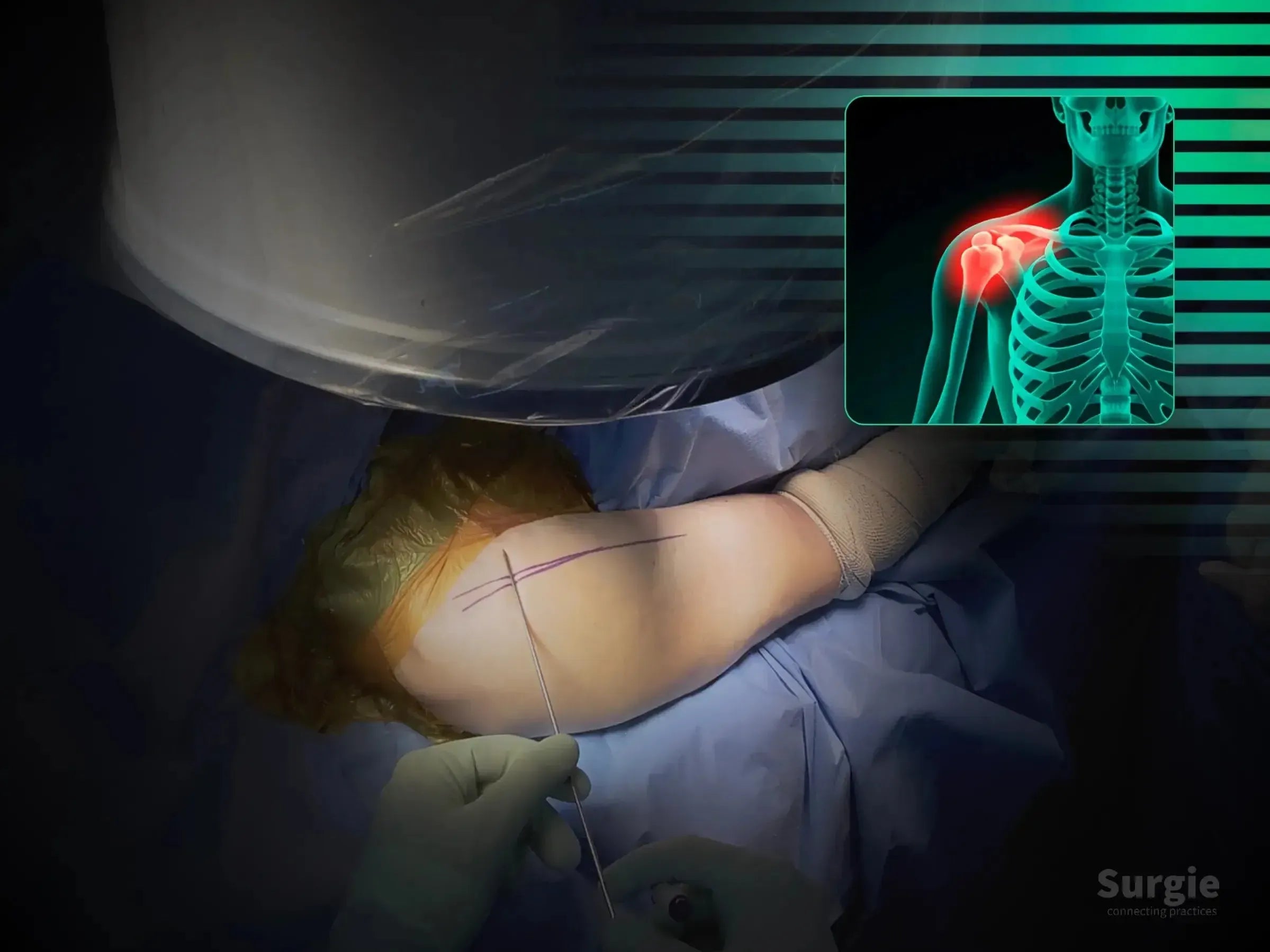
Long nail fixation with cuff preservation and fluoroscopic control
डॉ. सावीओ चामीऑर्थोपेडिस्ट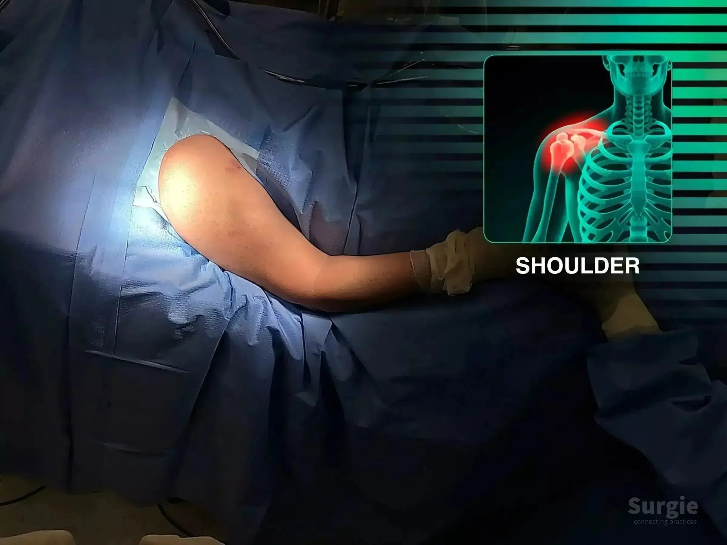
Percutaneous proximal and distal locking through a minimally invasive approach
डॉ. सावीओ चामीऑर्थोपेडिस्ट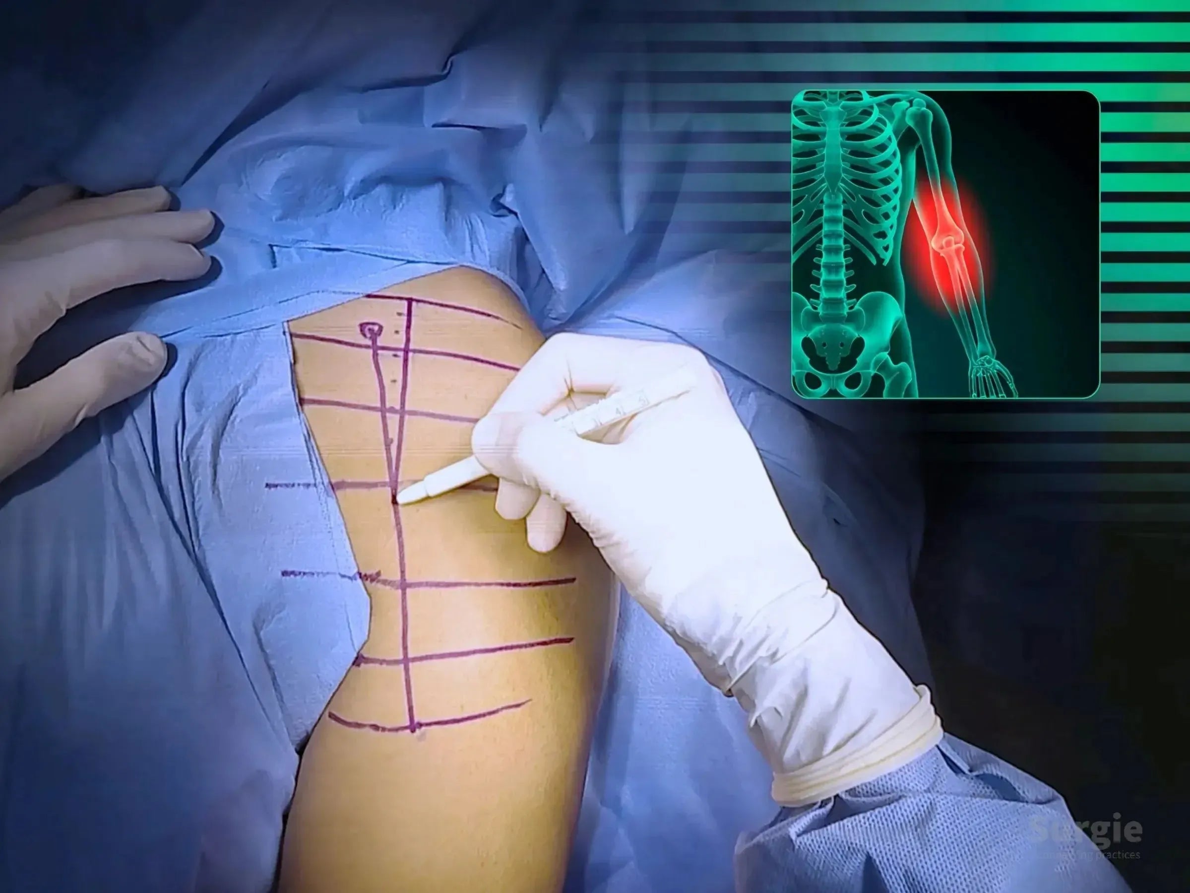
Medial anti-shear support with complementary lateral plating
डॉ. सावीओ चामीऑर्थोपेडिस्ट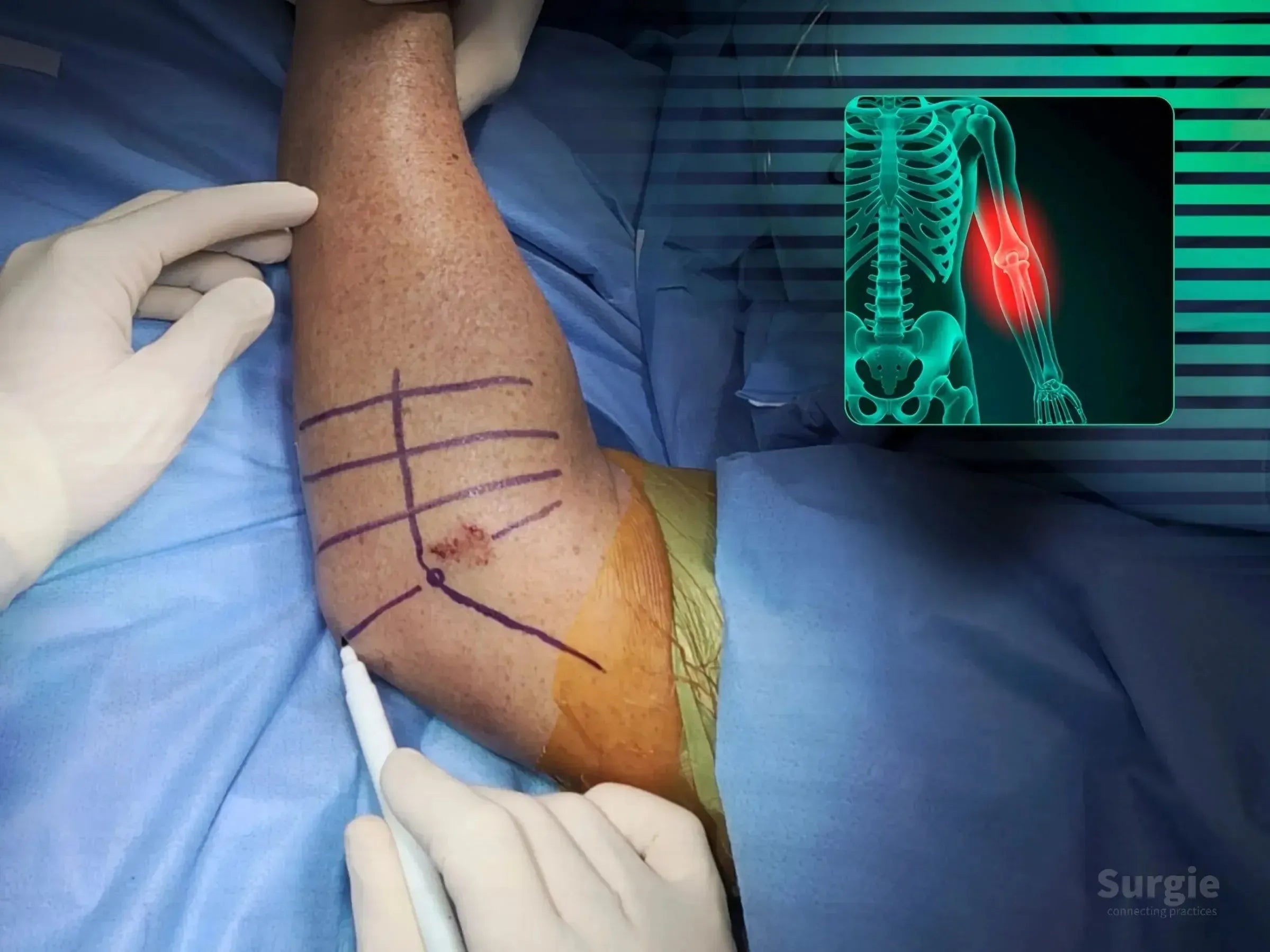
Tripod screw fixation with capsular and ligament reconstruction
डॉ. सावीओ चामीऑर्थोपेडिस्ट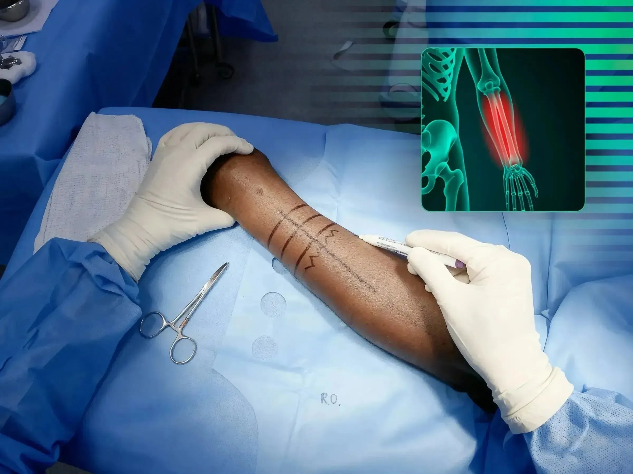
Thompson approach with rotational control and interfragmentary compression
डॉ. सावीओ चामीऑर्थोपेडिस्ट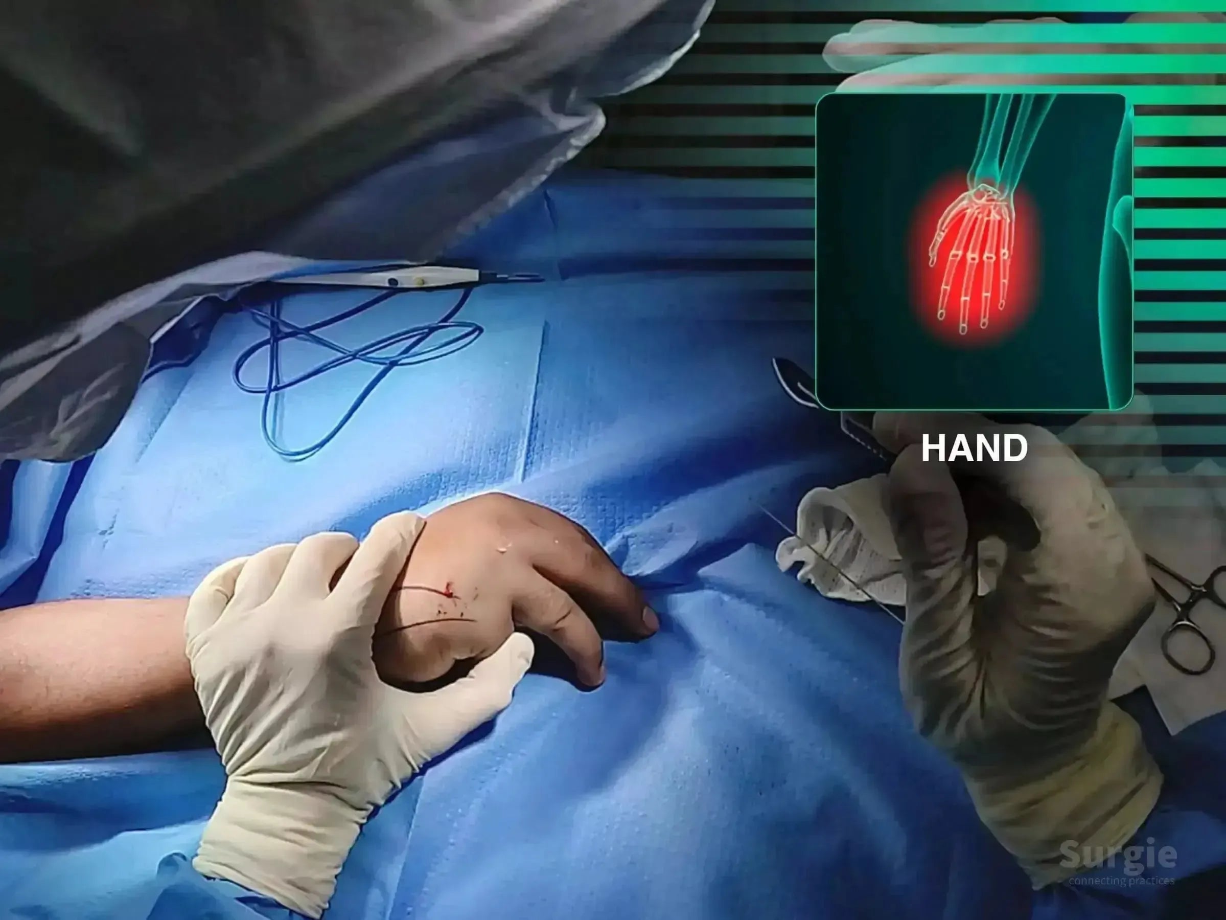
Anatomic forearm reconstruction through radial and ulnar plate fixation
डॉ. सावीओ चामीऑर्थोपेडिस्ट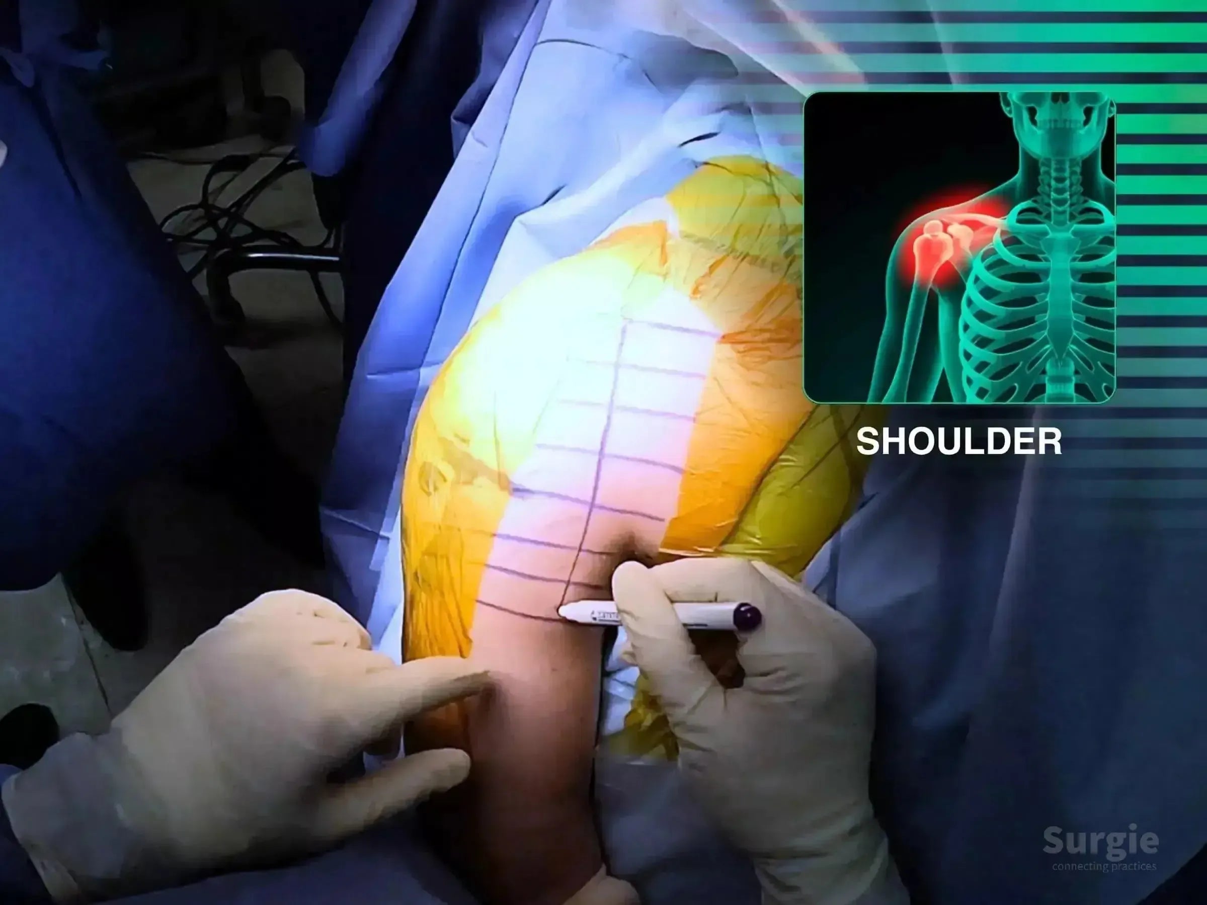
Single-approach reconstruction of humerus, glenoid, and coracoid injuries
डॉ. सावीओ चामीऑर्थोपेडिस्ट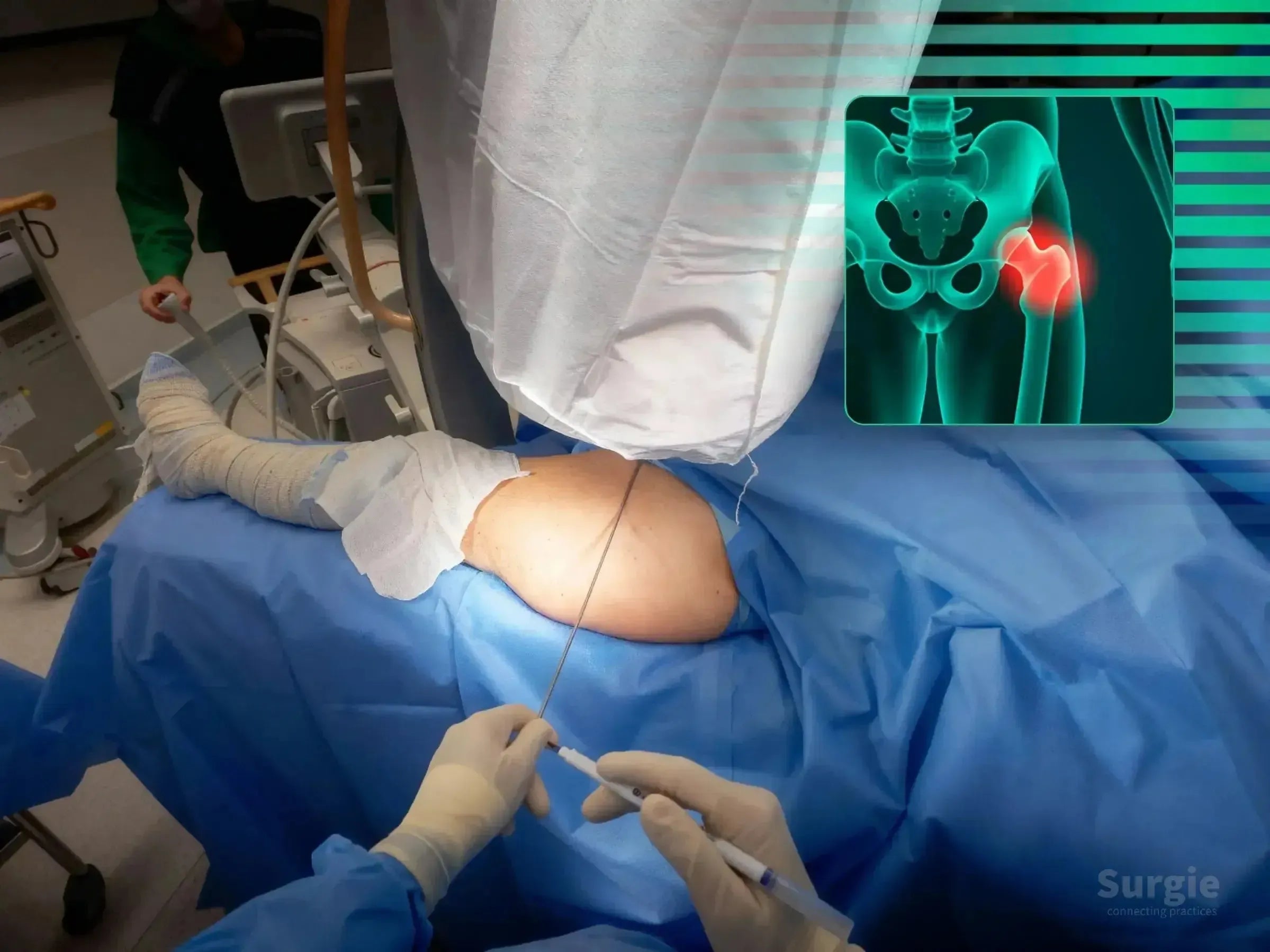
Percutaneous reduction, varus control, and cephalomedullary nailing
डॉ. सावीओ चामीऑर्थोपेडिस्ट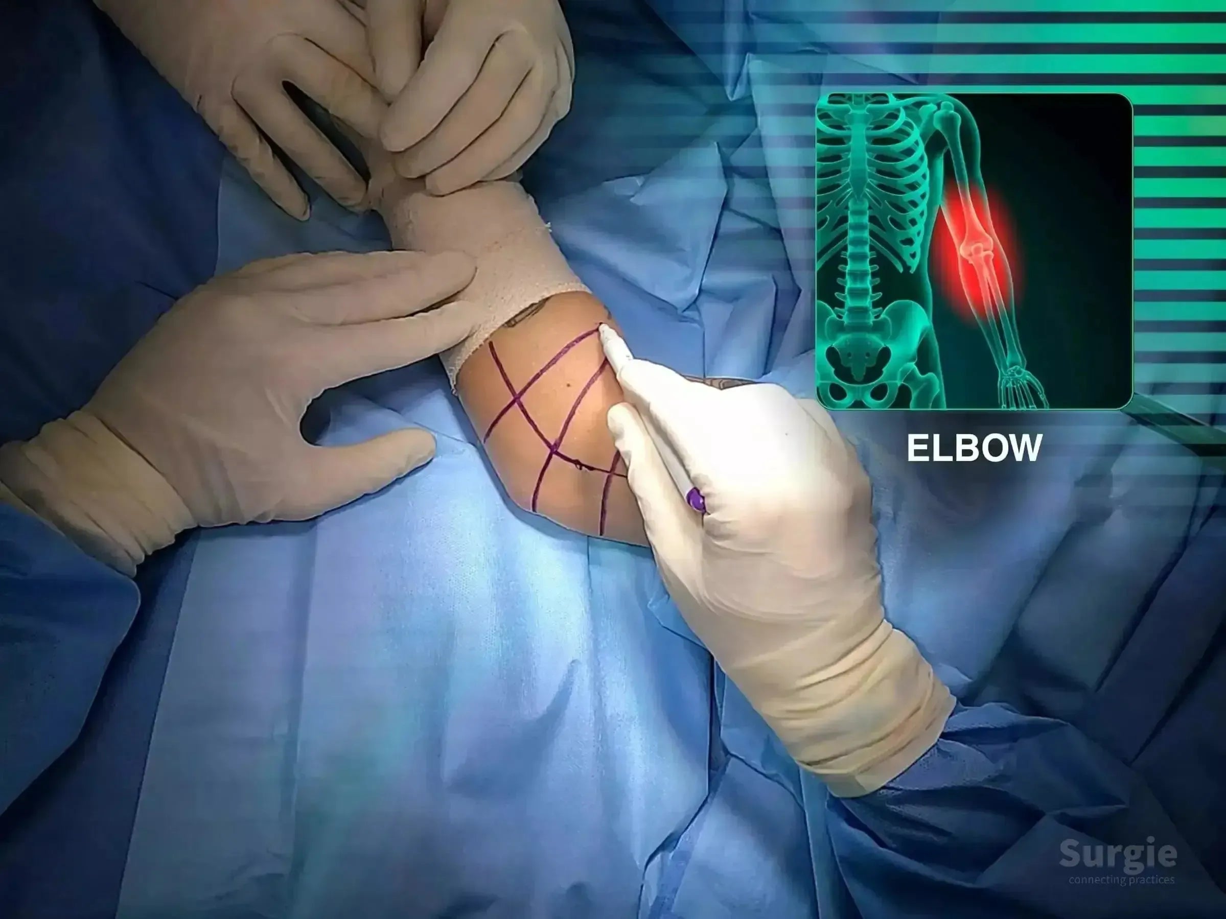
Radial head, coronoid, capsule, and lateral ligament reconstruction
डॉ. सावीओ चामीऑर्थोपेडिस्ट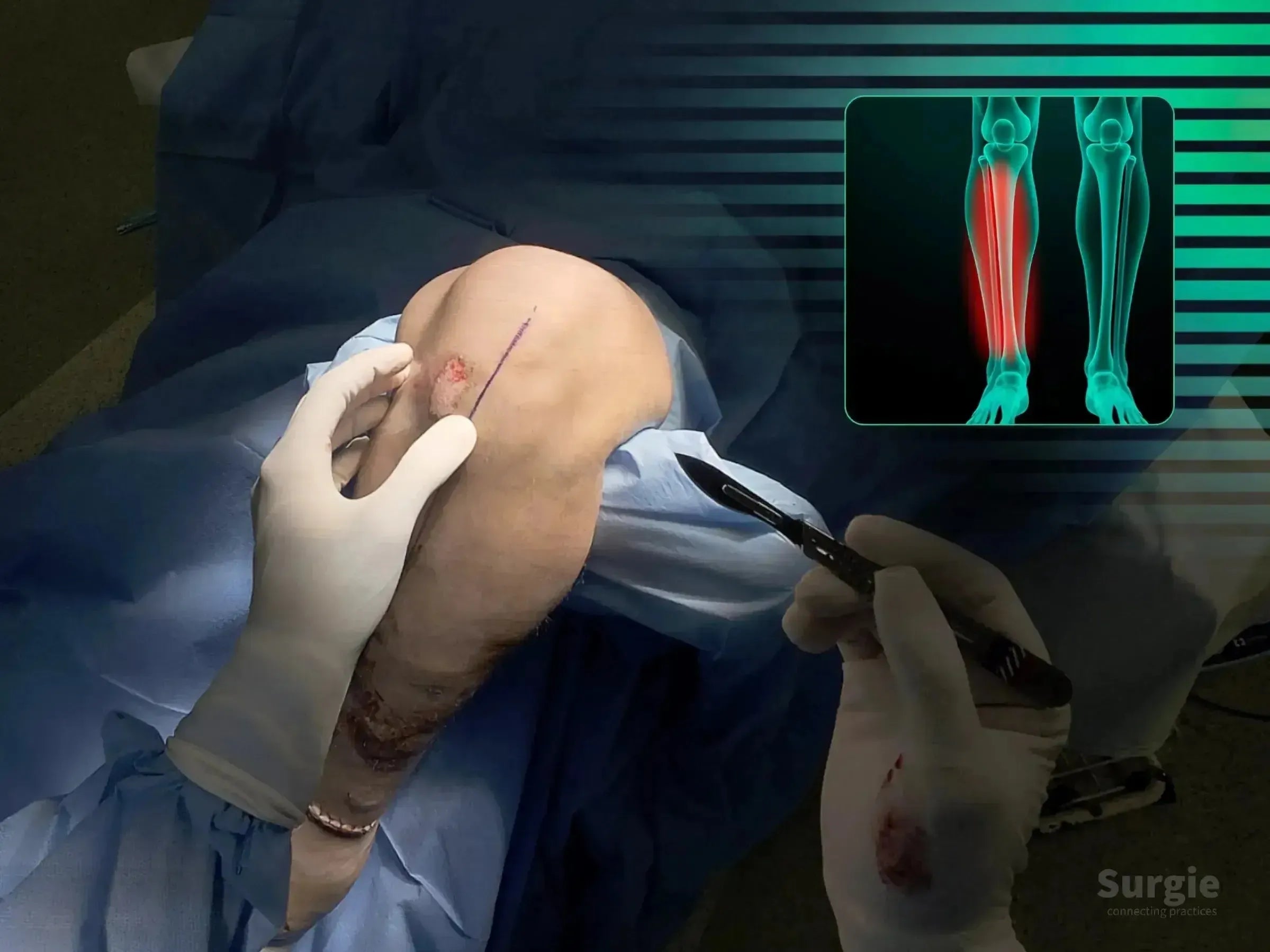
Infrapatellar entry with tendon preservation and fluoroscopic guidance
डॉ. सावीओ चामीऑर्थोपेडिस्ट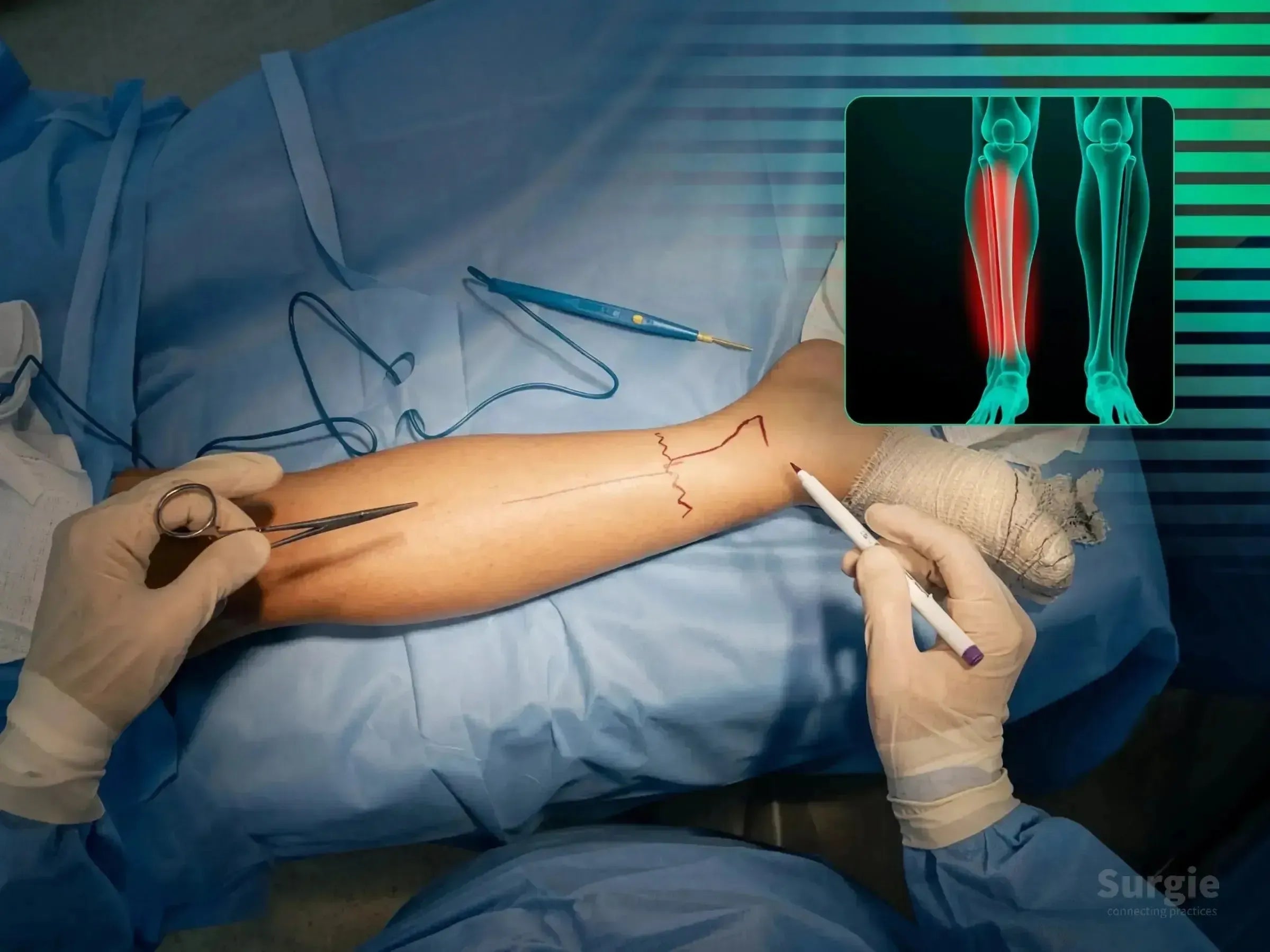
CT-guided dual-approach fixation of posteromedial pilon shear
डॉ. सावीओ चामीऑर्थोपेडिस्ट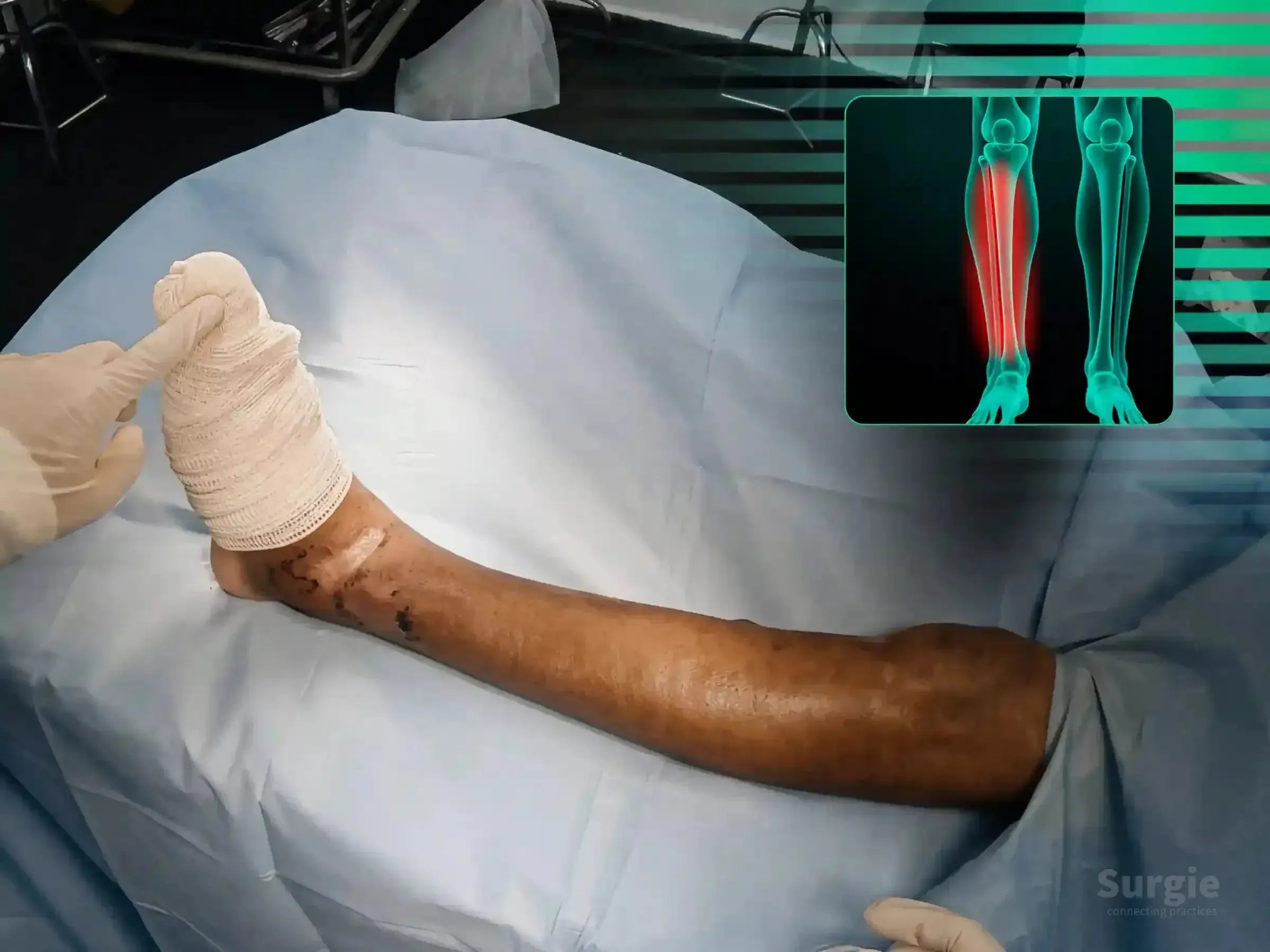
Staged dual-approach pilon reconstruction guided by CT morphology
डॉ. सावीओ चामीऑर्थोपेडिस्ट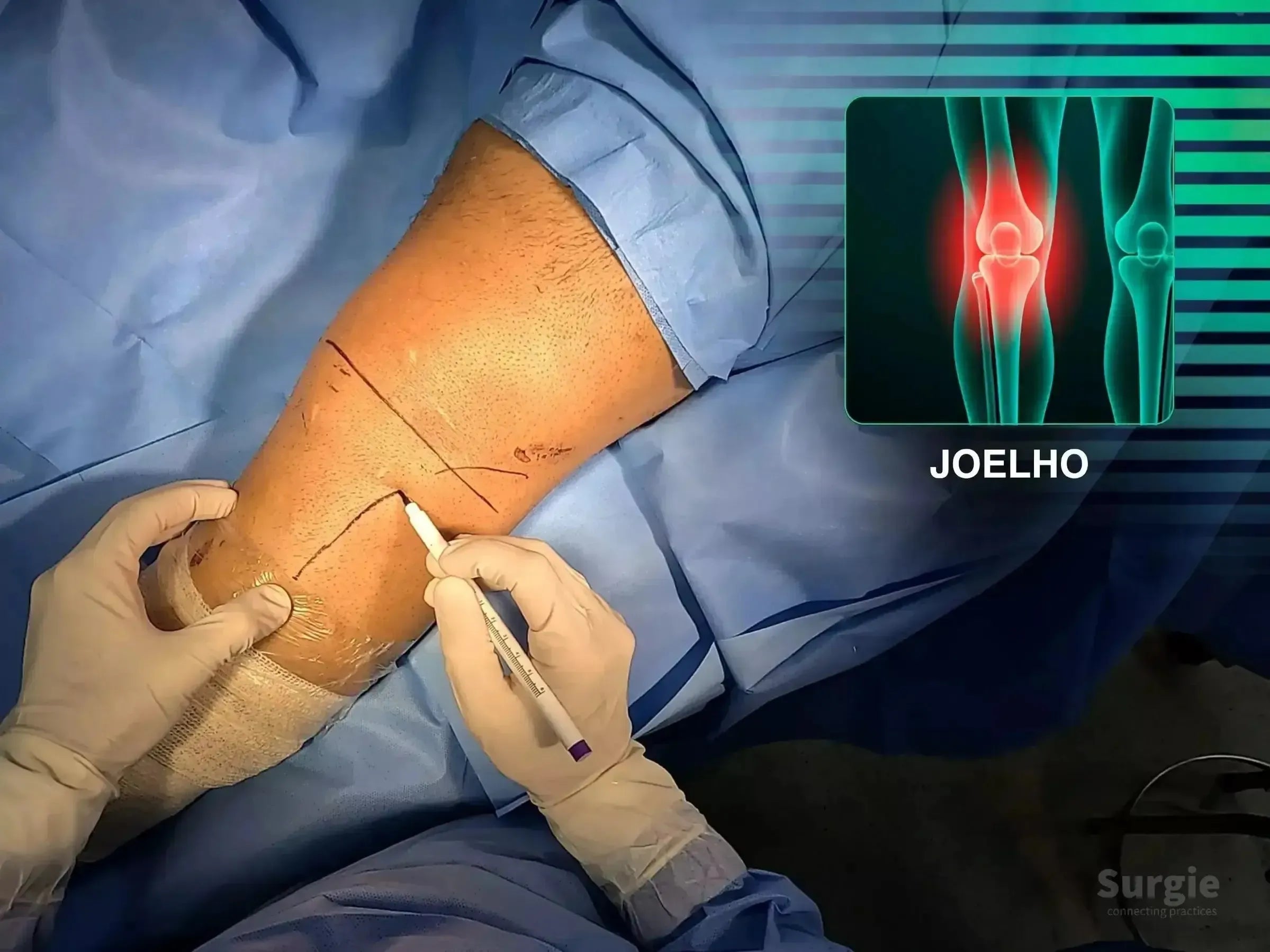
Anterolateral exposure with osteotomy, grafting, and subchondral support
डॉ. सावीओ चामीऑर्थोपेडिस्ट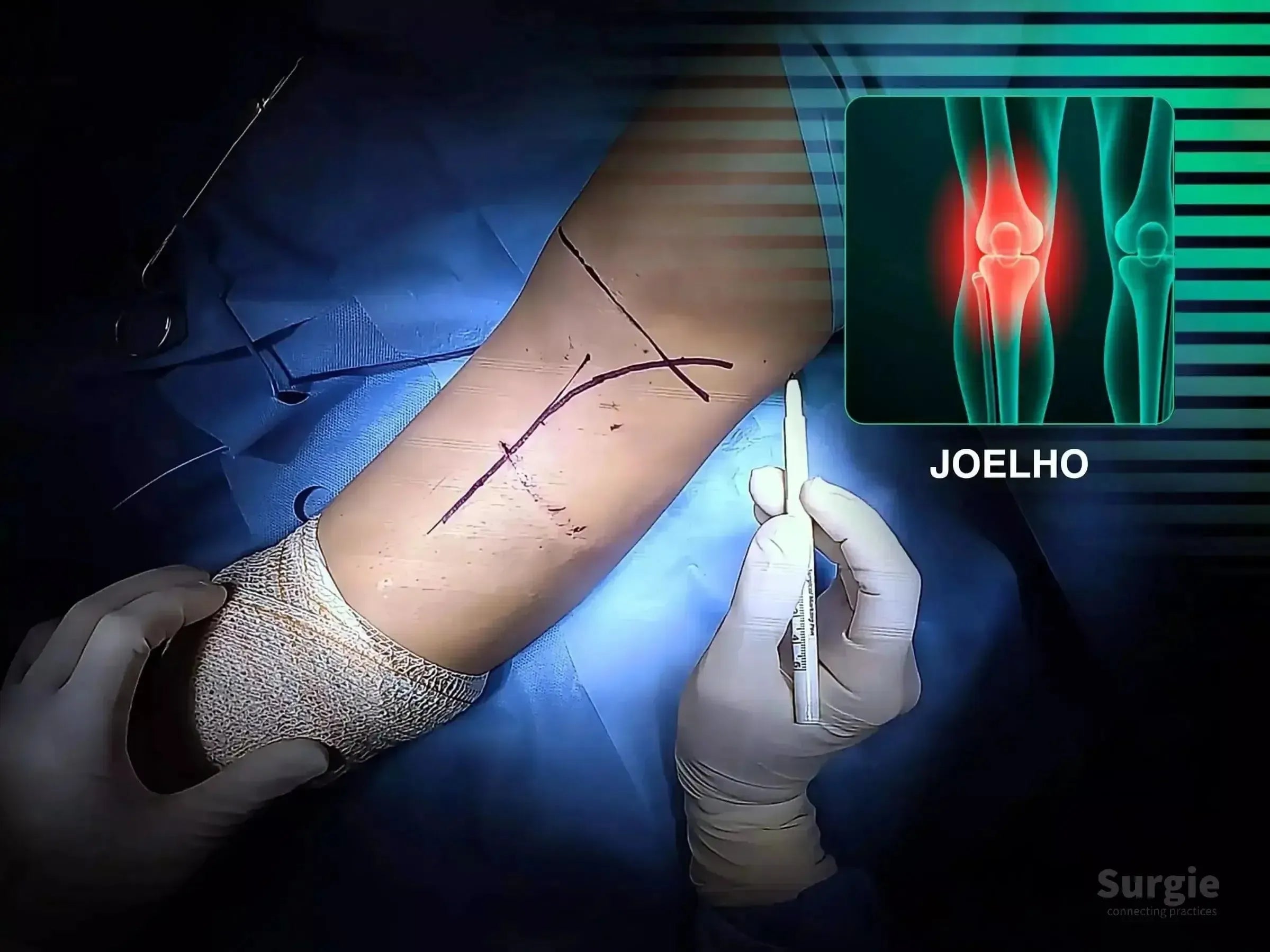
इलास्टिक ऑस्टियोटॉमी, ग्राफ्ट ऊँचाई, और सबकॉन्ड्रल फिक्सेशन
डॉ. सावीओ चामीऑर्थोपेडिस्ट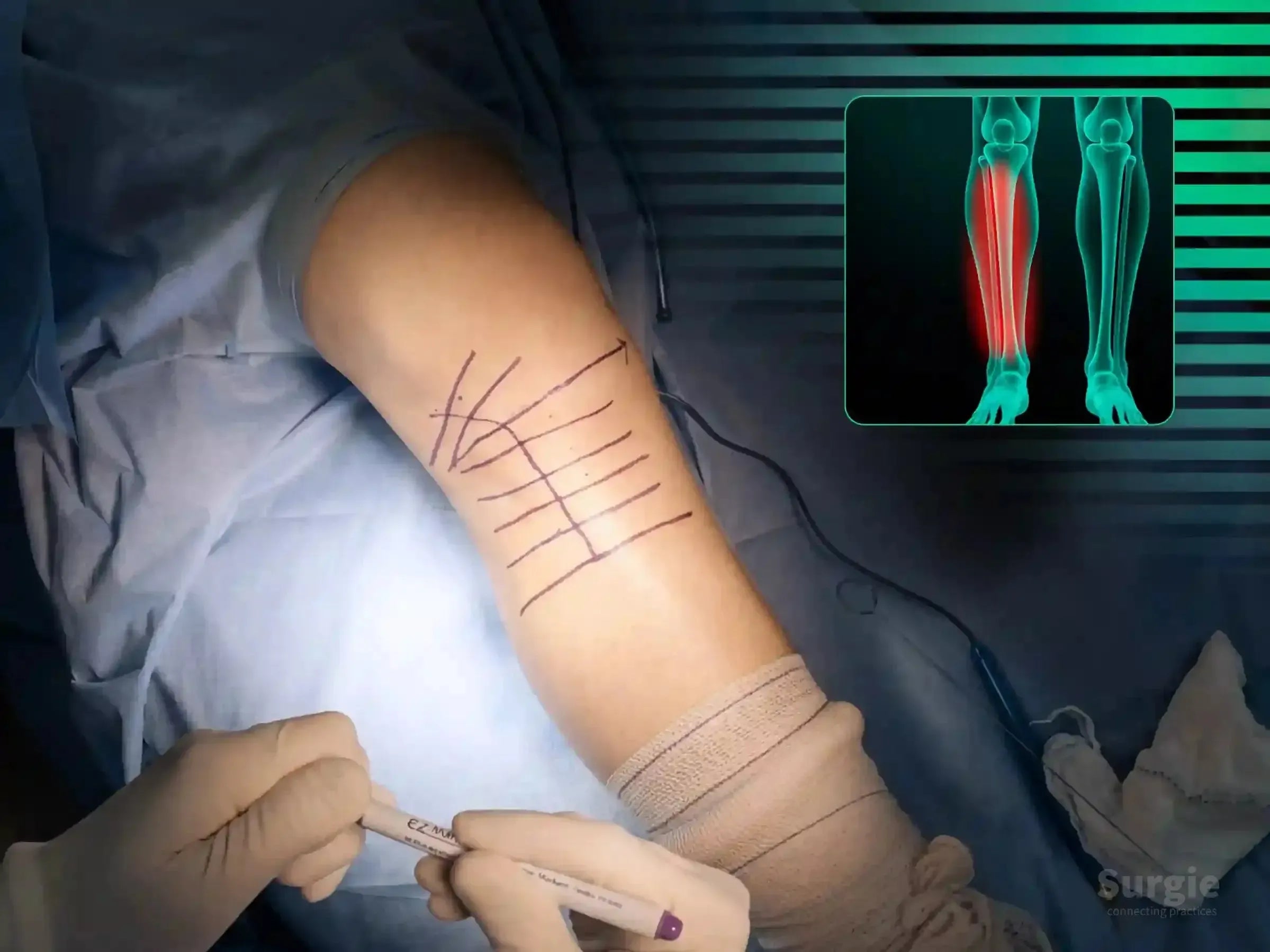
Posterolateral depression elevation with meniscal protection and fixation
डॉ. सावीओ चामीऑर्थोपेडिस्ट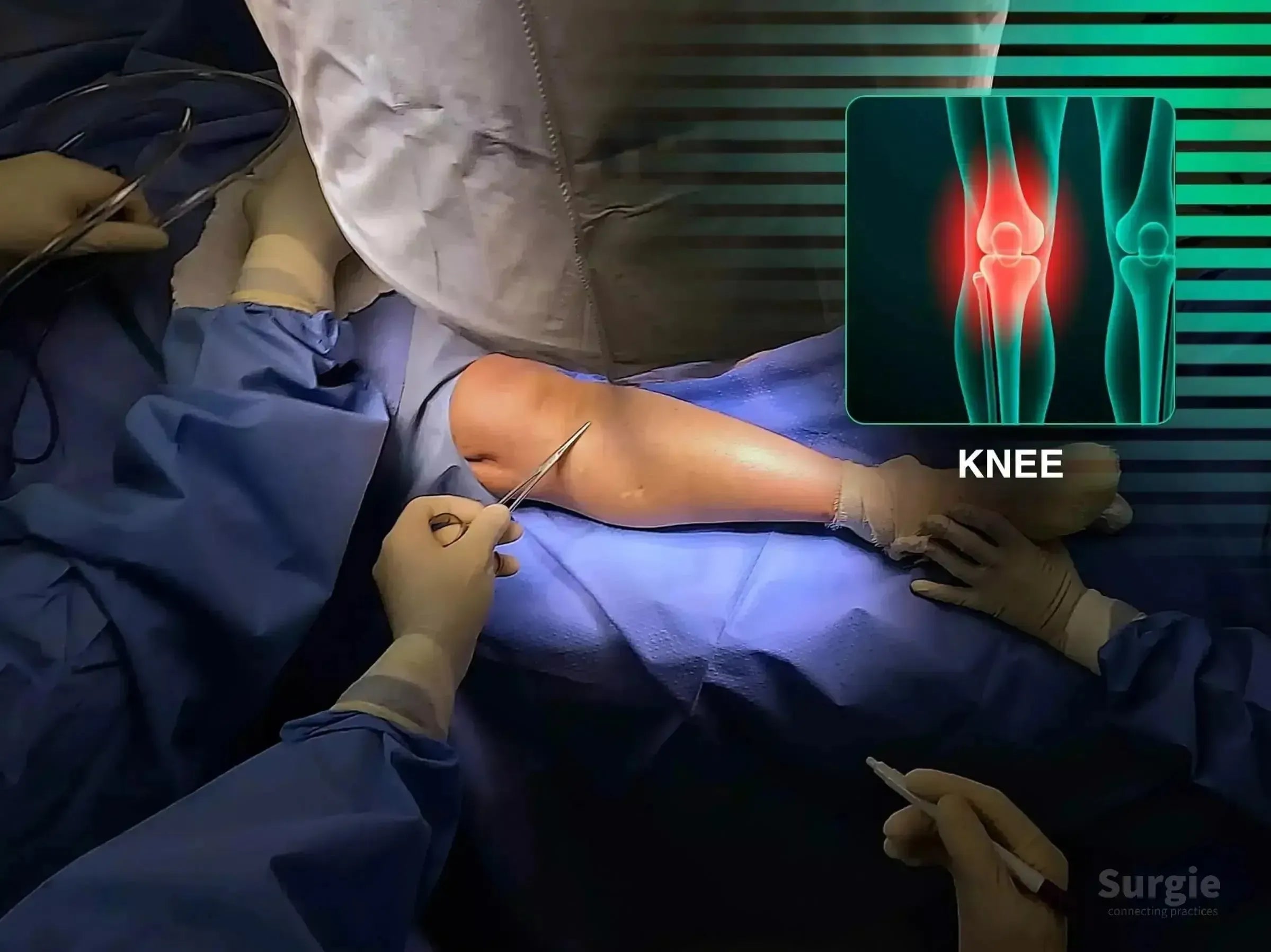
Dual-plate fixation for deformity, shear control, and osteopenic bone
डॉ. सावीओ चामीऑर्थोपेडिस्ट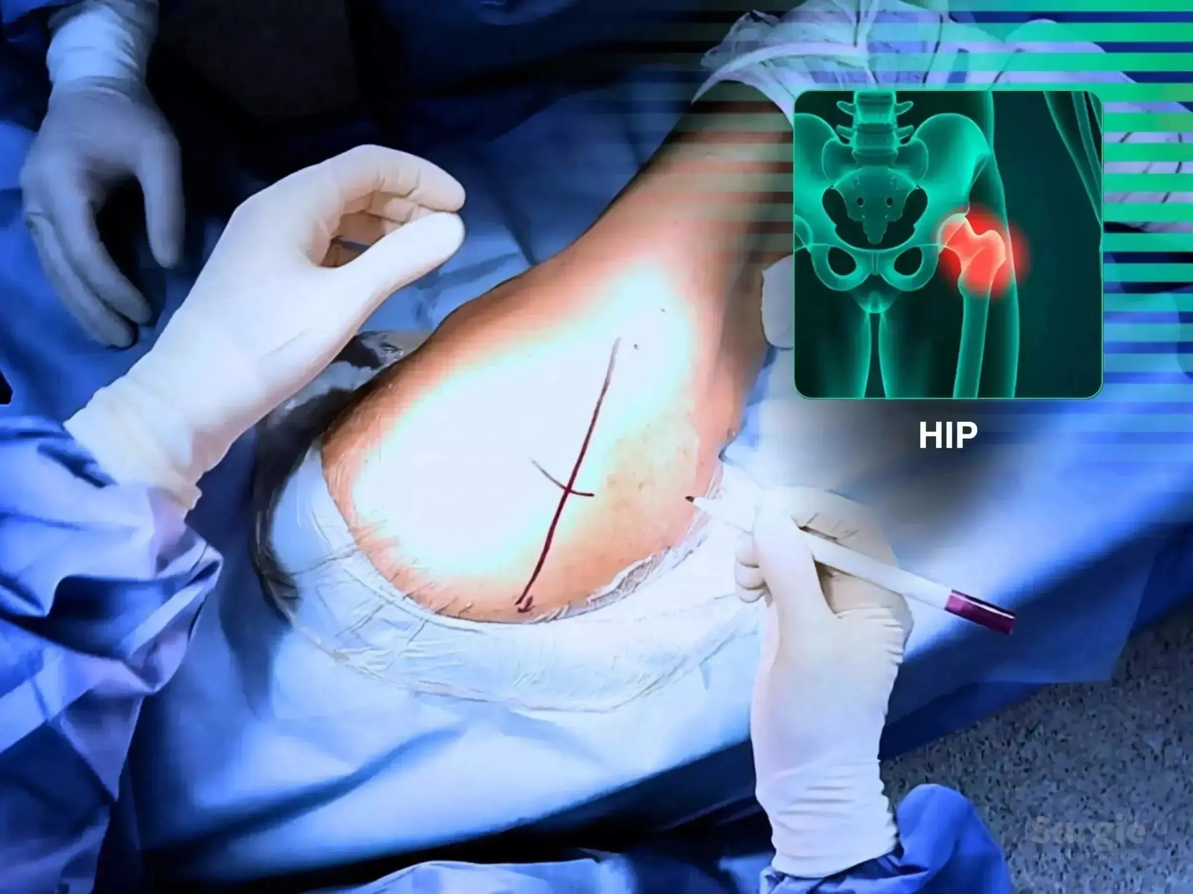
Posterior THA for coxarthrosis with soft-tissue and rotator management
डॉ. सावीओ चामीऑर्थोपेडिस्ट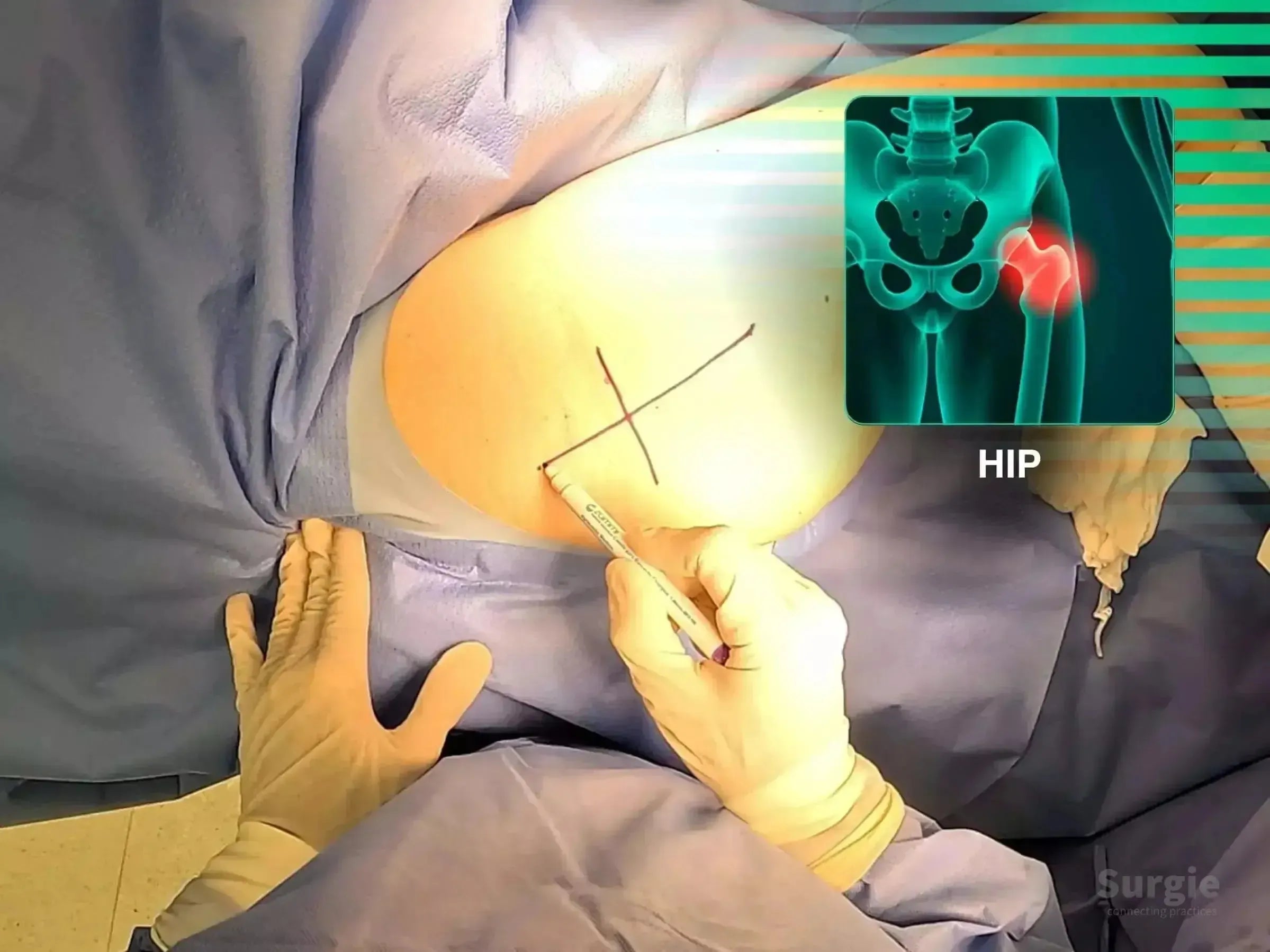
Posterior THA with component positioning and soft-tissue reconstruction
डॉ. सावीओ चामीऑर्थोपेडिस्ट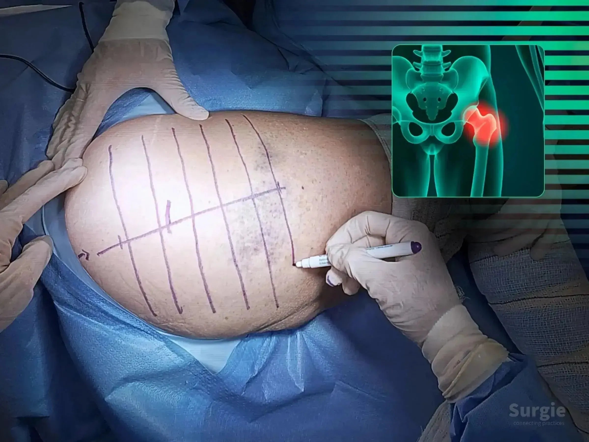
Posterior THA with piriformis preservation and press-fit components
डॉ. सावीओ चामीऑर्थोपेडिस्ट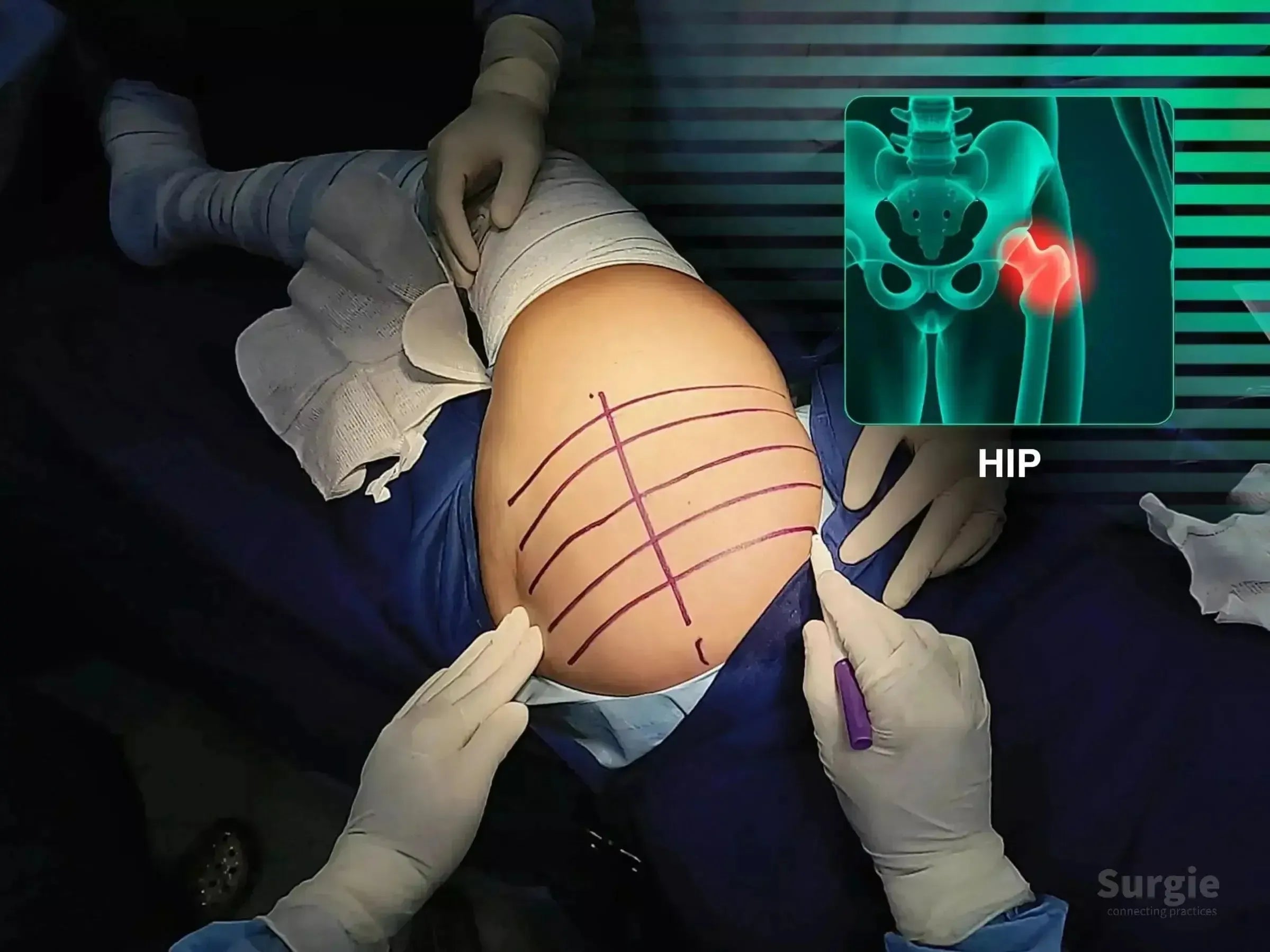
Acetabular and femoral preparation with stability-focused implant testing
डॉ. सावीओ चामीऑर्थोपेडिस्ट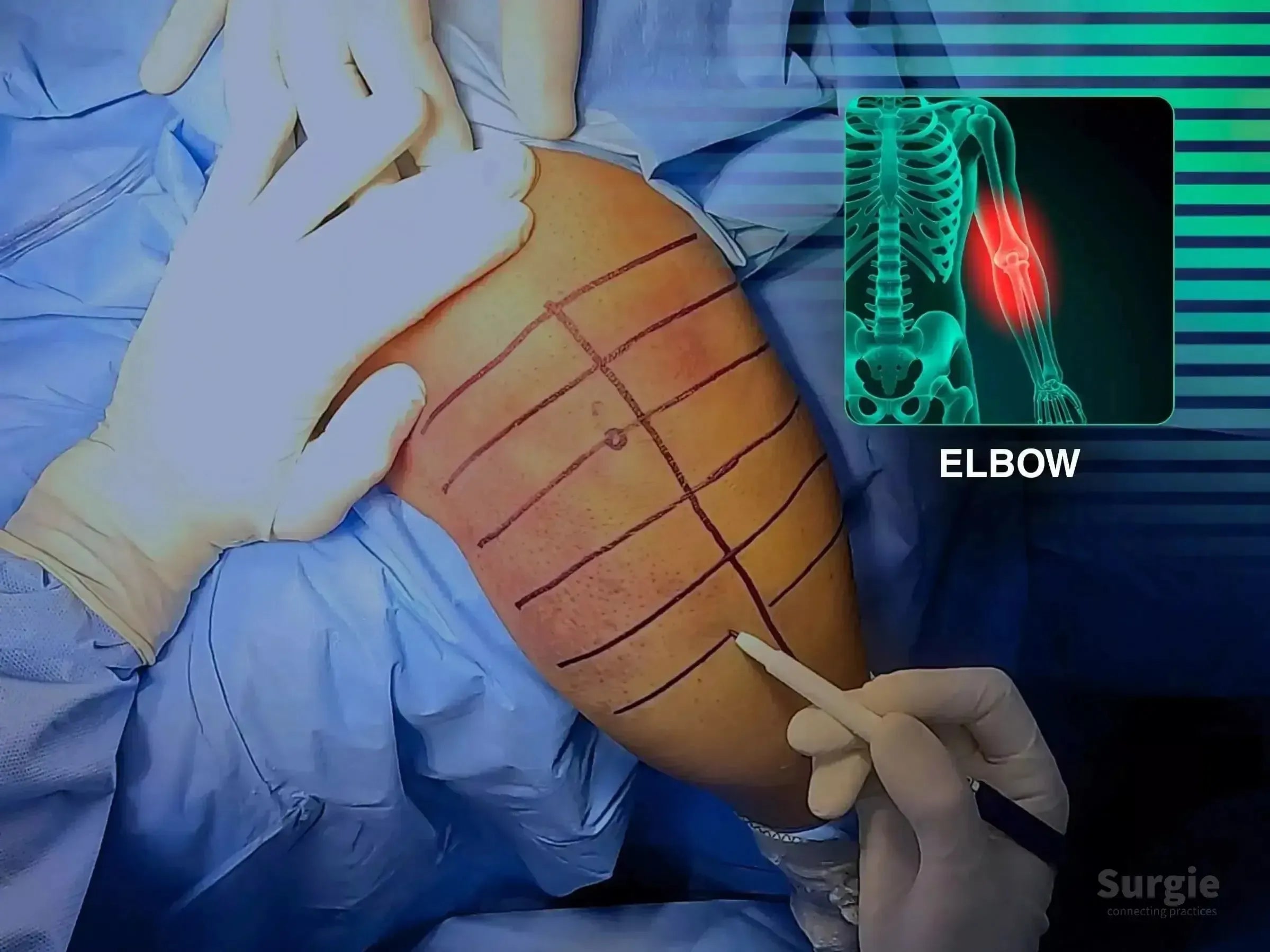
Pull-out capsular repair with proximal ulna reconstruction
डॉ. सावीओ चामीऑर्थोपेडिस्ट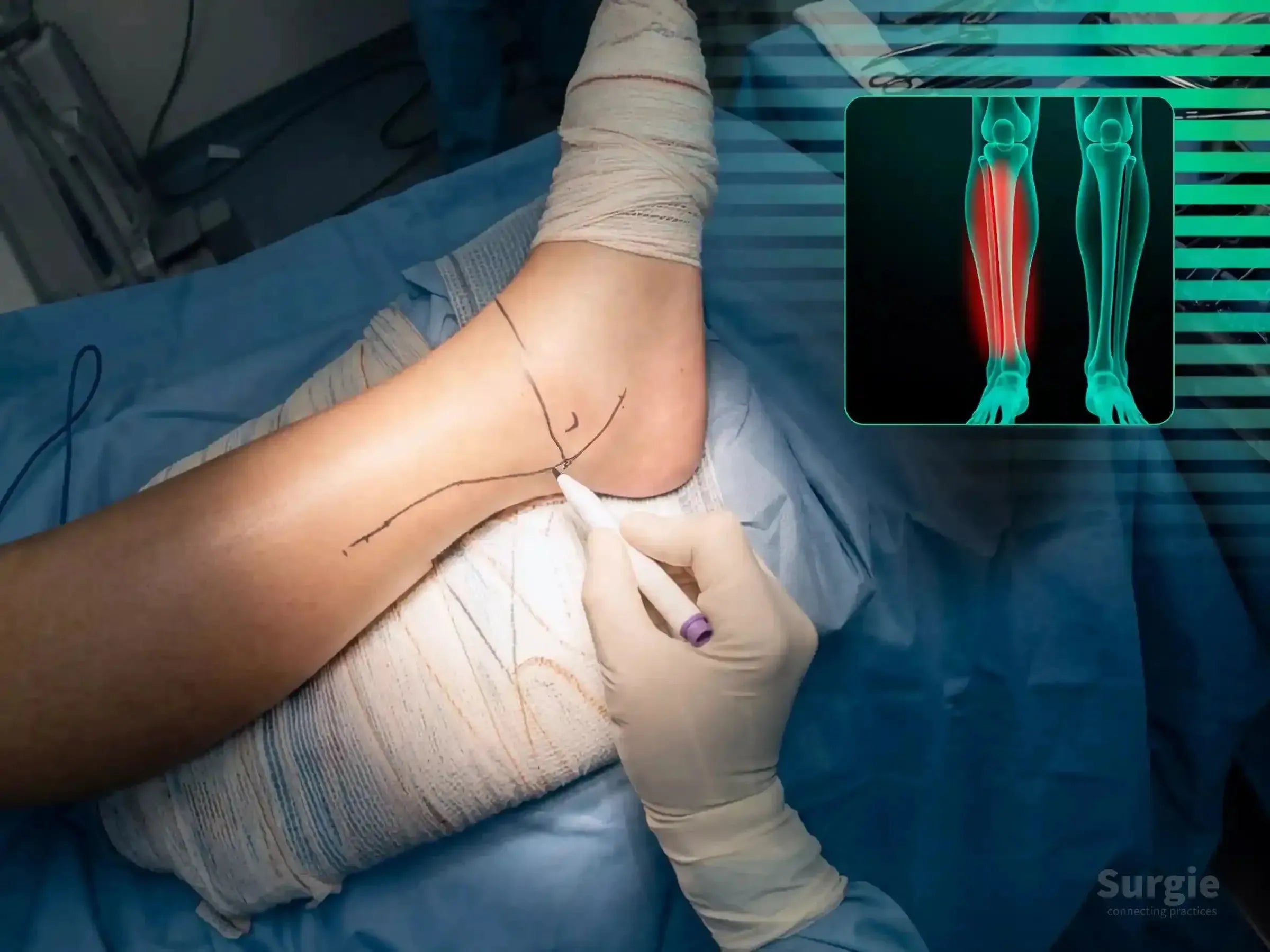
Posterolateral ankle fixation with staged malleolar reconstruction
डॉ. सावीओ चामीऑर्थोपेडिस्ट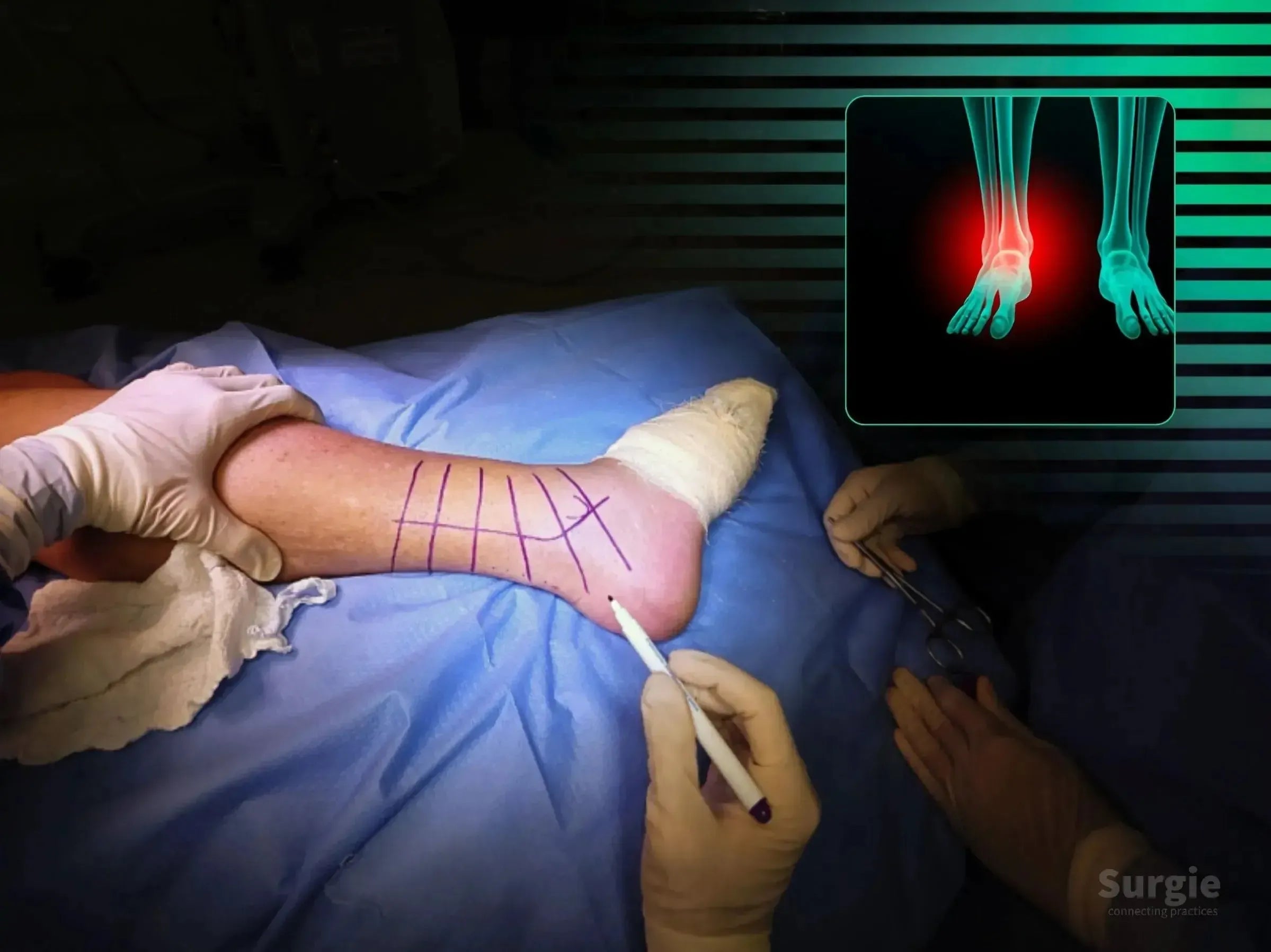
Sequential posterior, lateral, and medial fixation in lateral decubitus
डॉ. सावीओ चामीऑर्थोपेडिस्ट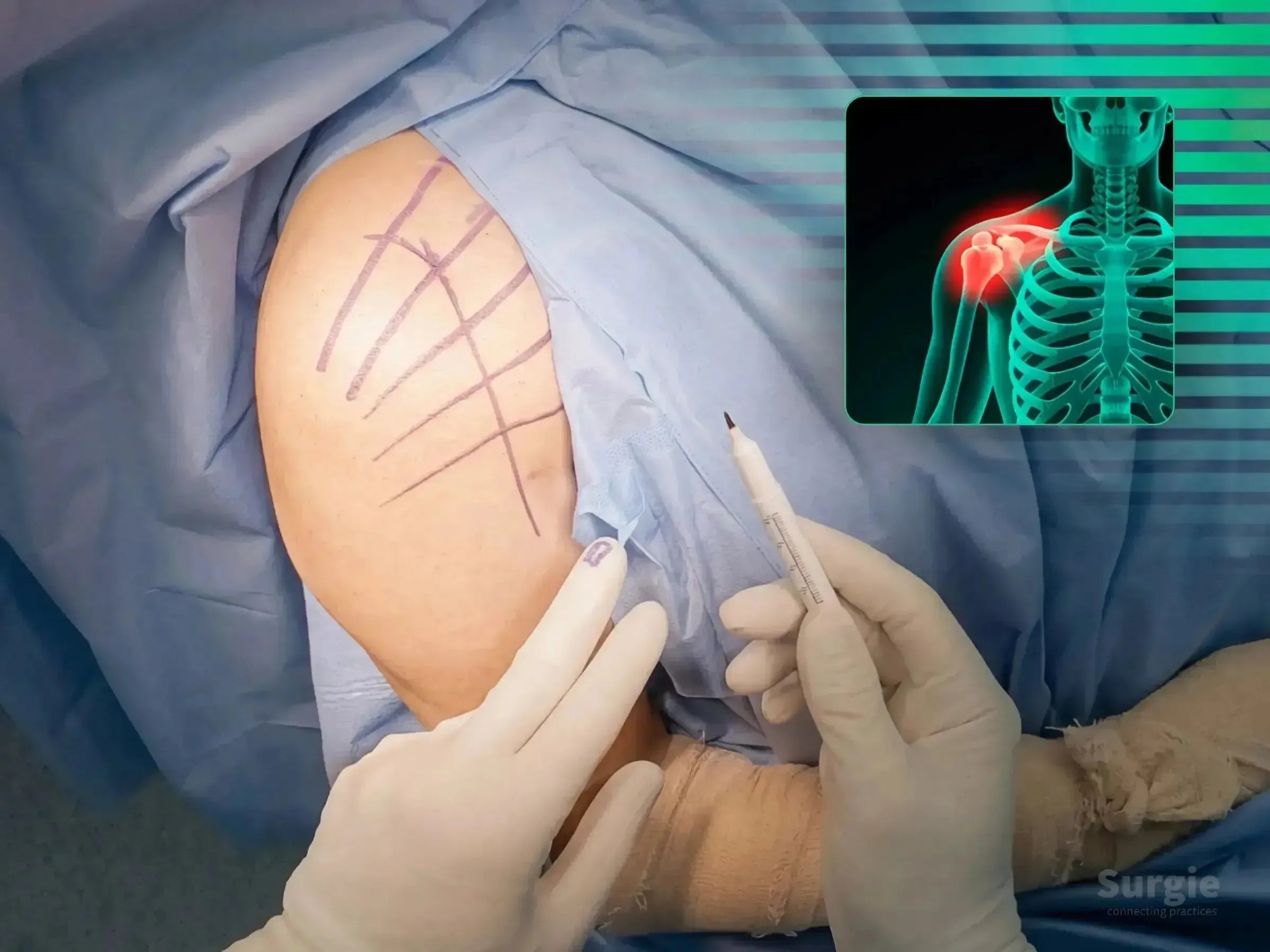
Varus-impacted two-part fracture reduction with plate fixation
डॉ. सावीओ चामीऑर्थोपेडिस्ट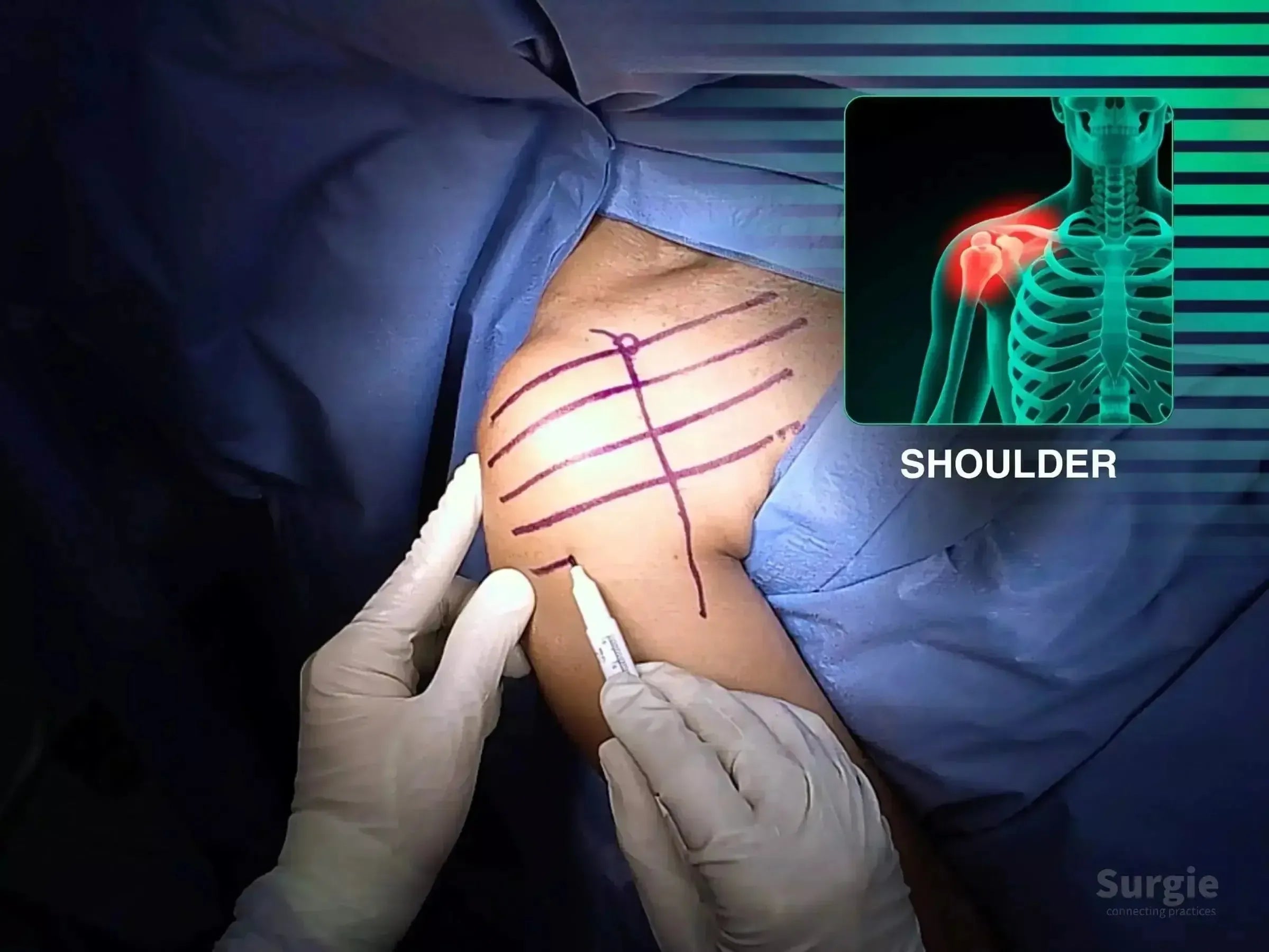
Two-part fracture reduction with anatomic plate positioning
डॉ. सावीओ चामीऑर्थोपेडिस्ट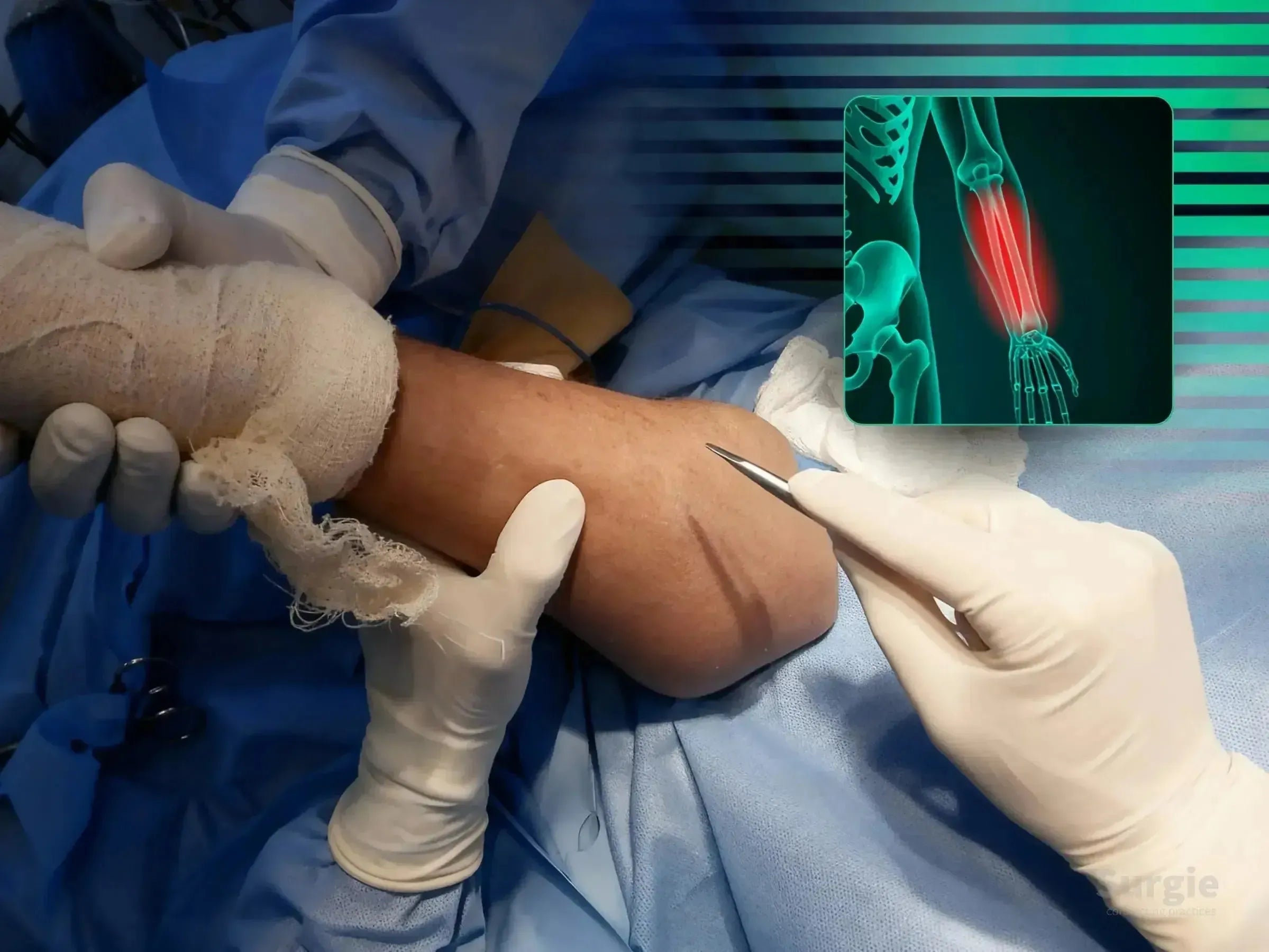
Muscle-sparing ulnar exposure with compression plate fixation
डॉ. सावीओ चामीऑर्थोपेडिस्ट