
Filters
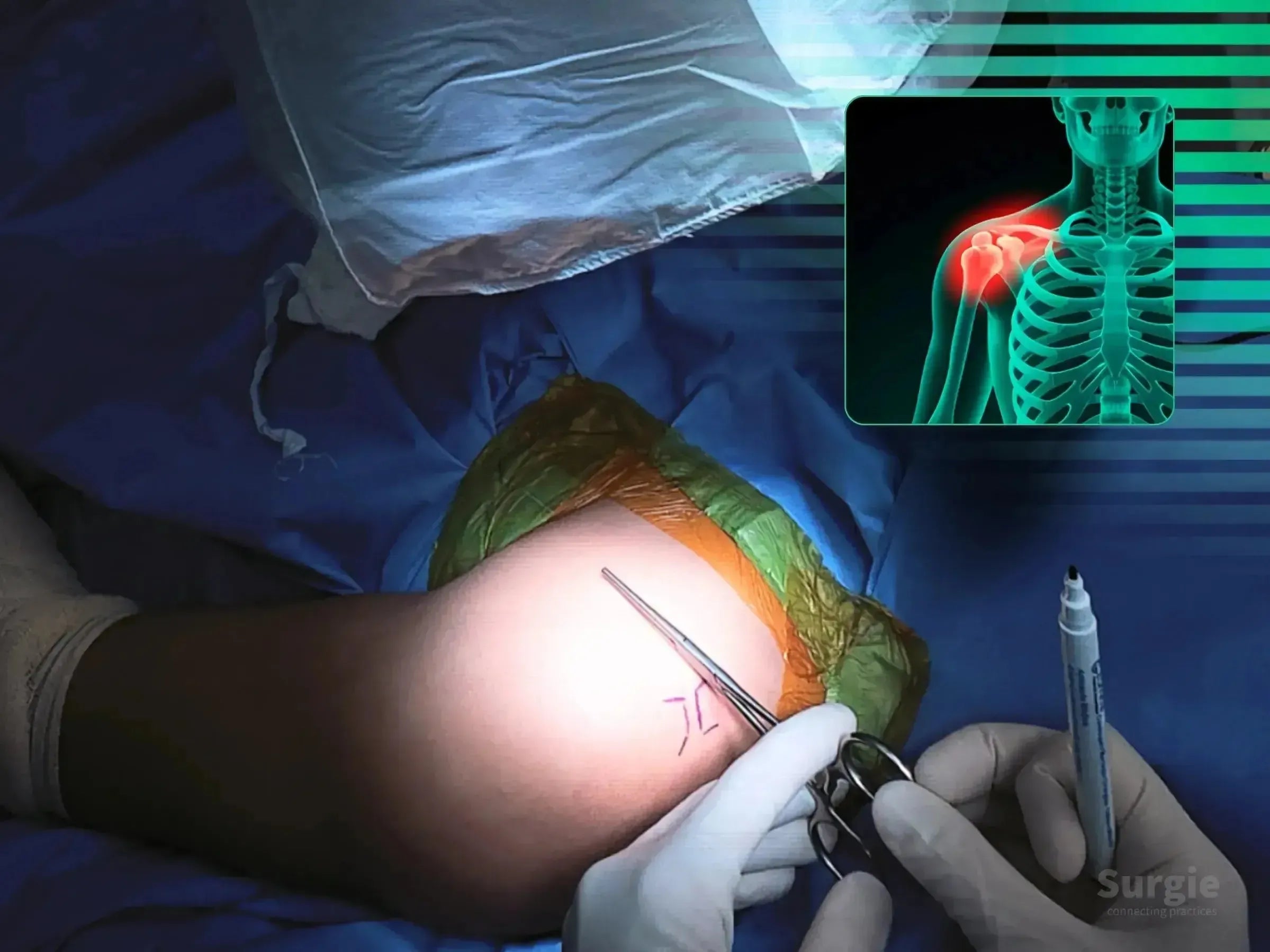
Precise clavicle and coracoid tunneling for TightRope fixation
 डॉ. सावीओ चामीऑर्थोपेडिस्ट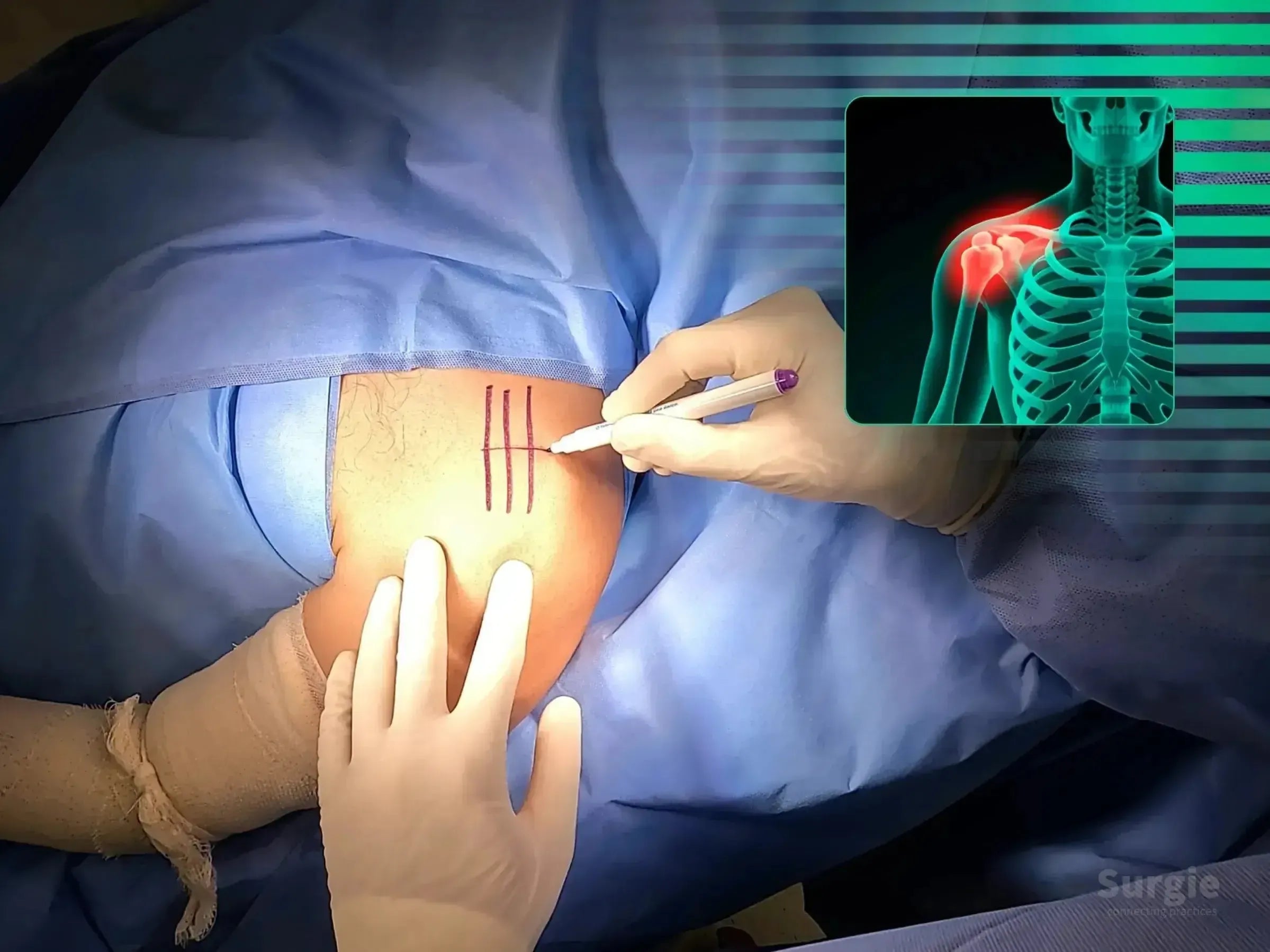
Open coracoclavicular tunnel creation and TightRope stabilization
डॉ. सावीओ चामीऑर्थोपेडिस्ट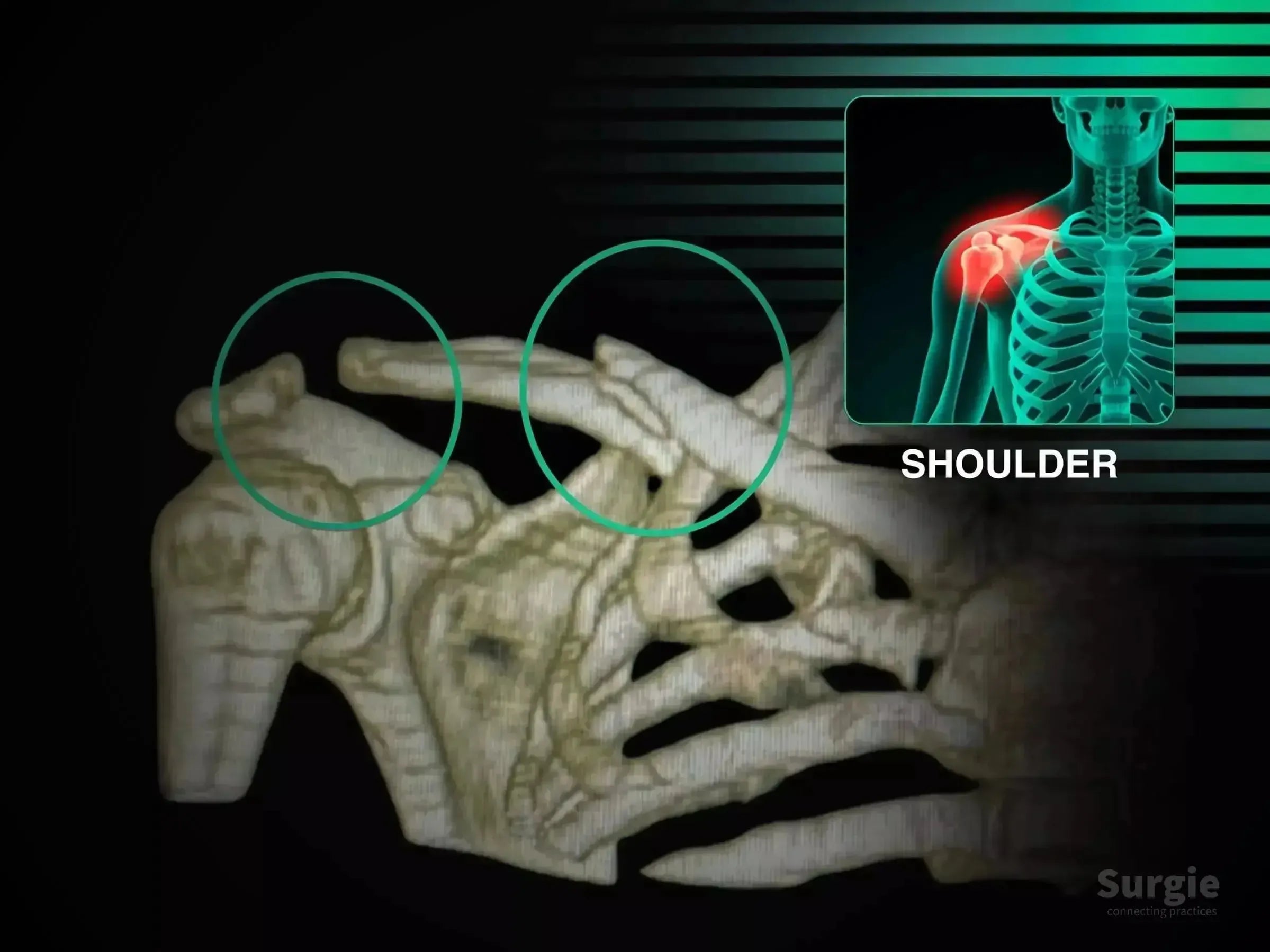
Combined clavicle fixation and multidirectional AC joint stabilization
डॉ. सावीओ चामीऑर्थोपेडिस्ट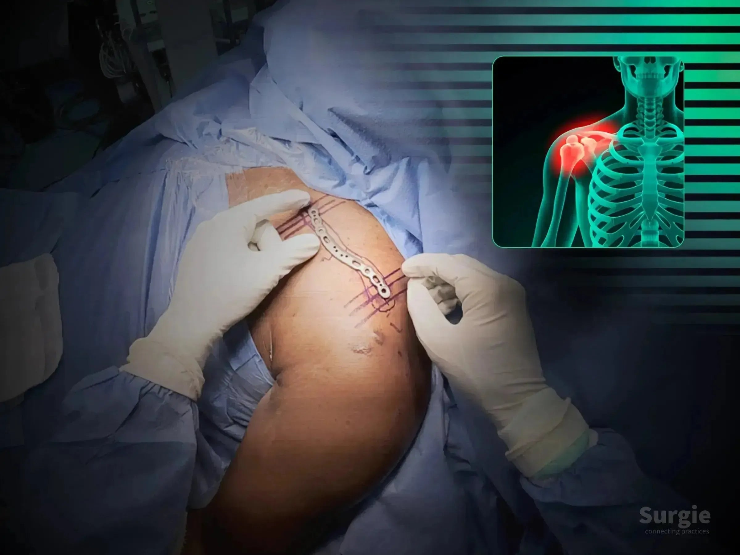
Two-portal clavicle fixation with fluoroscopic reduction
डॉ. सावीओ चामीऑर्थोपेडिस्ट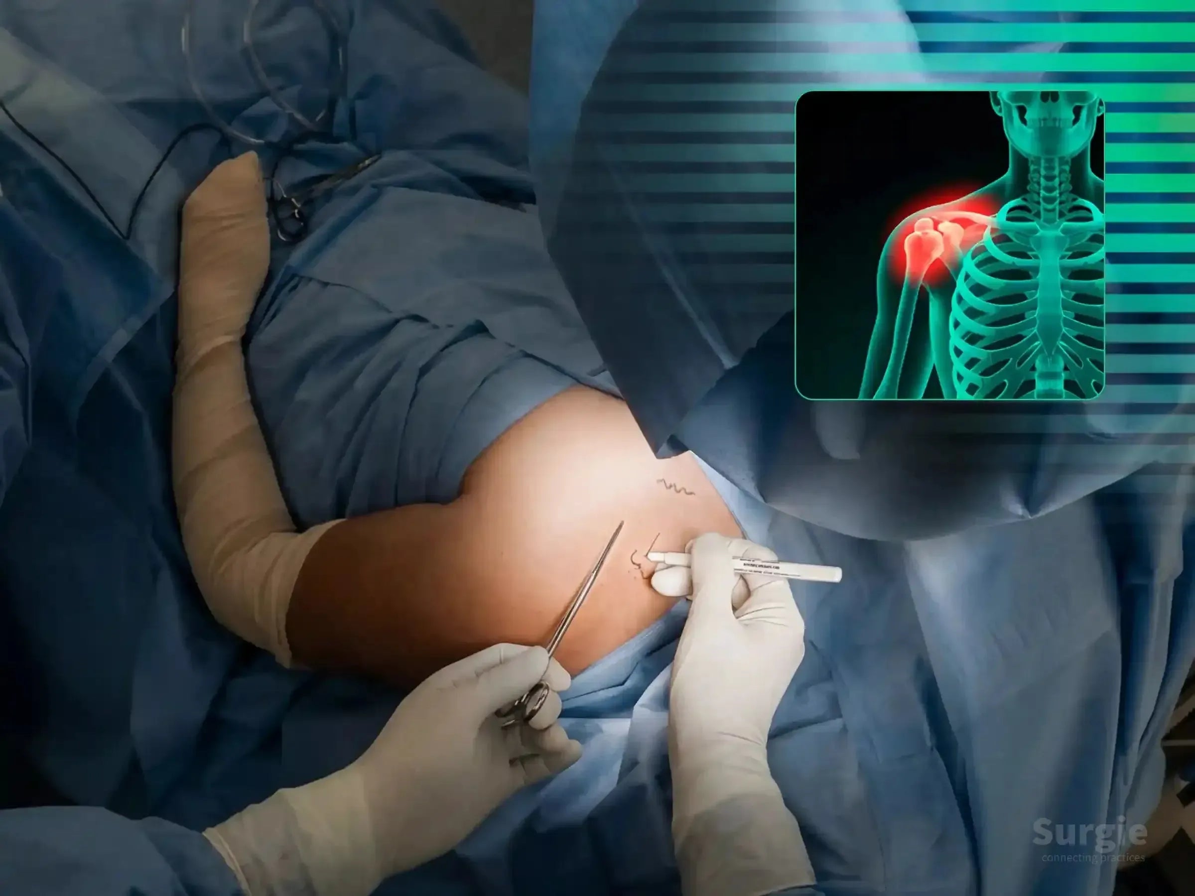
Two-portal percutaneous plating with fluoroscopic guidance
डॉ. सावीओ चामीऑर्थोपेडिस्ट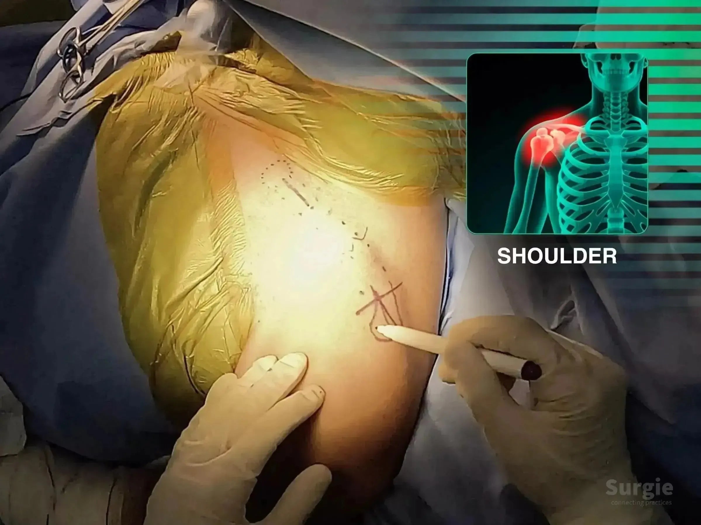
Two-portal bridge plating with soft-tissue preservation
डॉ. सावीओ चामीऑर्थोपेडिस्ट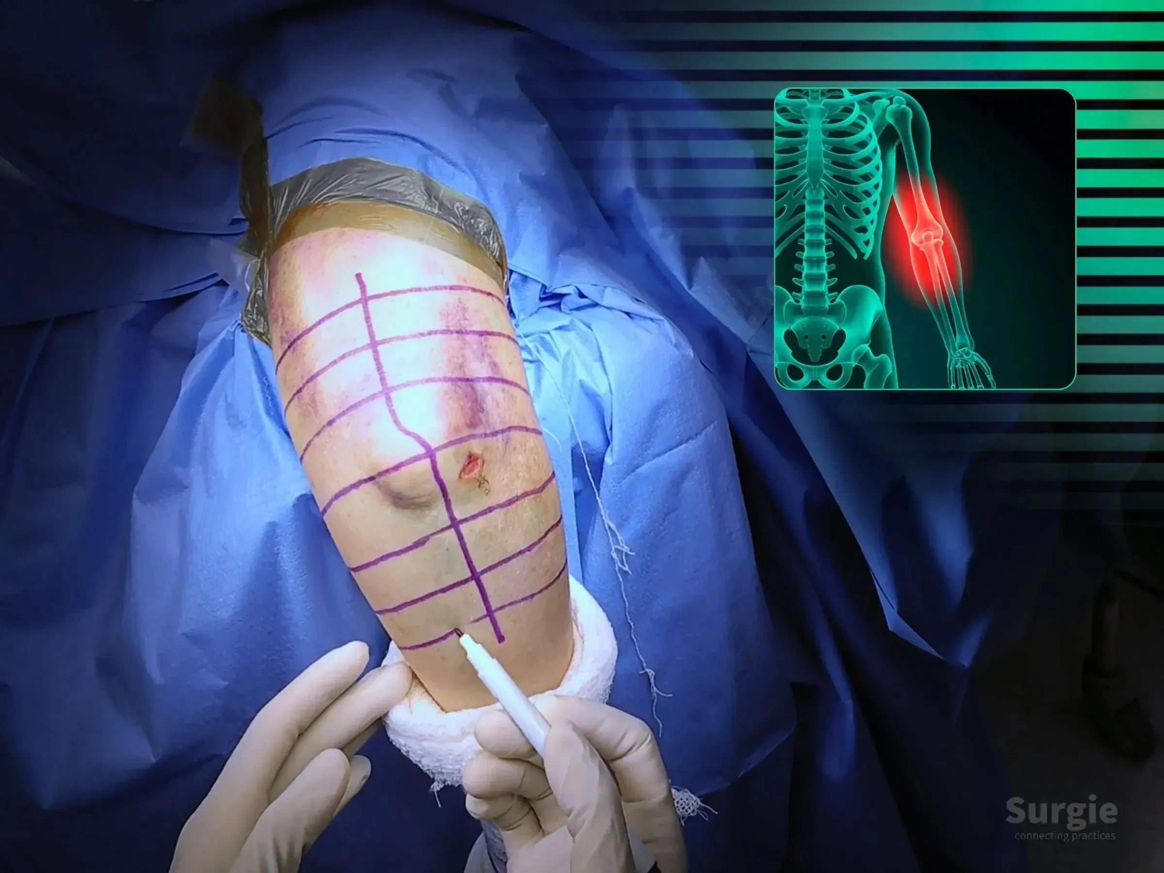
Olecranon osteotomy, articular reduction, and dual-column fixation
डॉ. सावीओ चामीऑर्थोपेडिस्ट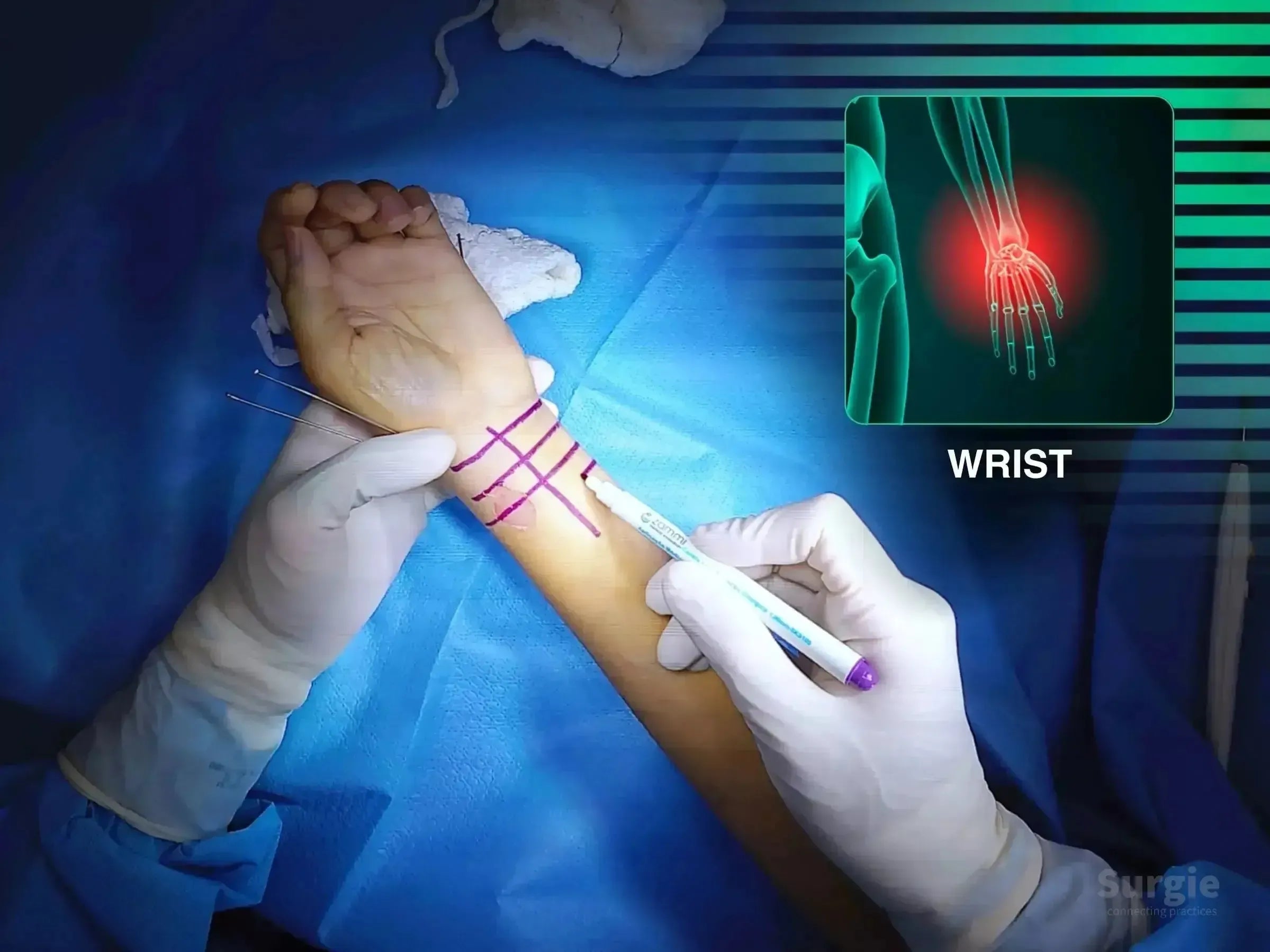
Restore radial length and articular congruity through volar plating
डॉ. सावीओ चामीऑर्थोपेडिस्ट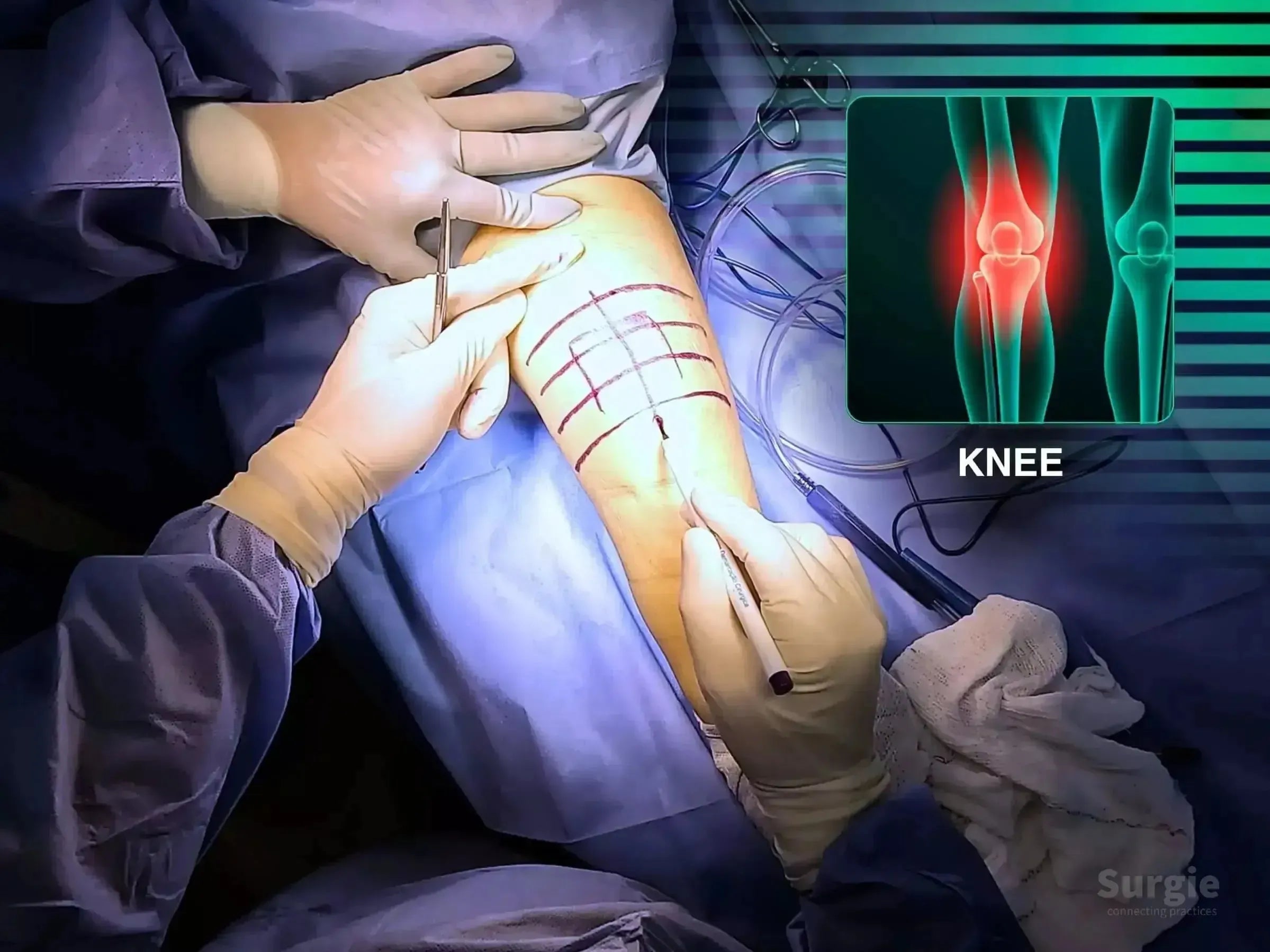
Cannulated screws and cerclage for extensor mechanism reconstruction
डॉ. सावीओ चामीऑर्थोपेडिस्ट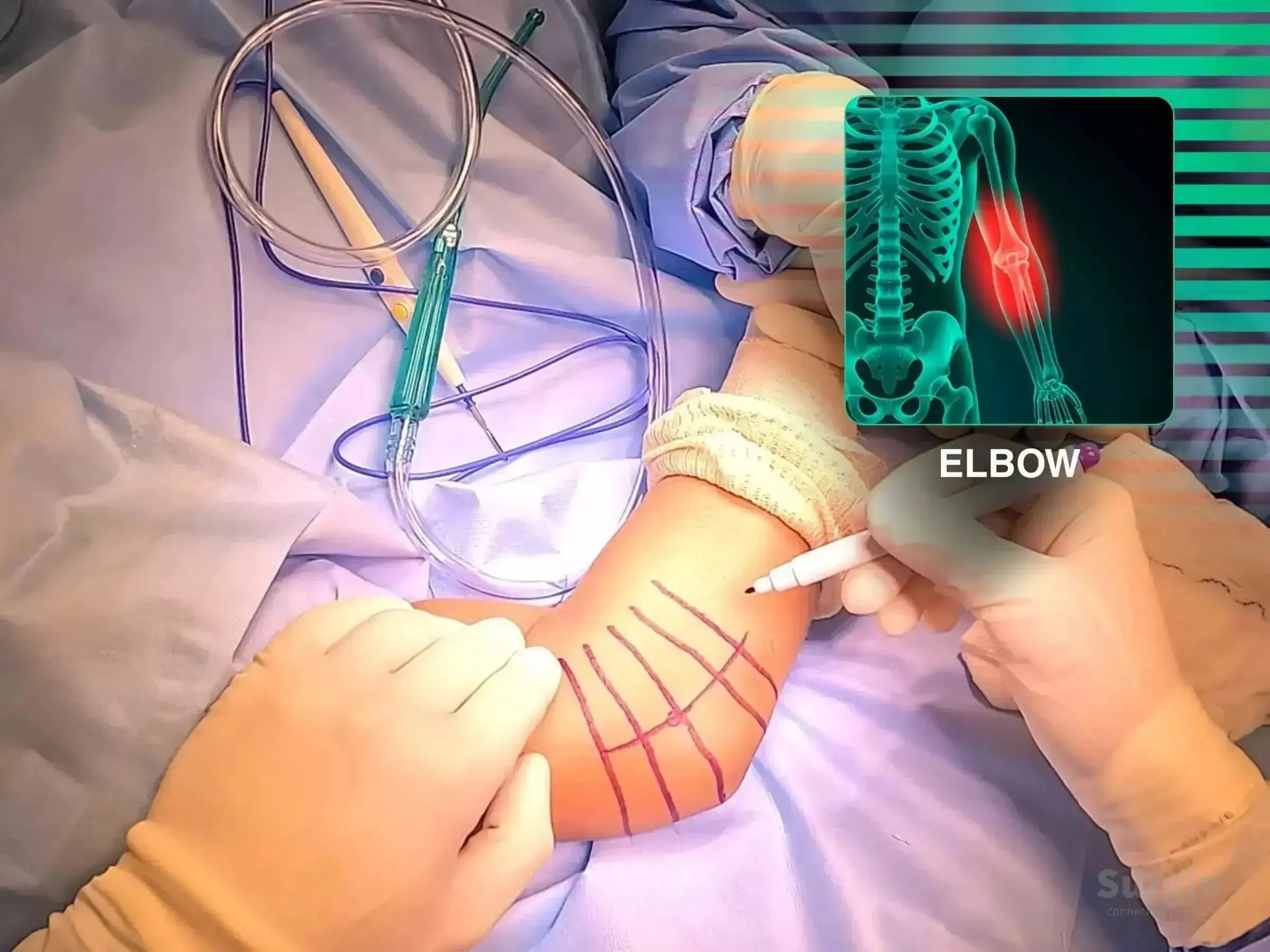
Radial head replacement with ligament repair and stability testing
डॉ. सावीओ चामीऑर्थोपेडिस्ट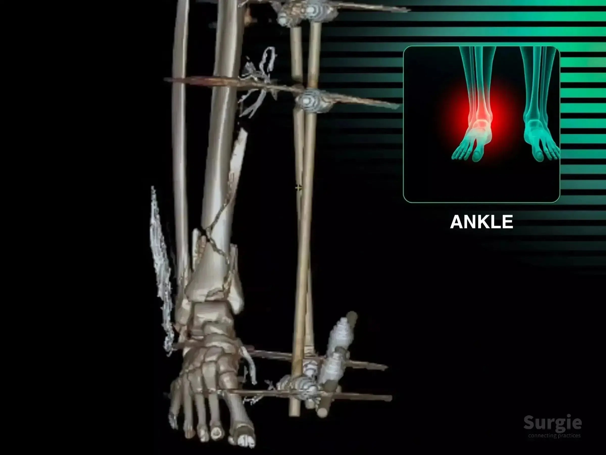
Staged pilon reconstruction with articular reduction and multiplanar fixation
डॉ. सावीओ चामीऑर्थोपेडिस्ट
Achilles, posterior tibial tendon, deltoid, and syndesmosis reconstruction
डॉ. सावीओ चामीऑर्थोपेडिस्ट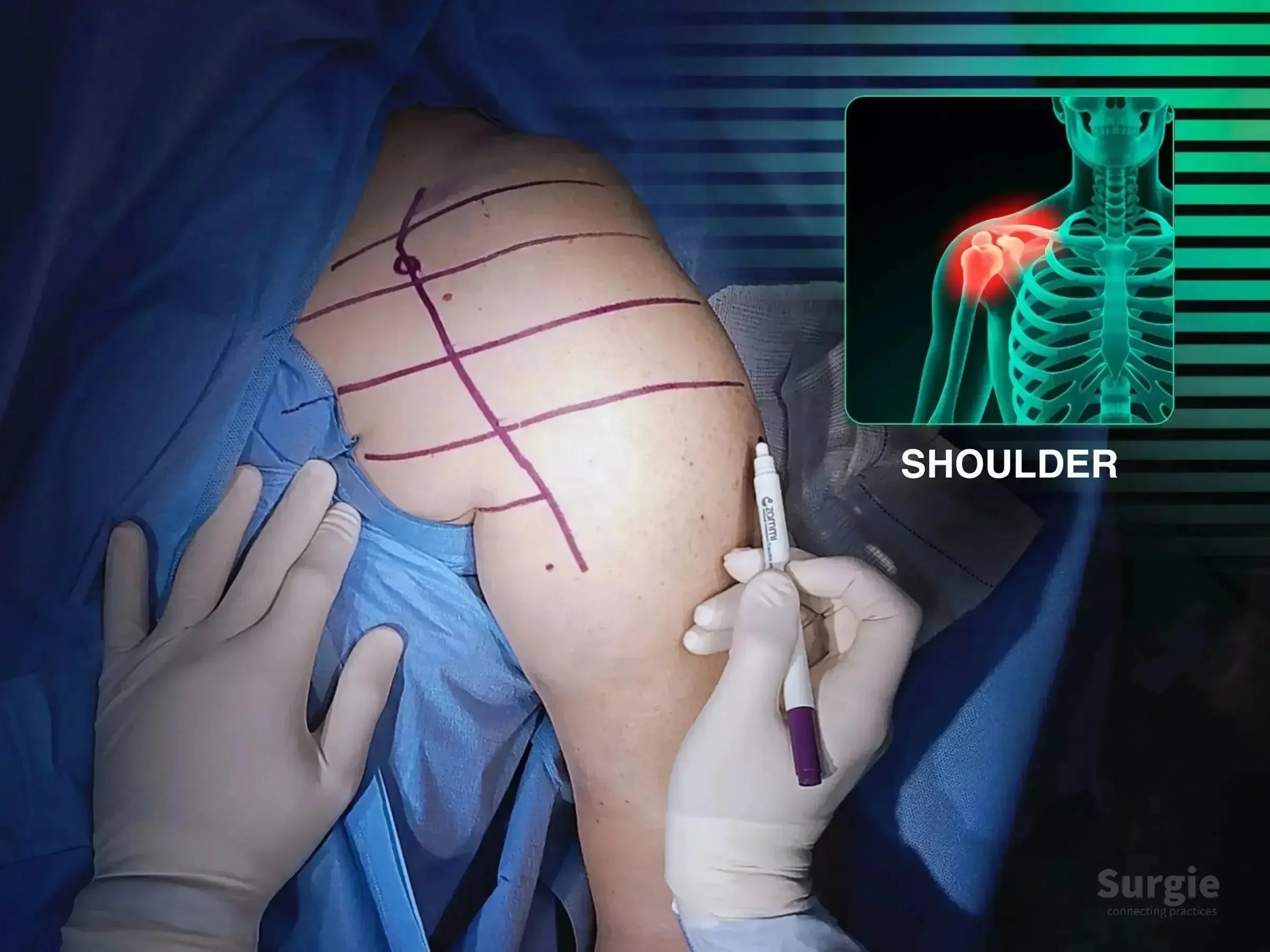
Four-part reconstruction with valgus correction, plate, and suture fixation
डॉ. सावीओ चामीऑर्थोपेडिस्ट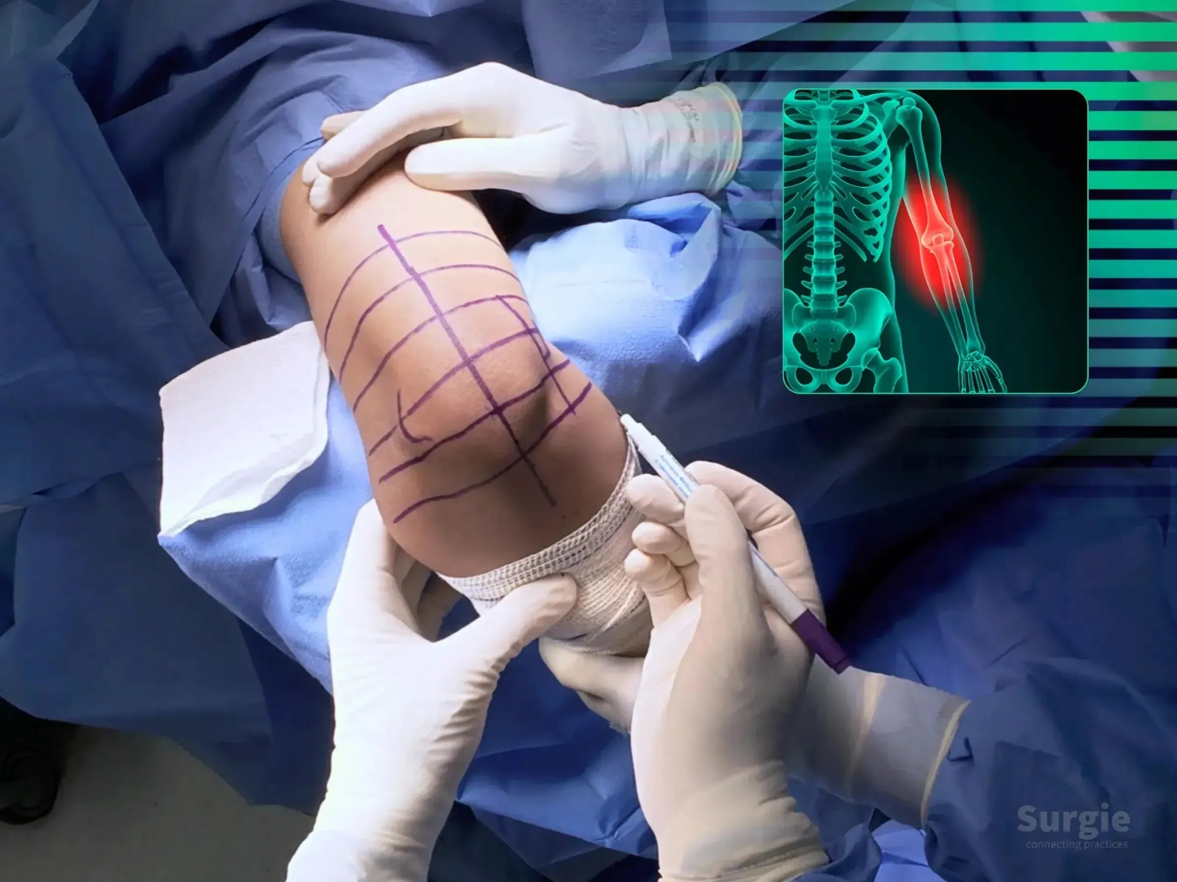
3D-guided corrective osteotomy with ulnar nerve transposition
डॉ. सावीओ चामीऑर्थोपेडिस्ट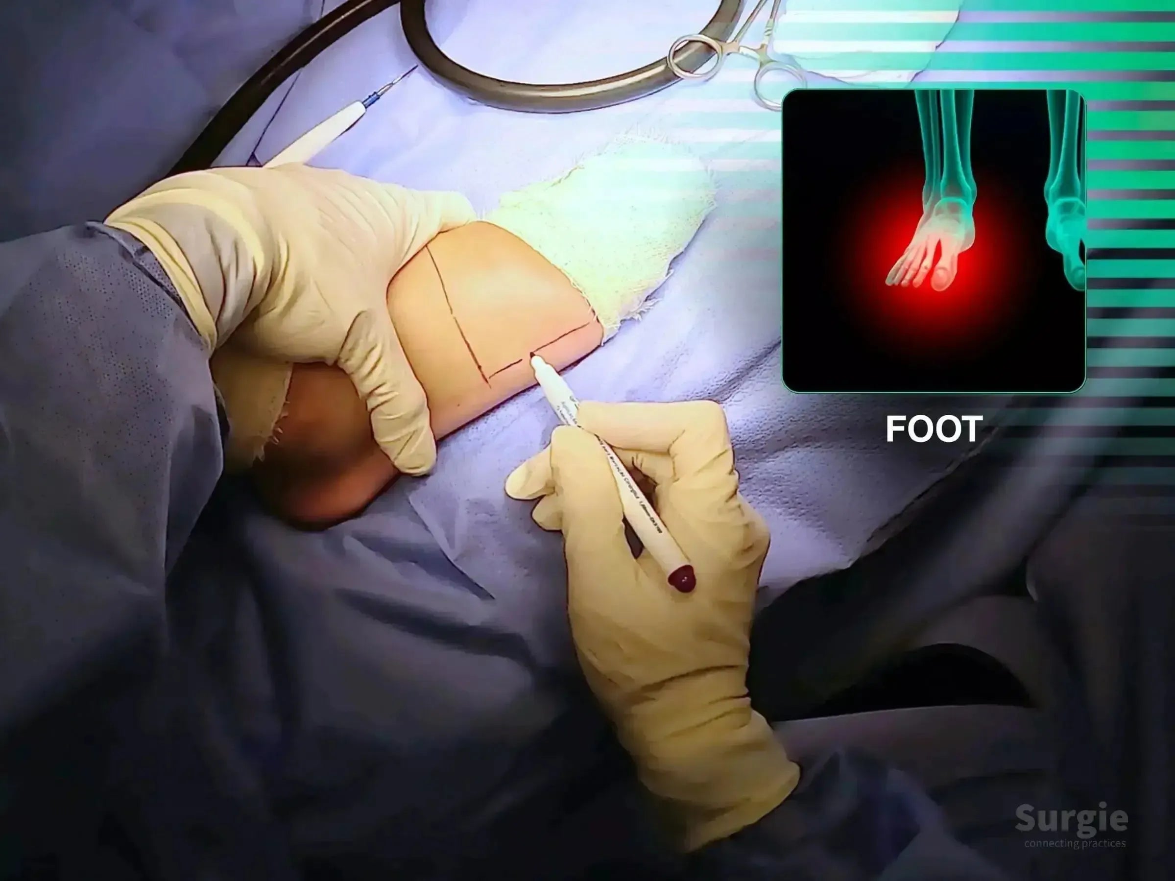
Anatomic reduction and low-profile plate fixation of the fifth metatarsal
डॉ. सावीओ चामीऑर्थोपेडिस्ट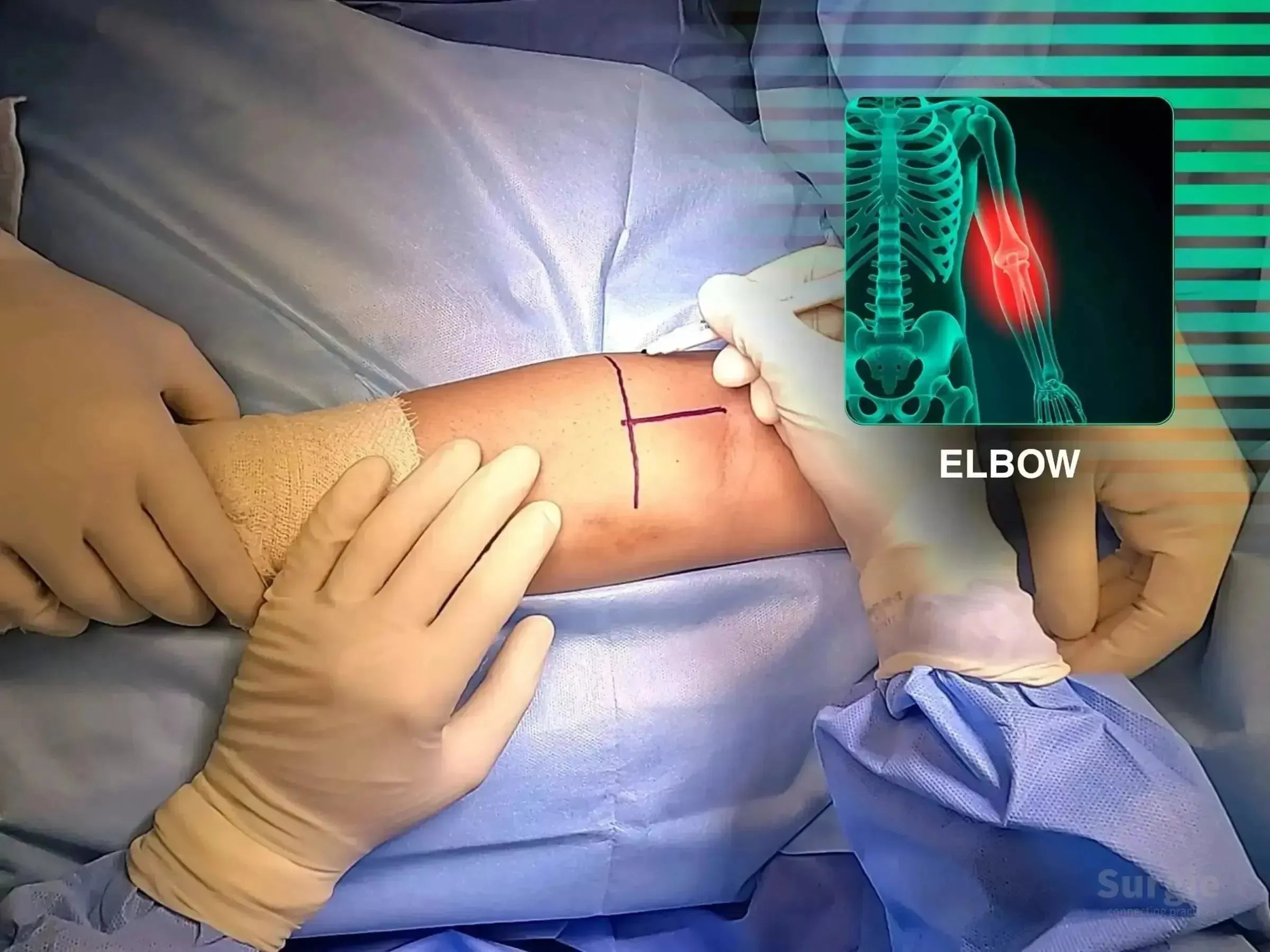
TightRope reinsertion at the radial tuberosity with tissue preservation
डॉ. सावीओ चामीऑर्थोपेडिस्ट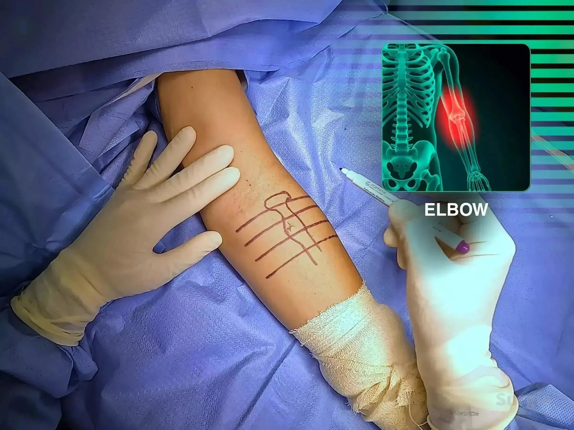
TightRope tendon repair with focused neurovascular protection
डॉ. सावीओ चामीऑर्थोपेडिस्ट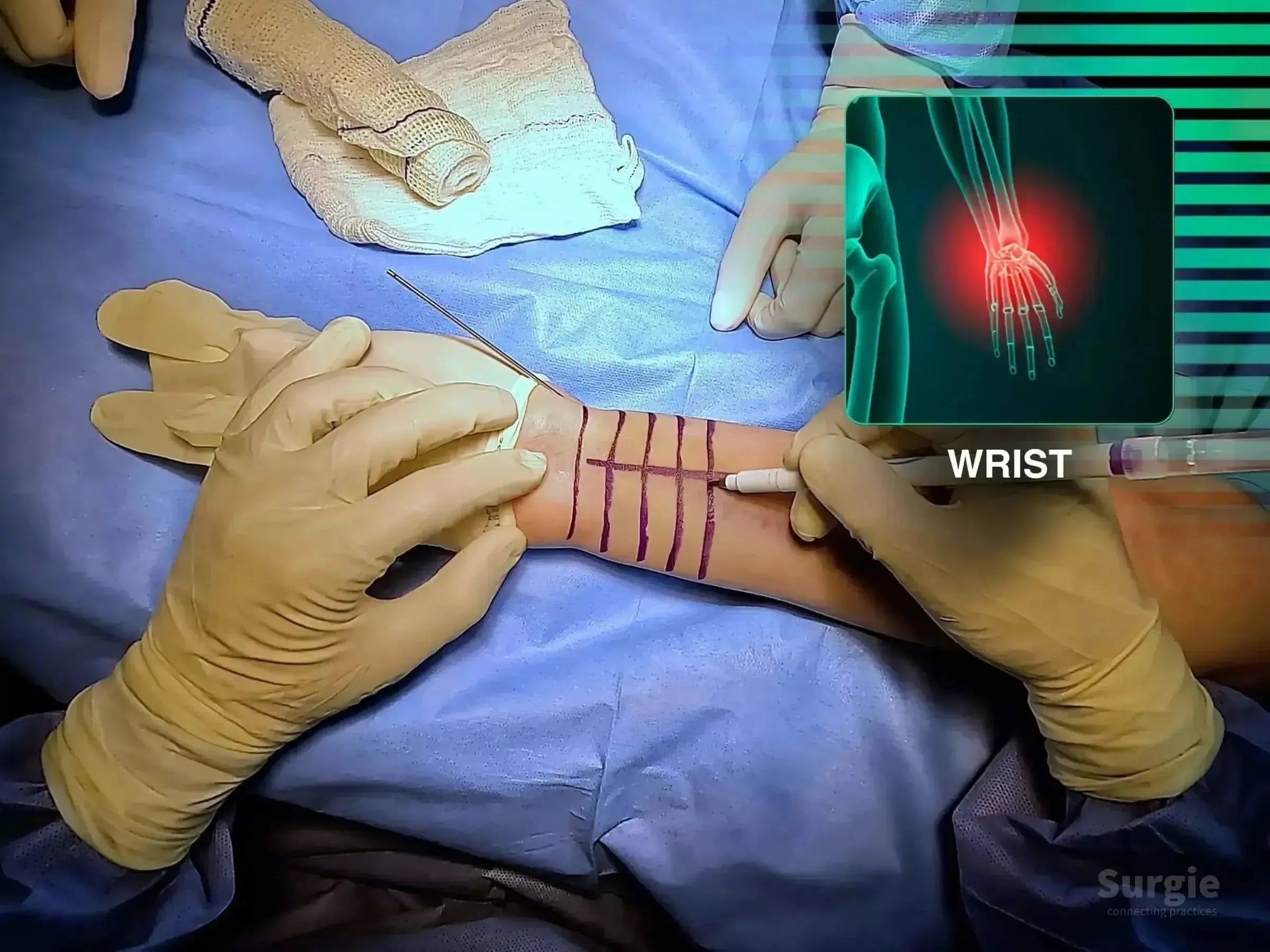
Volar exposure, provisional reduction, and plate stabilization
डॉ. सावीओ चामीऑर्थोपेडिस्ट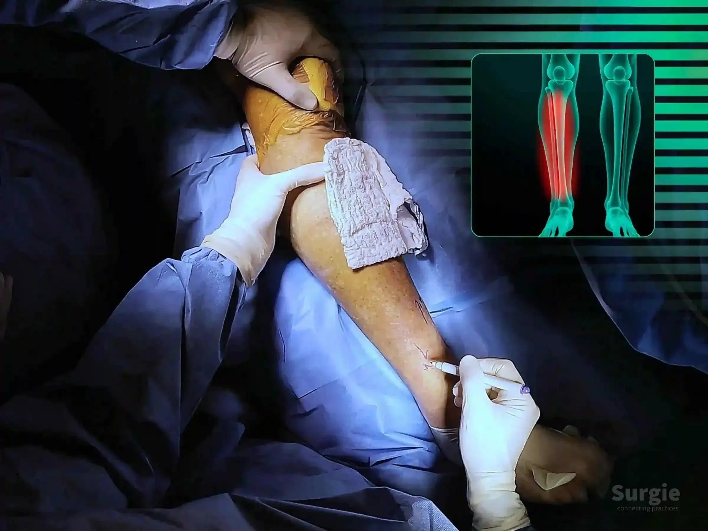
Percutaneous reduction and minimally invasive locked plate fixation
डॉ. सावीओ चामीऑर्थोपेडिस्ट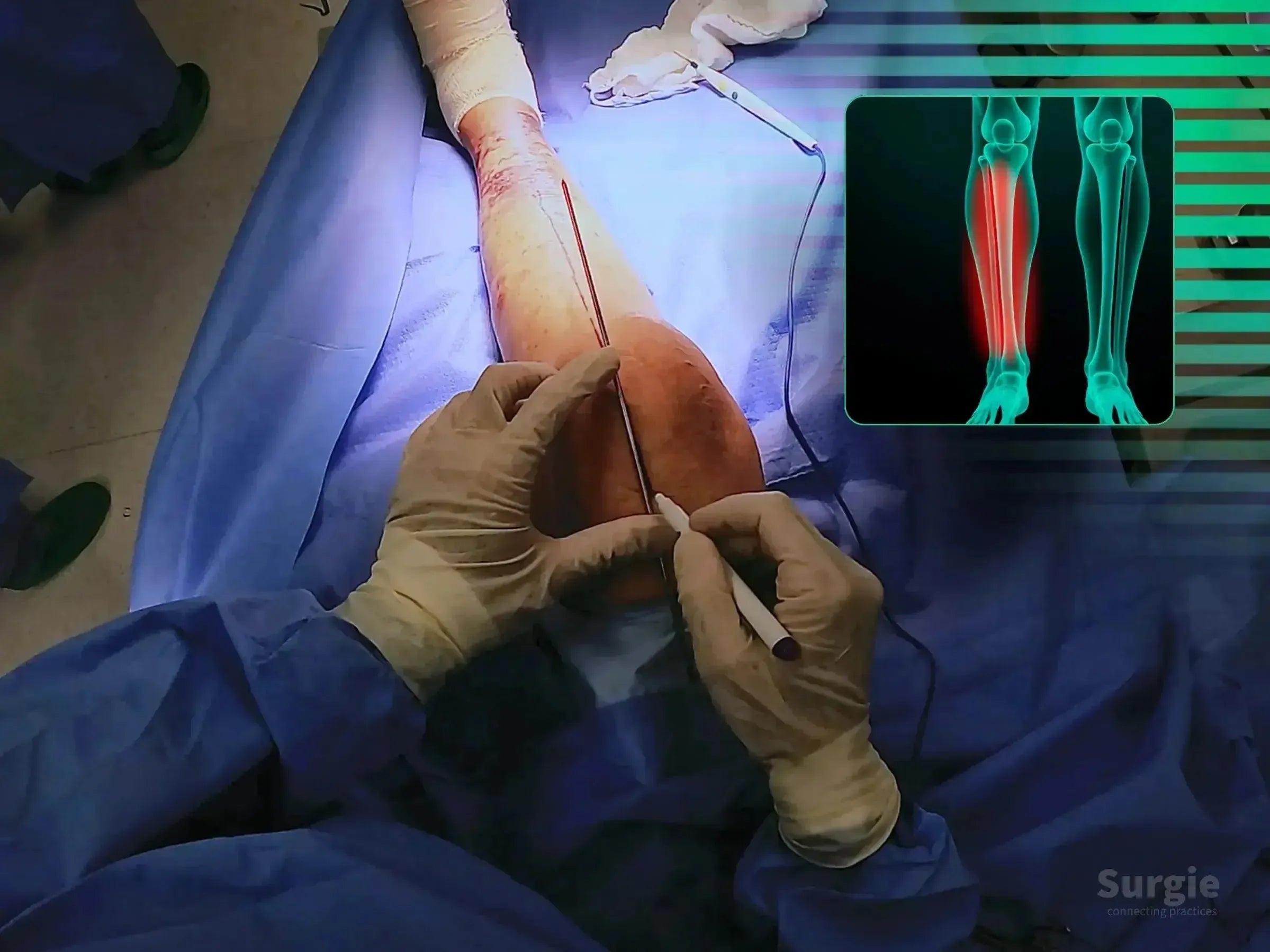
Suprapatellar nailing with alignment control and soft-tissue protection
डॉ. सावीओ चामीऑर्थोपेडिस्ट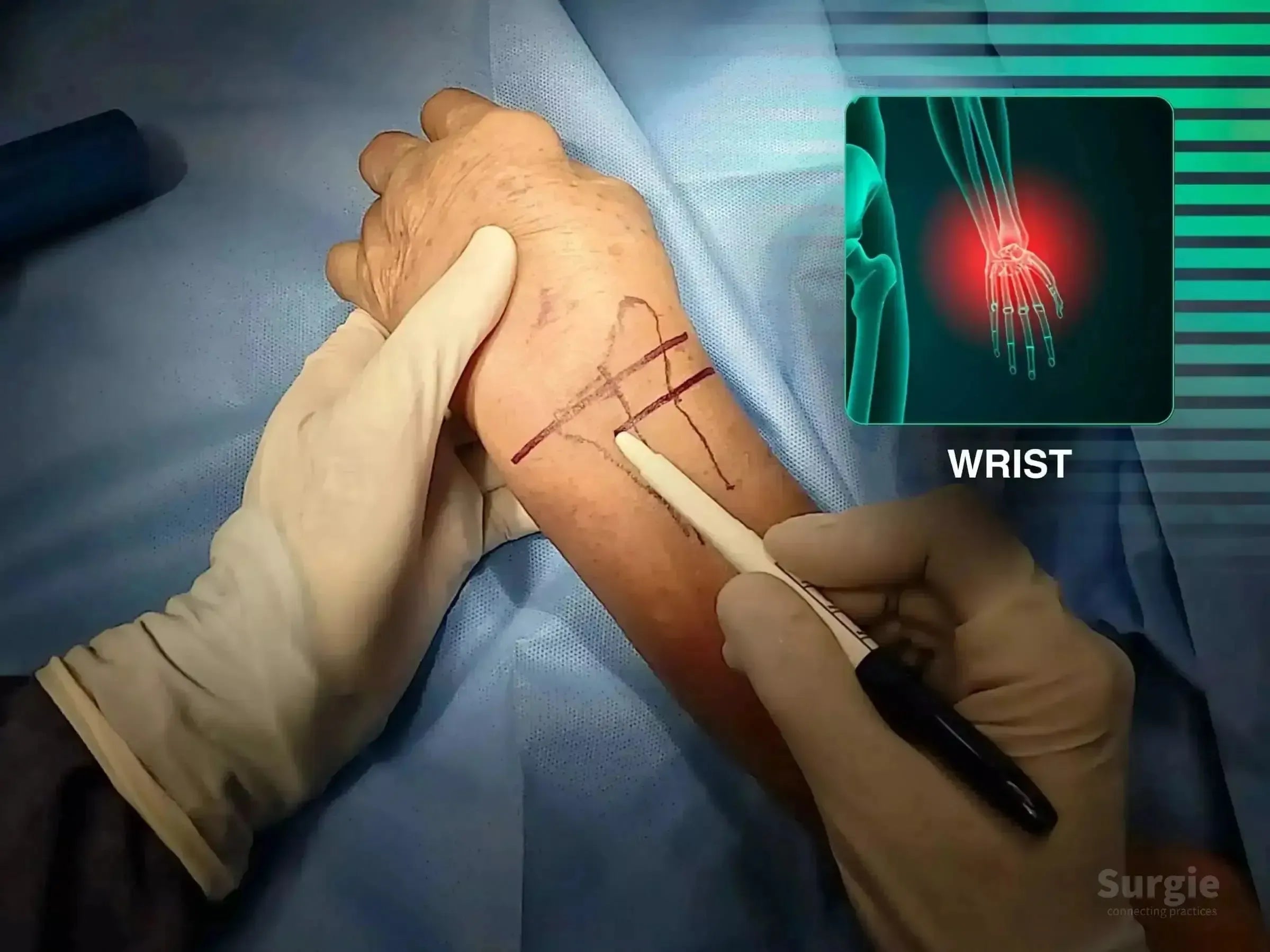
Dorsal fragment reduction with targeted radius fixation
डॉ. सावीओ चामीऑर्थोपेडिस्ट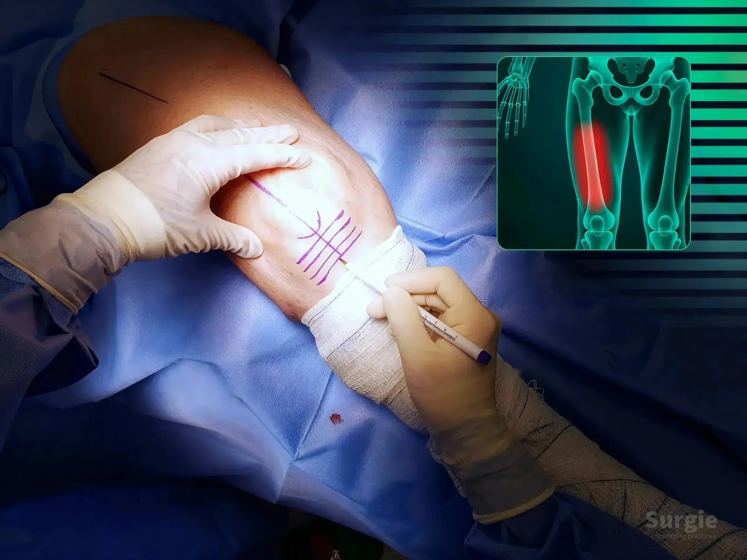
Retrograde femoral nailing with rotational and fluoroscopic control
डॉ. सावीओ चामीऑर्थोपेडिस्ट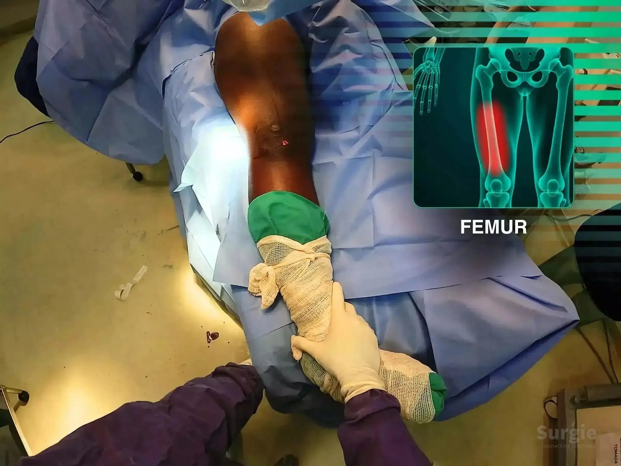
Retrograde nailing with free-limb reduction and fluoroscopic control
डॉ. सावीओ चामीऑर्थोपेडिस्ट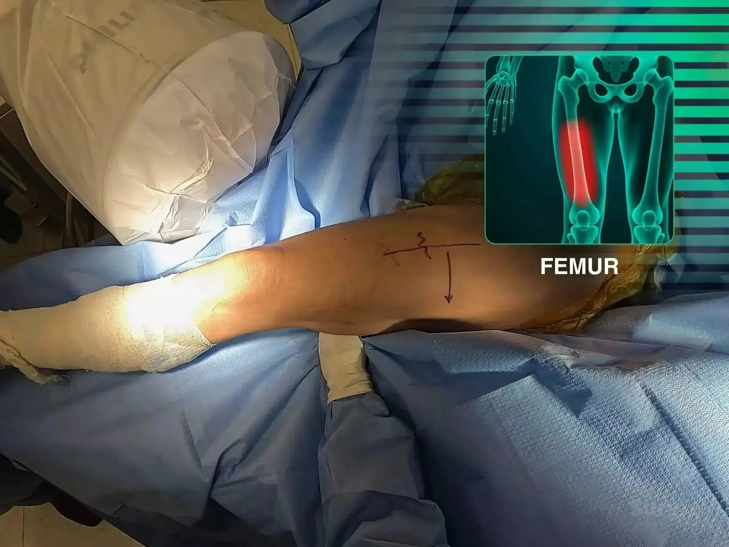
Antegrade nailing without a traction table for femoral shaft refracture
डॉ. सावीओ चामीऑर्थोपेडिस्ट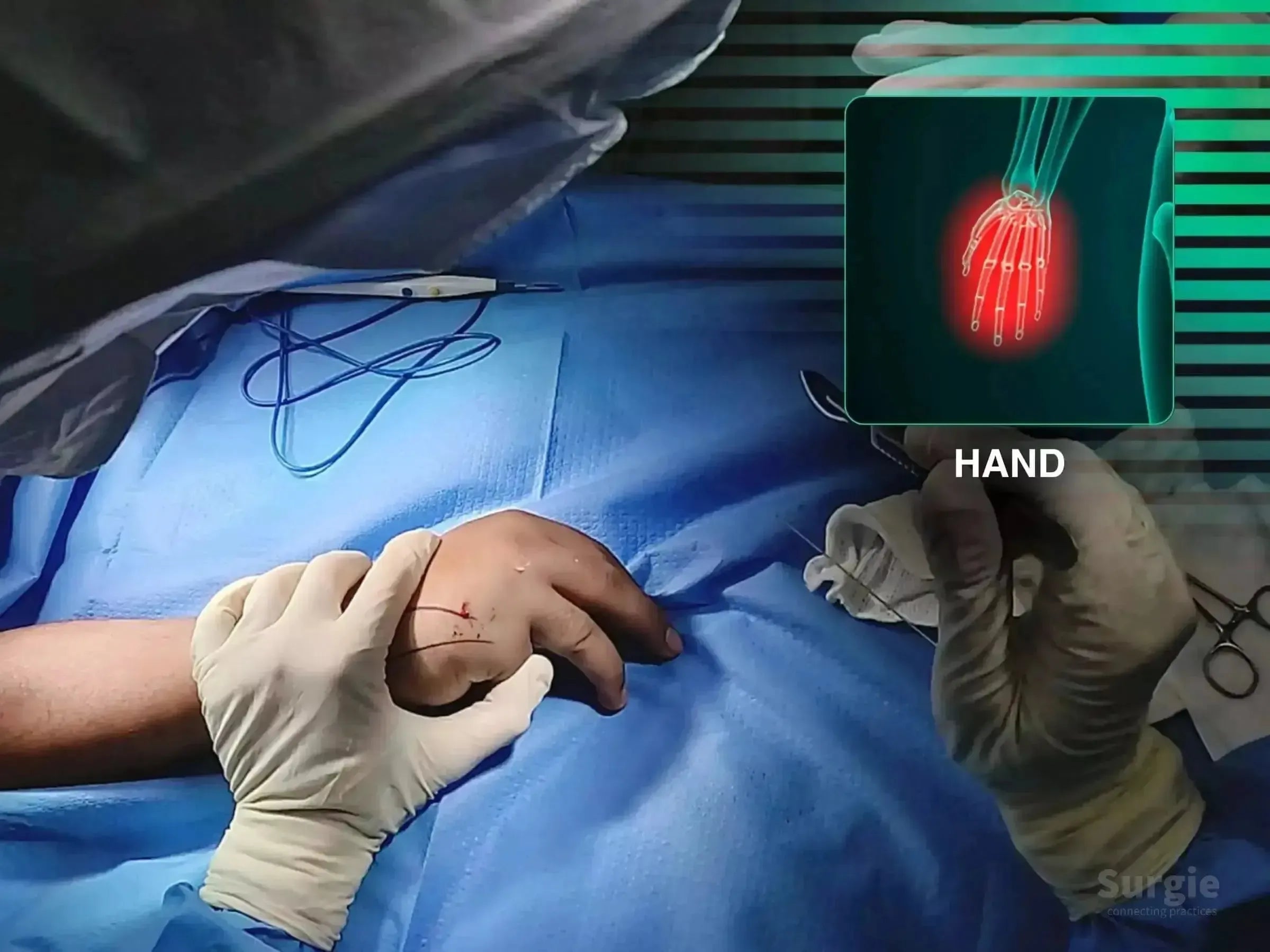
Minimally invasive double-compression fixation of the fifth metacarpal
डॉ. सावीओ चामीऑर्थोपेडिस्ट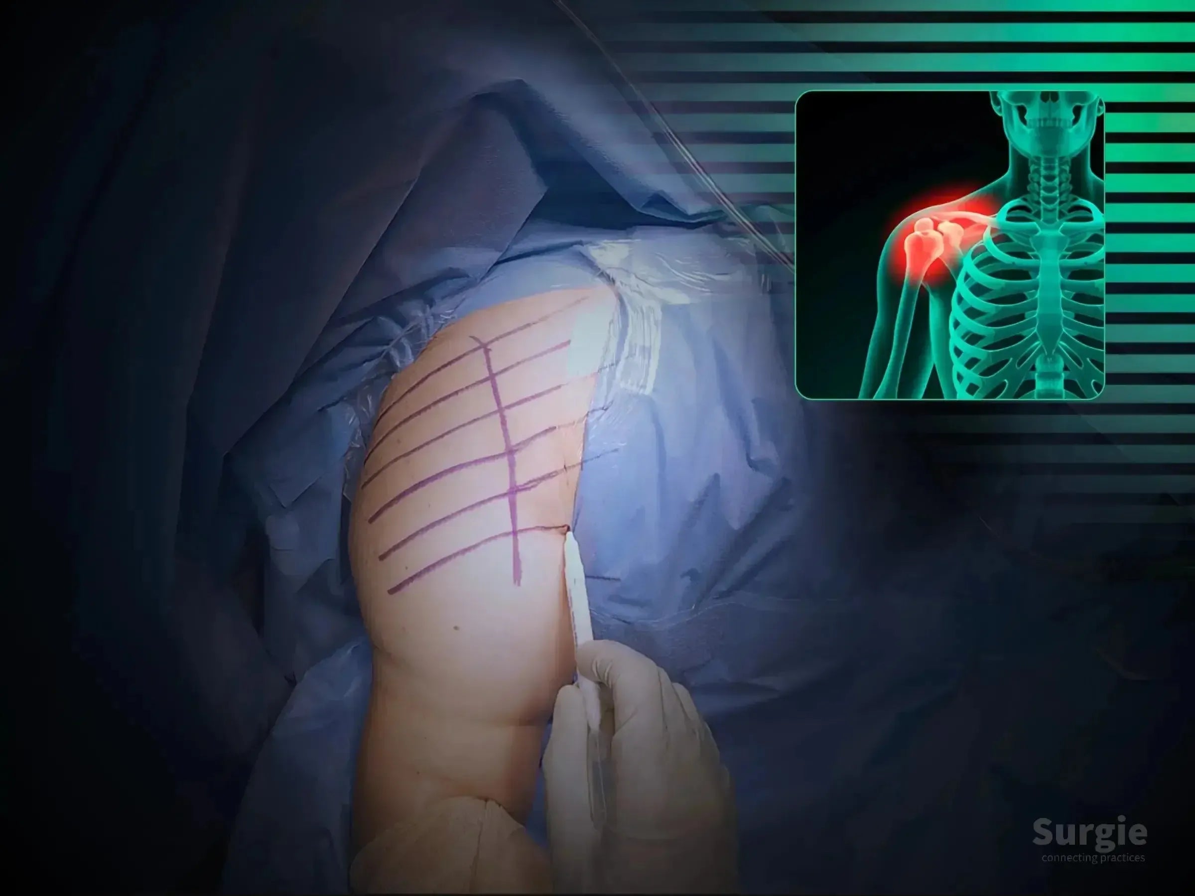
Valgus-impacted head reduction with plate, calcar screws, and FiberTape
डॉ. सावीओ चामीऑर्थोपेडिस्ट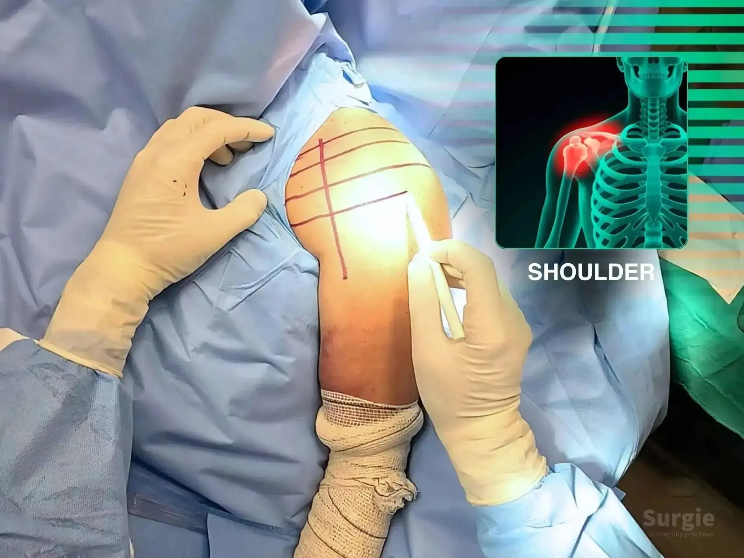
Tuberosity reconstruction with plate-and-screw fixation
डॉ. सावीओ चामीऑर्थोपेडिस्ट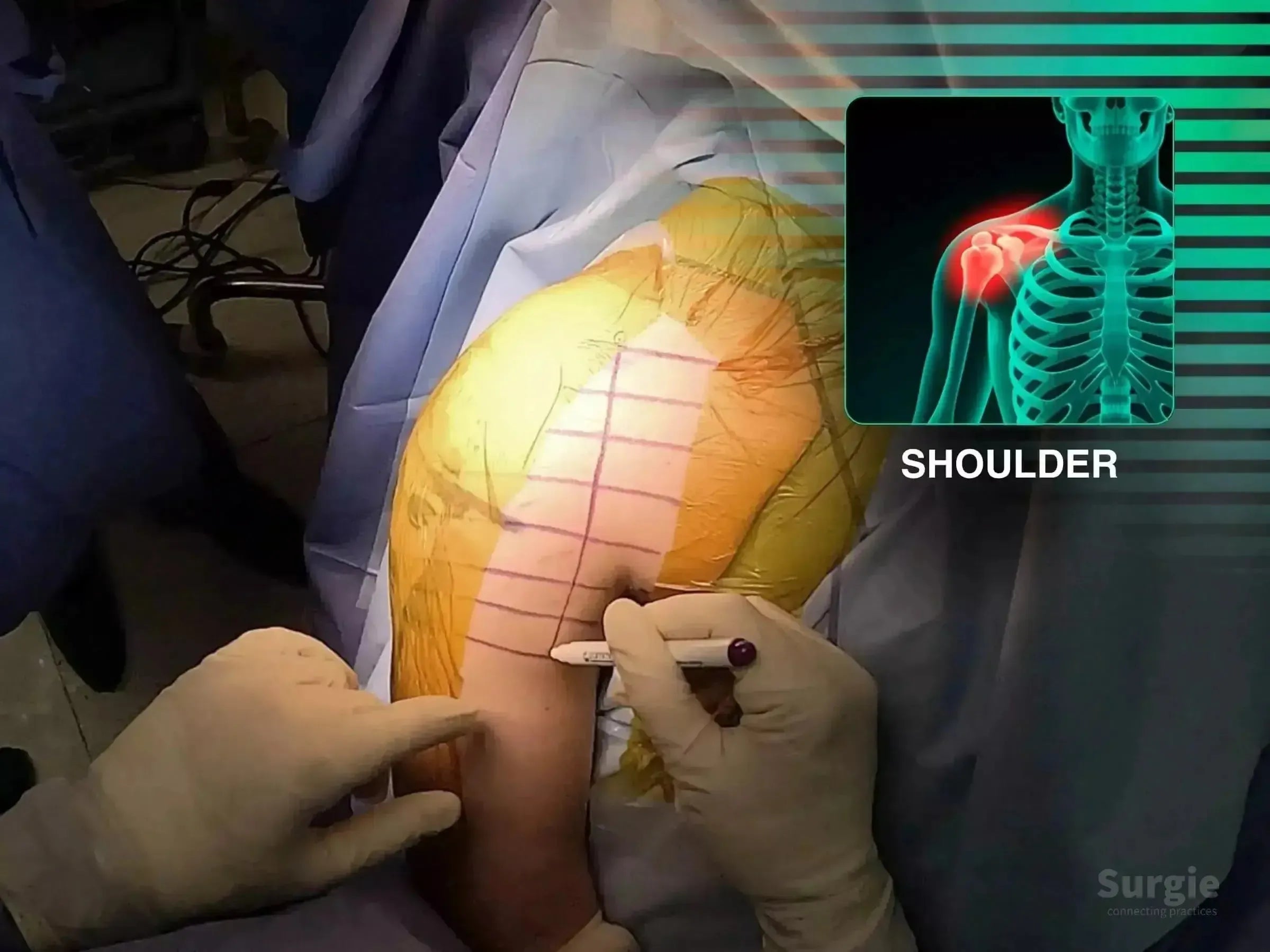
Anterior glenoid exposure with compression screw fixation
डॉ. सावीओ चामीऑर्थोपेडिस्ट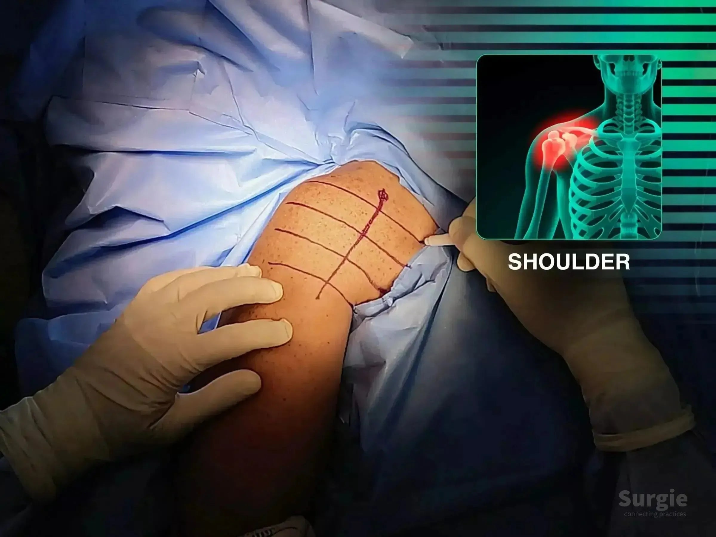
Direct tuberosity reduction with locking plate and suture reinforcement
डॉ. सावीओ चामीऑर्थोपेडिस्ट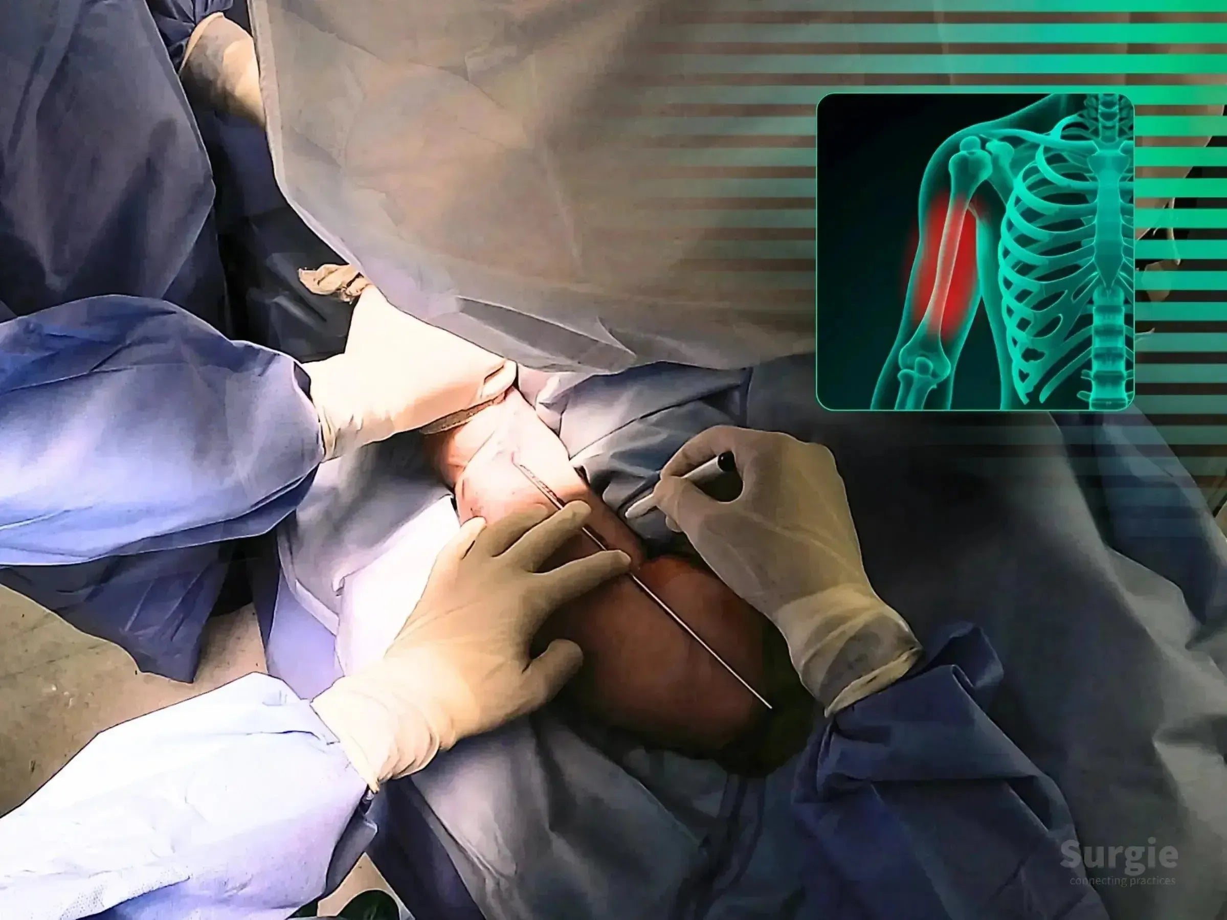
Joystick-assisted reduction with proximal and distal nail locking
डॉ. सावीओ चामीऑर्थोपेडिस्ट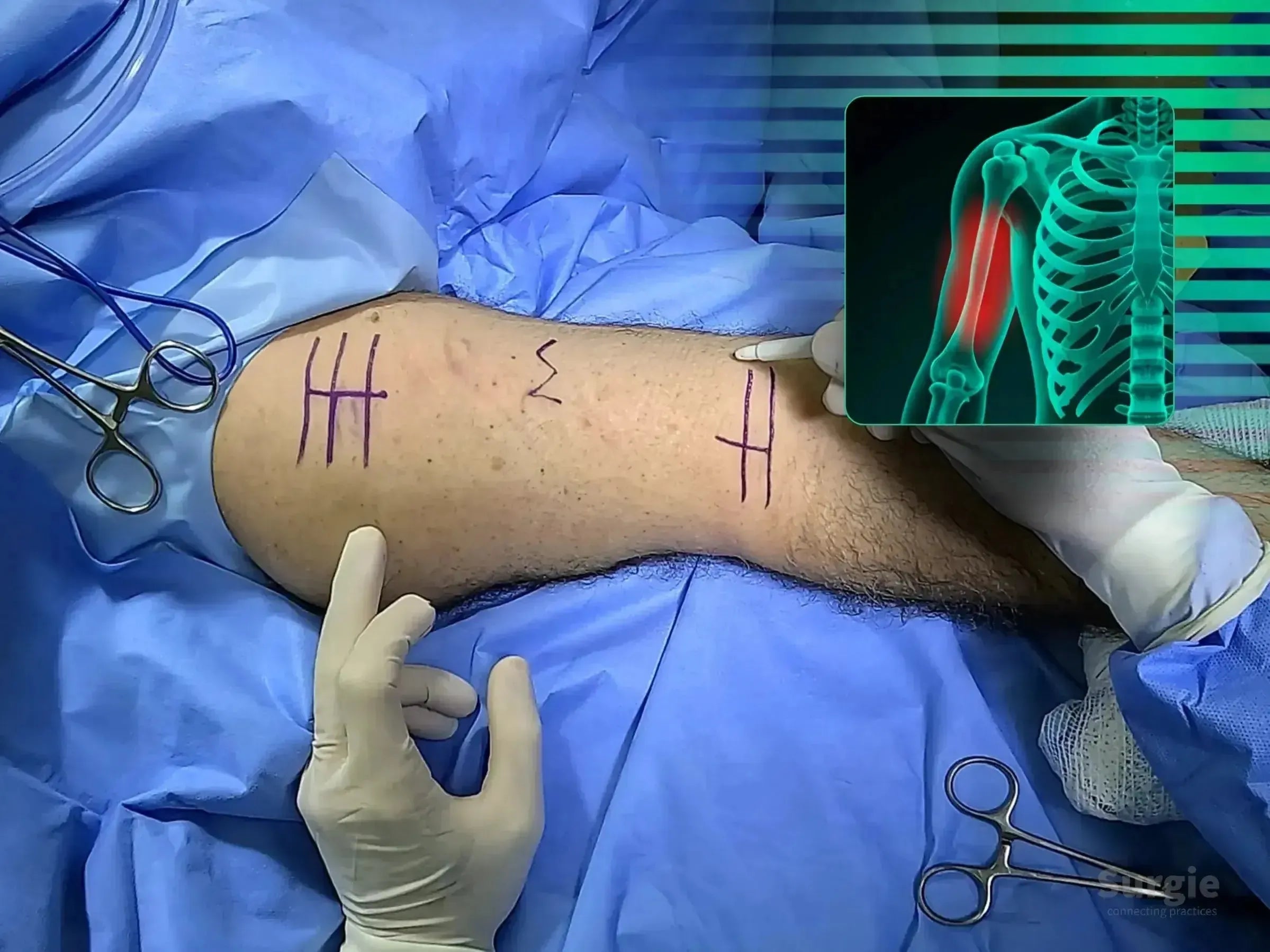
Submuscular bridge plating with radial nerve protection
डॉ. सावीओ चामीऑर्थोपेडिस्ट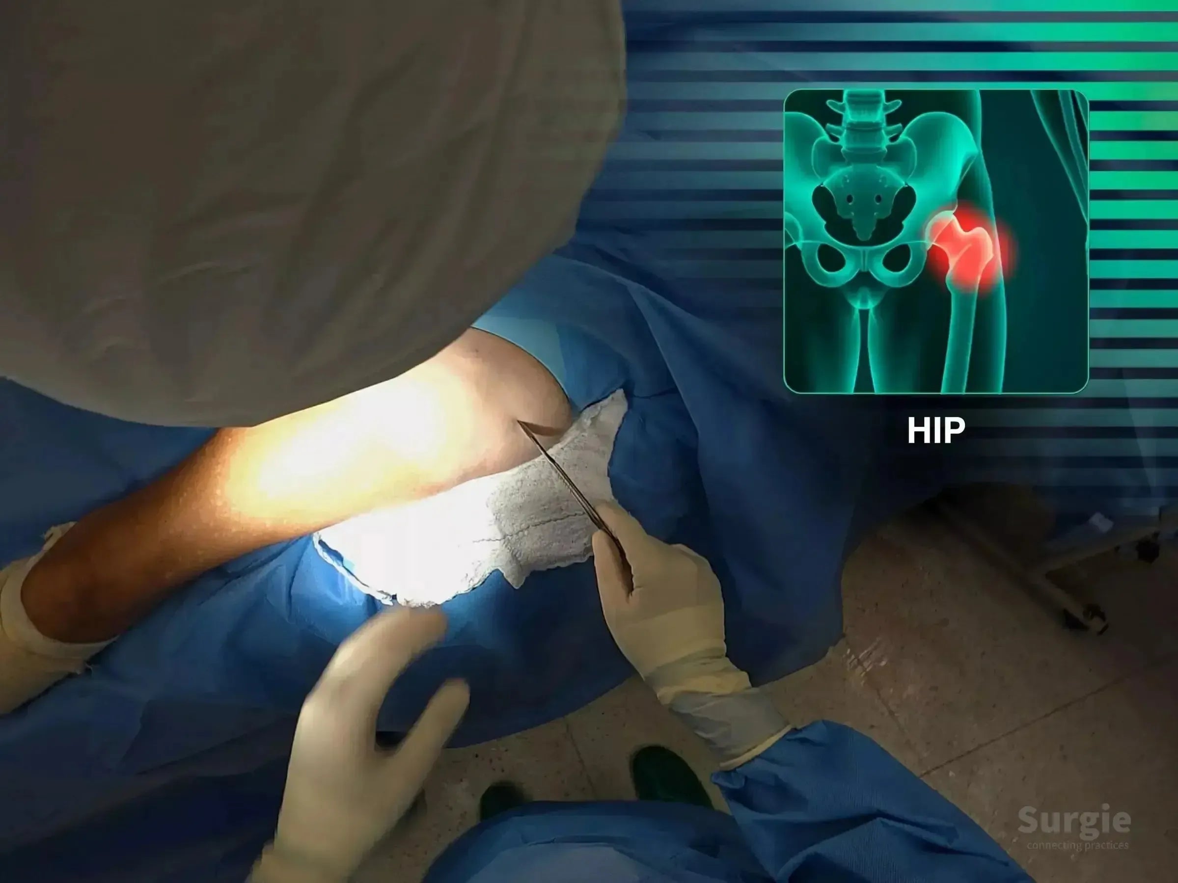
Guidewire precision and intramedullary fixation without traction
डॉ. सावीओ चामीऑर्थोपेडिस्ट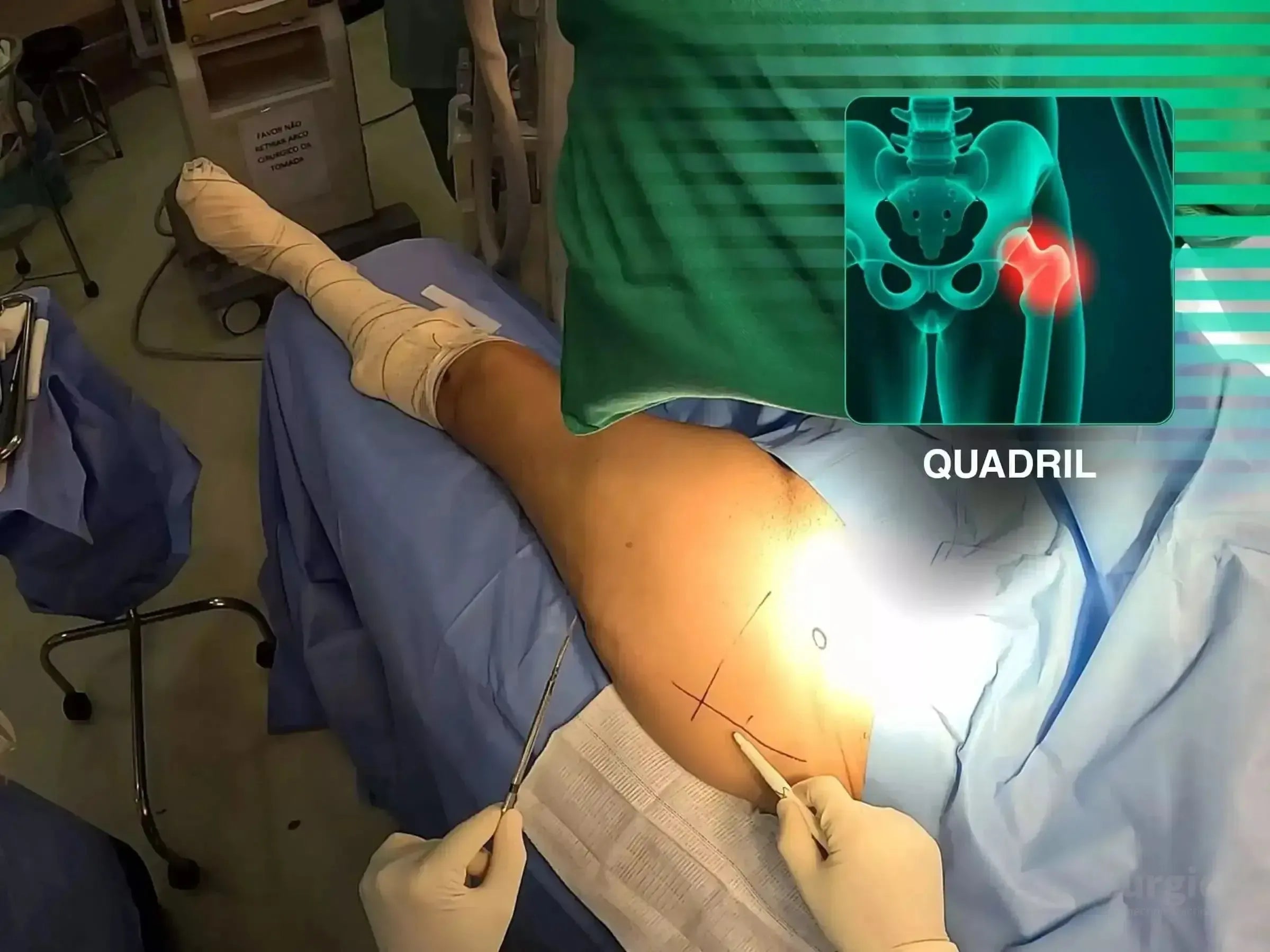
Intertrochanteric nailing without a traction table
डॉ. सावीओ चामीऑर्थोपेडिस्ट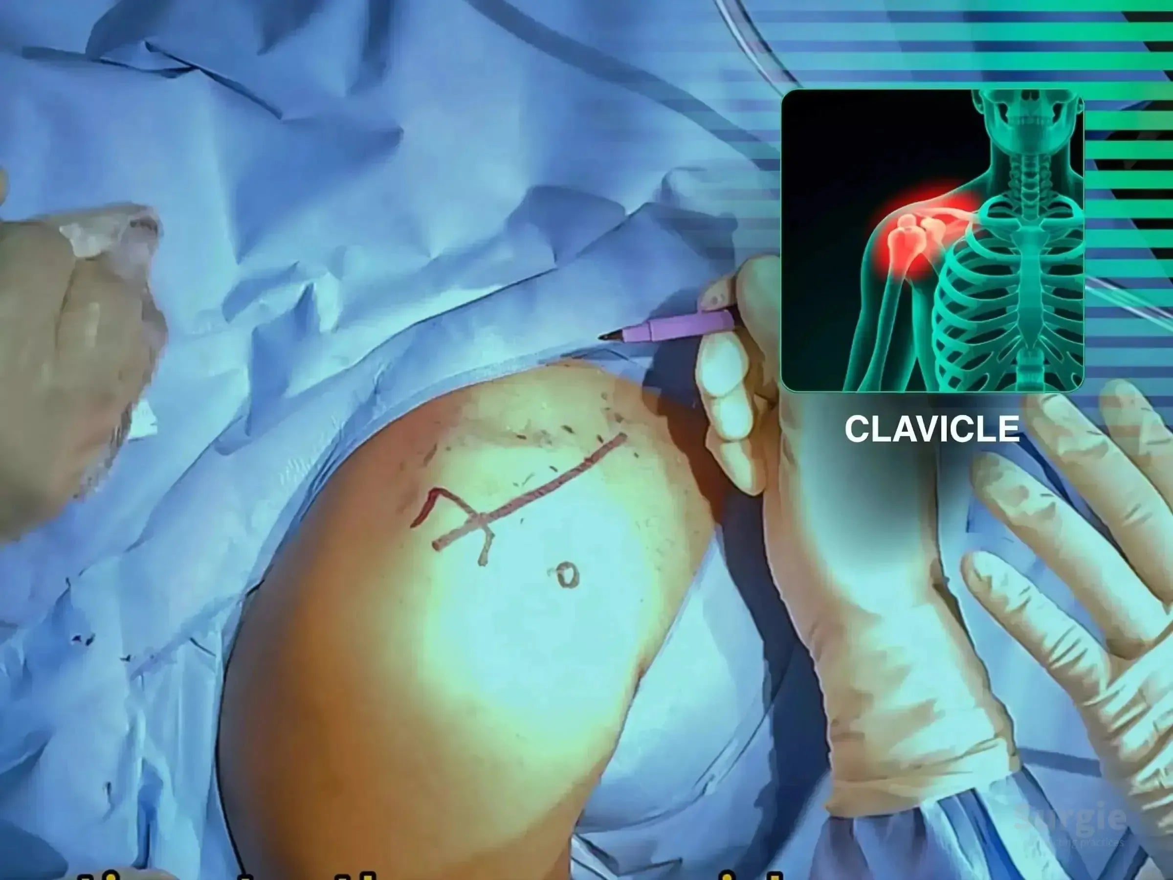
Lateral clavicle reconstruction with coracoclavicular TightRope fixation
डॉ. सावीओ चामीऑर्थोपेडिस्ट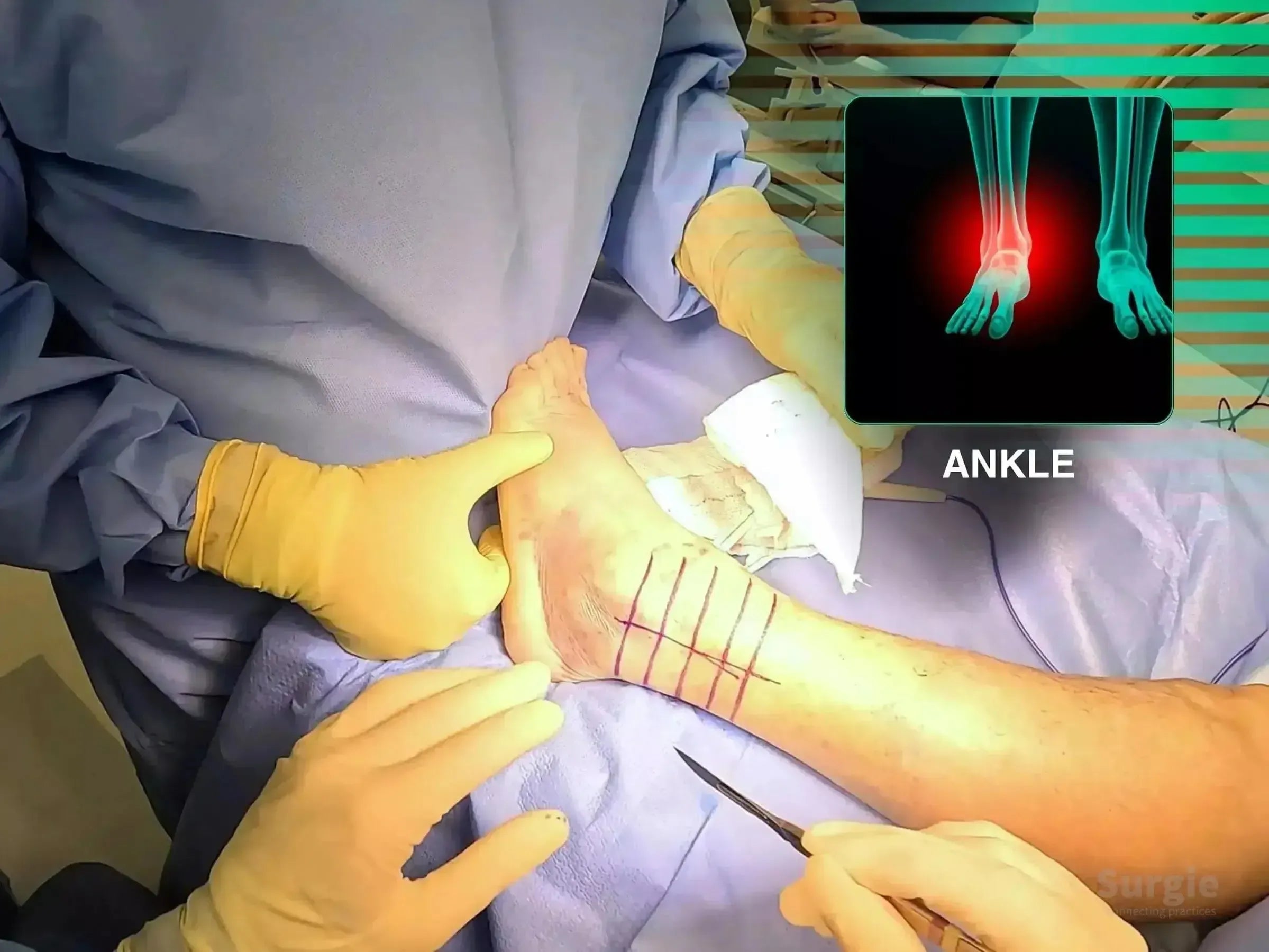
Wagstaffe fragment compression with locked lateral malleolus fixation
डॉ. सावीओ चामीऑर्थोपेडिस्ट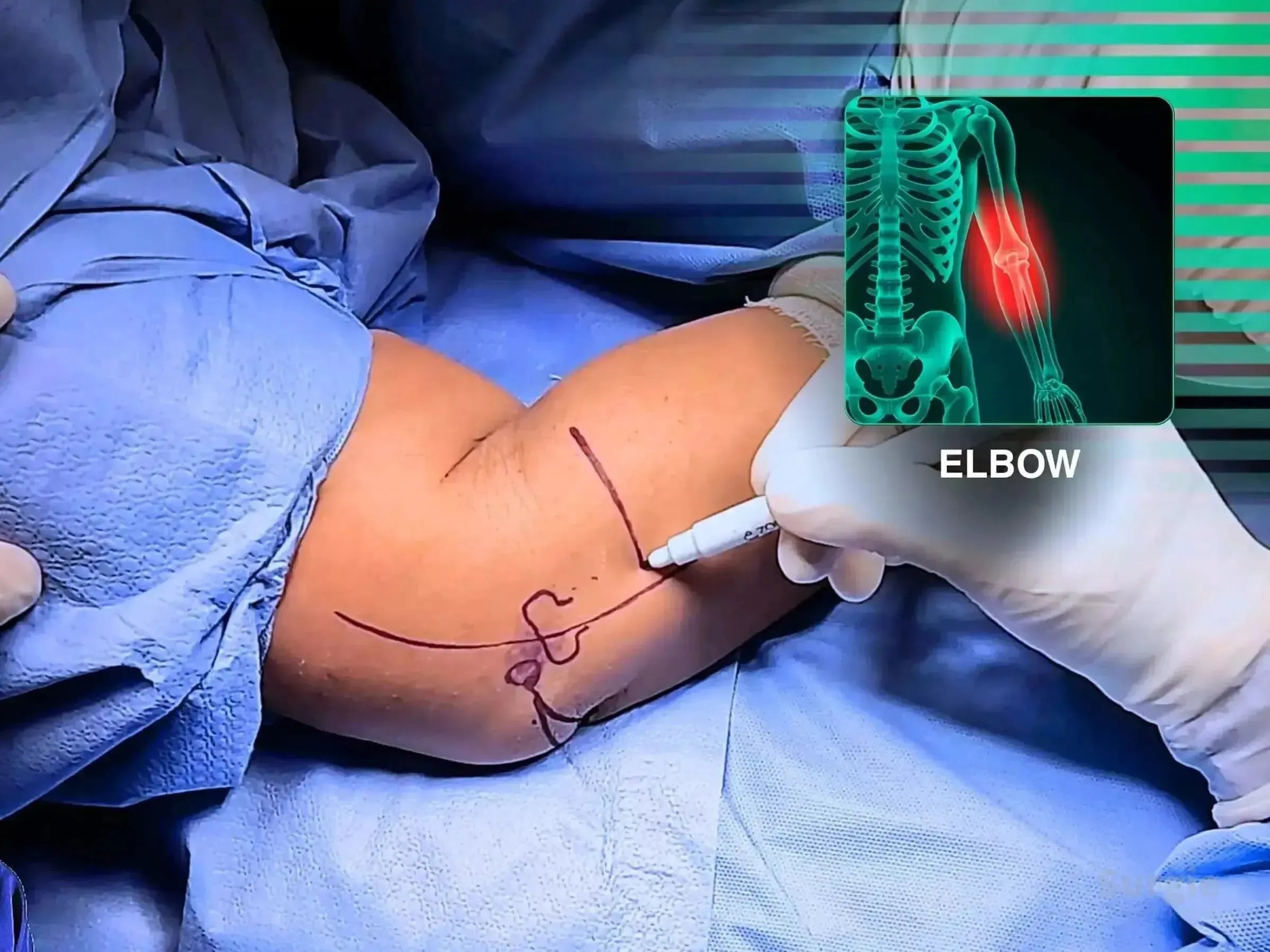
Mini-fragment fixation with coronoid and ligament reconstruction
डॉ. सावीओ चामीऑर्थोपेडिस्ट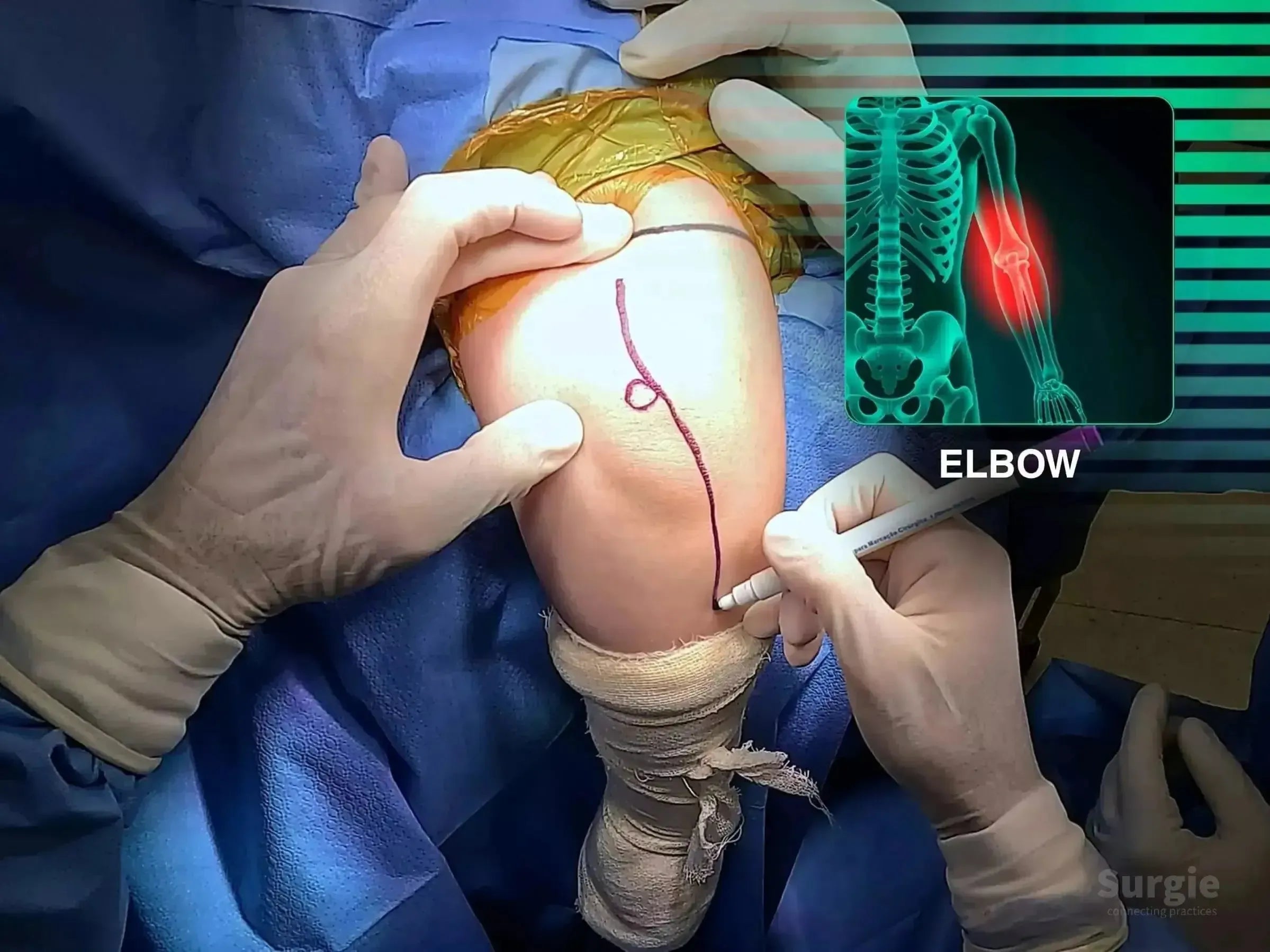
Combined ulna and radial head fixation for Monteggia instability
डॉ. सावीओ चामीऑर्थोपेडिस्ट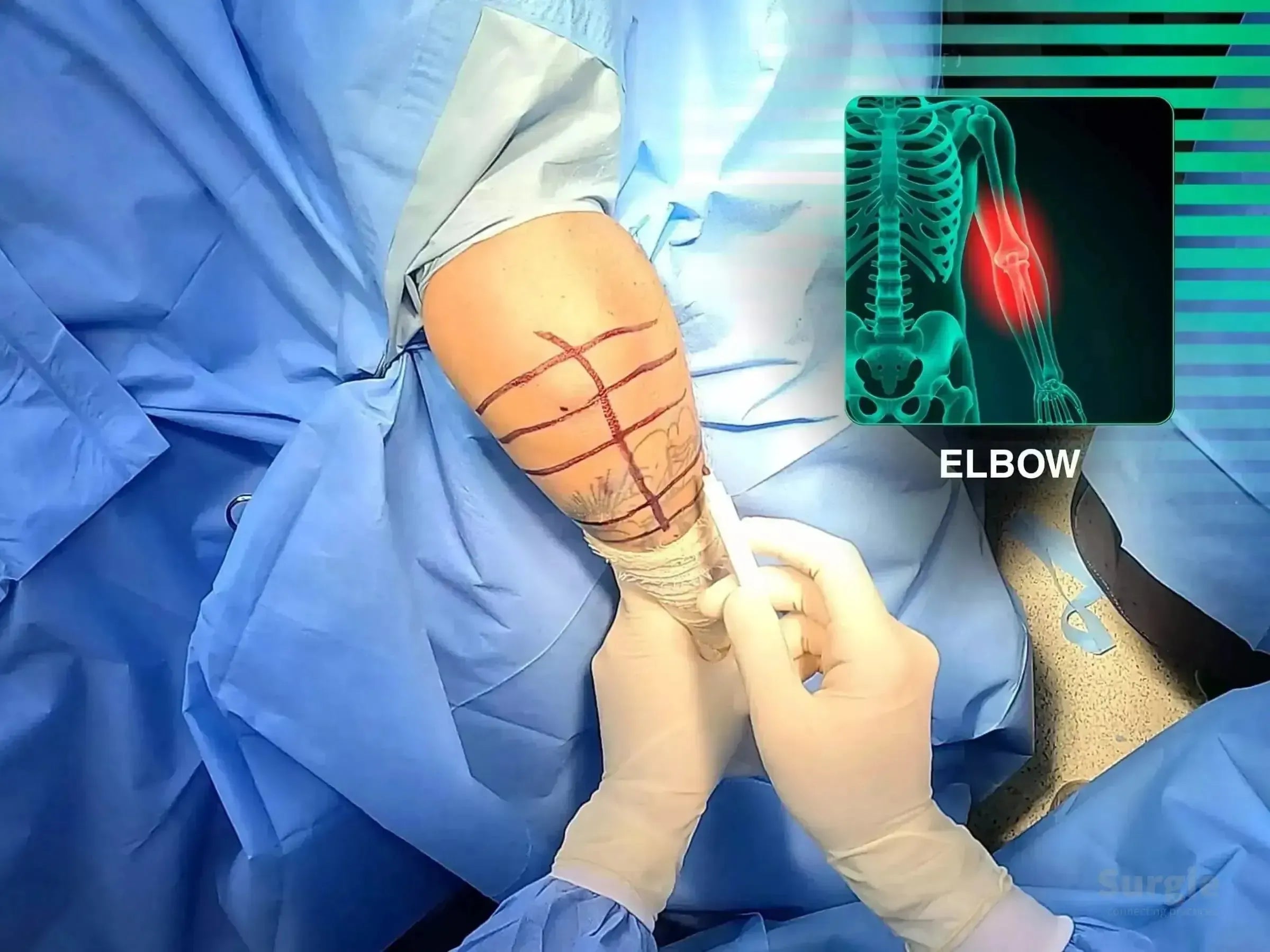
Minimally invasive olecranon plating with anatomic joint restoration
डॉ. सावीओ चामीऑर्थोपेडिस्ट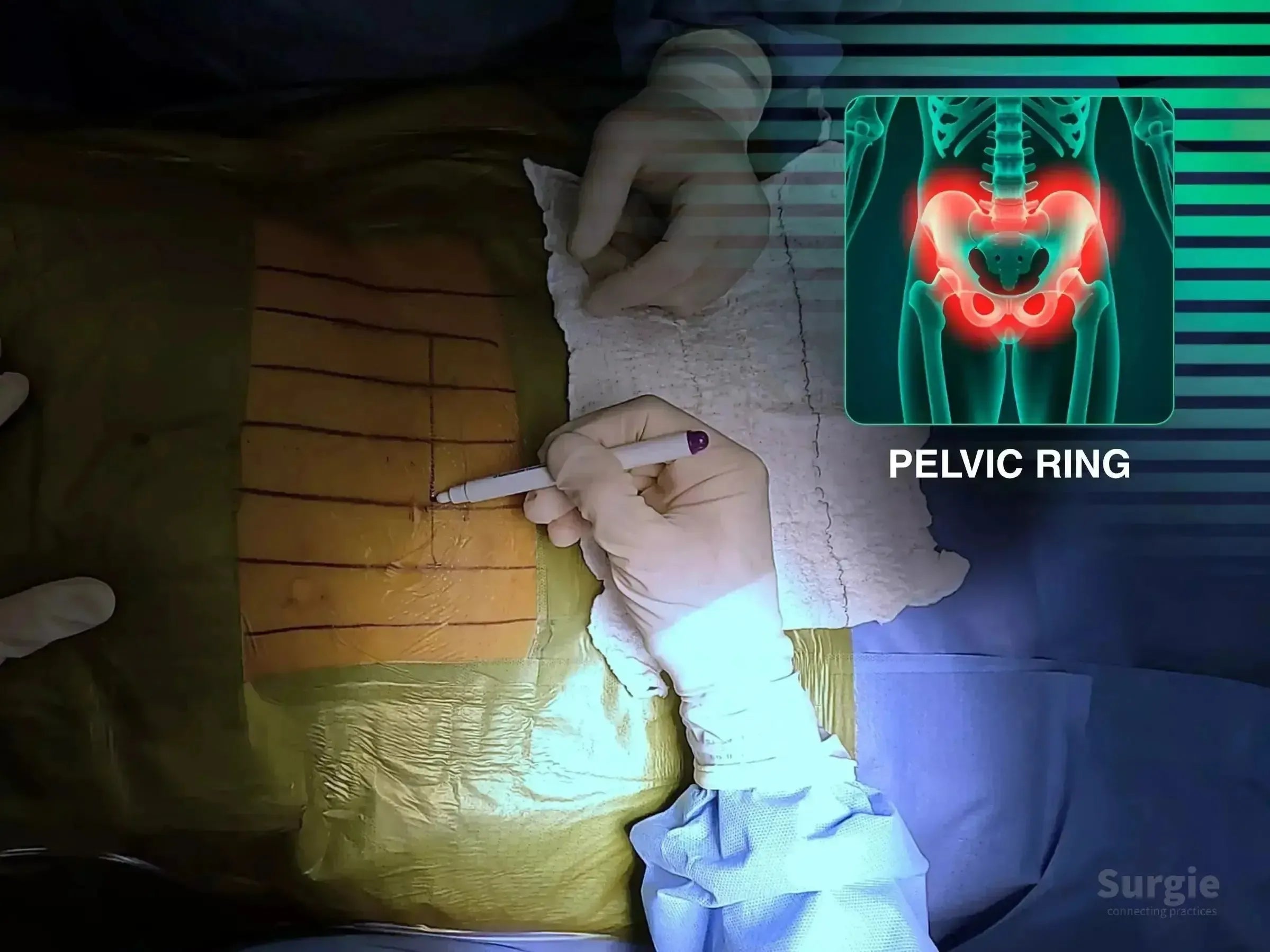
Controlled symphyseal reduction with anterior pelvic ring fixation
डॉ. सावीओ चामीऑर्थोपेडिस्ट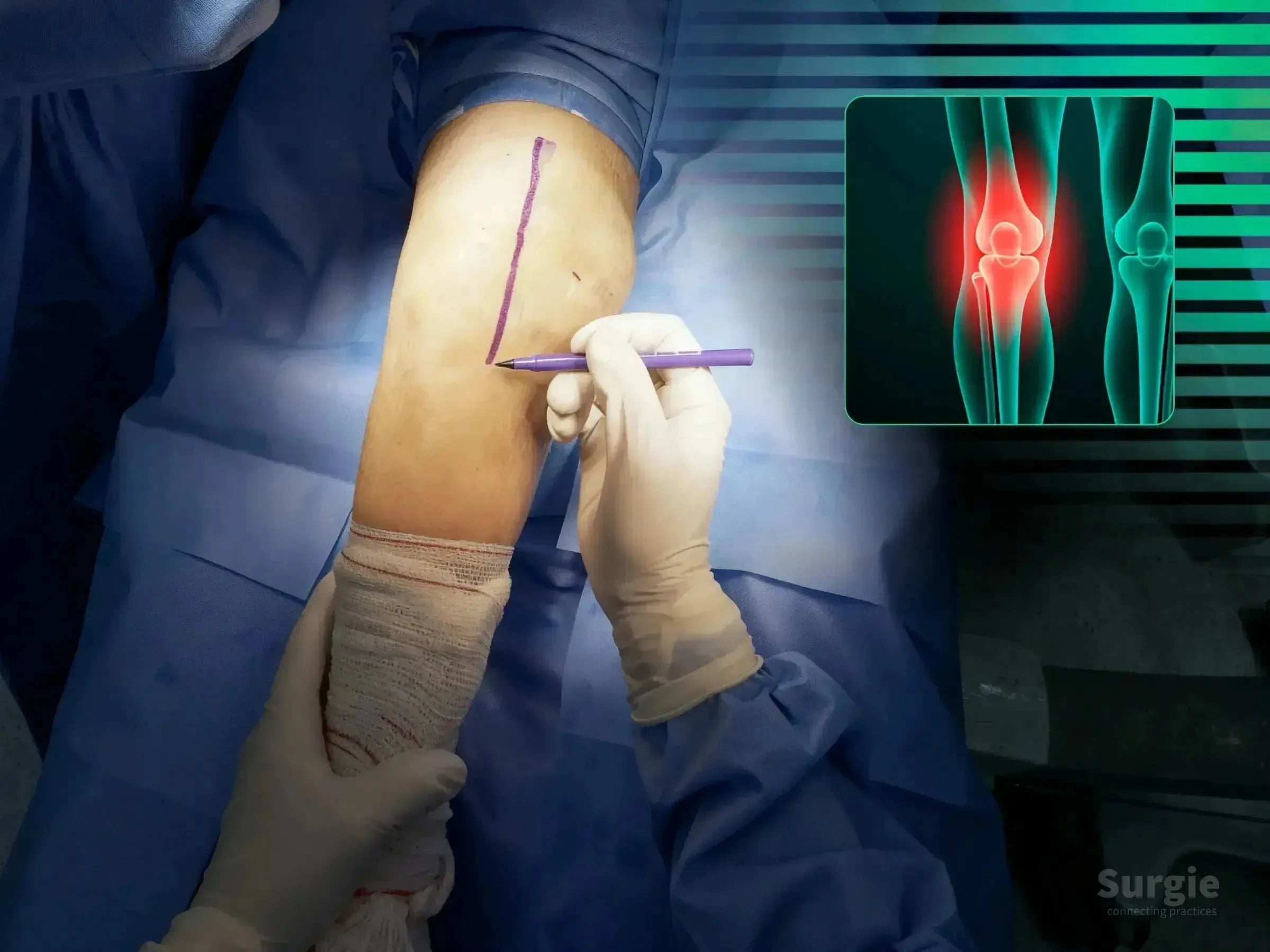
Cannulated screws and cerclage with extensor mechanism preservation
डॉ. सावीओ चामीऑर्थोपेडिस्ट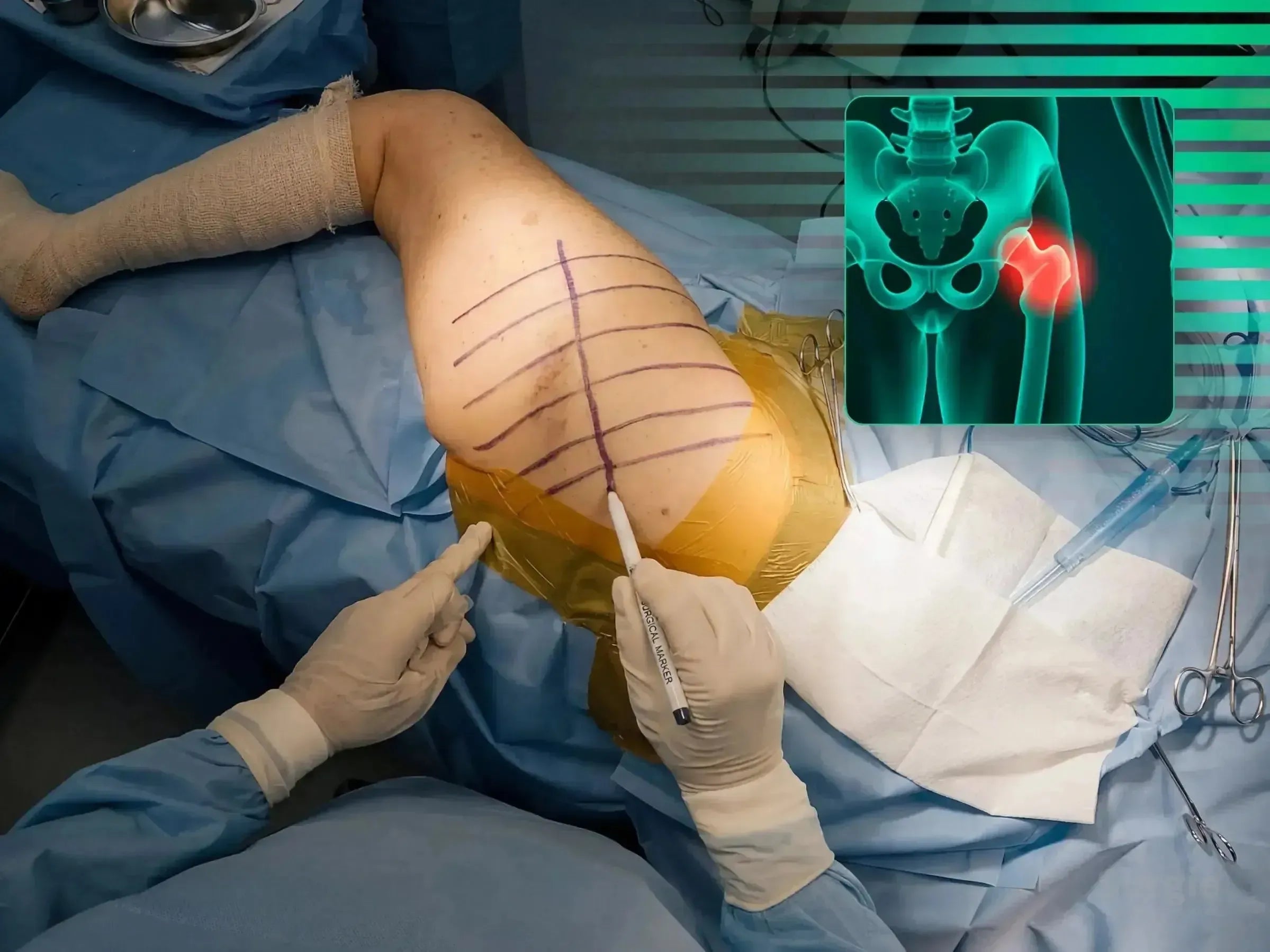
Trochanteric plating and cerclage without femoral component revision
डॉ. सावीओ चामीऑर्थोपेडिस्ट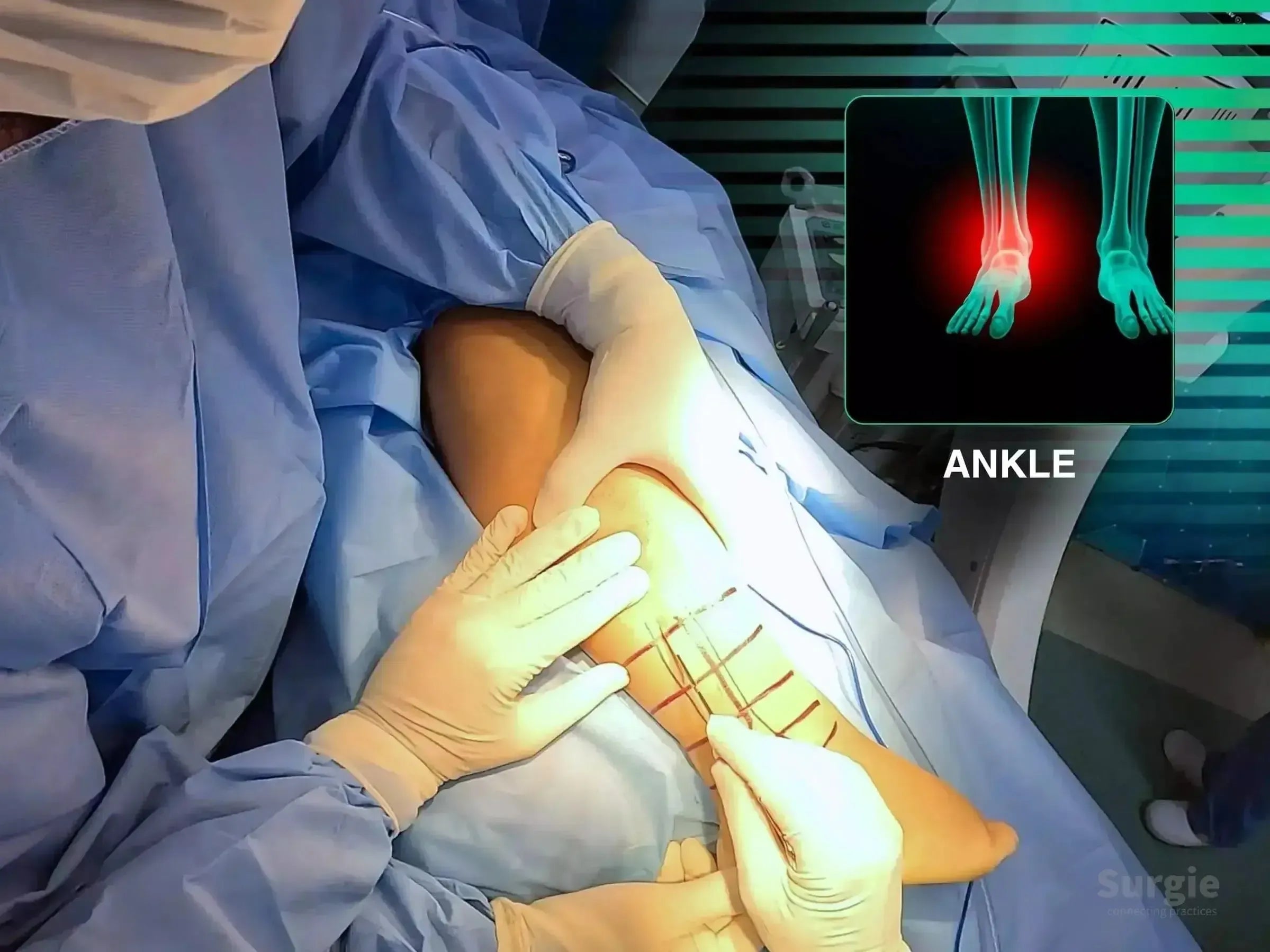
Single posterolateral exposure for posterior and lateral malleolus fixation
डॉ. सावीओ चामीऑर्थोपेडिस्ट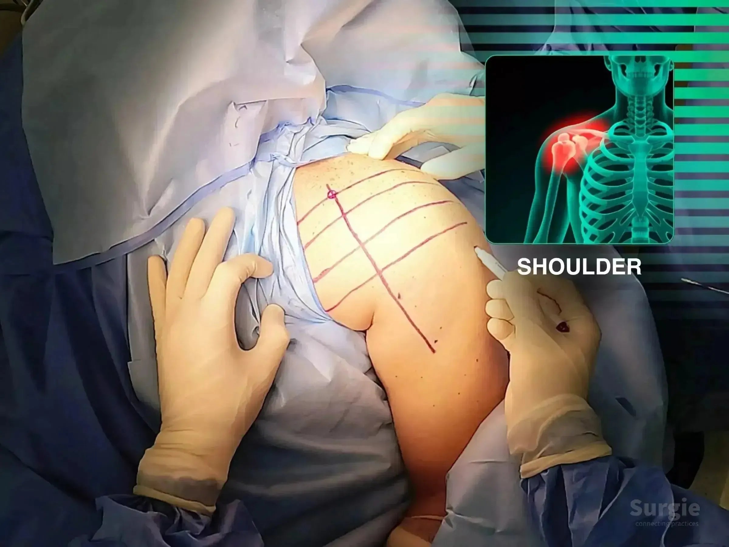
Reduction of a locked posterior dislocation with carbon-fiber plating
डॉ. सावीओ चामीऑर्थोपेडिस्ट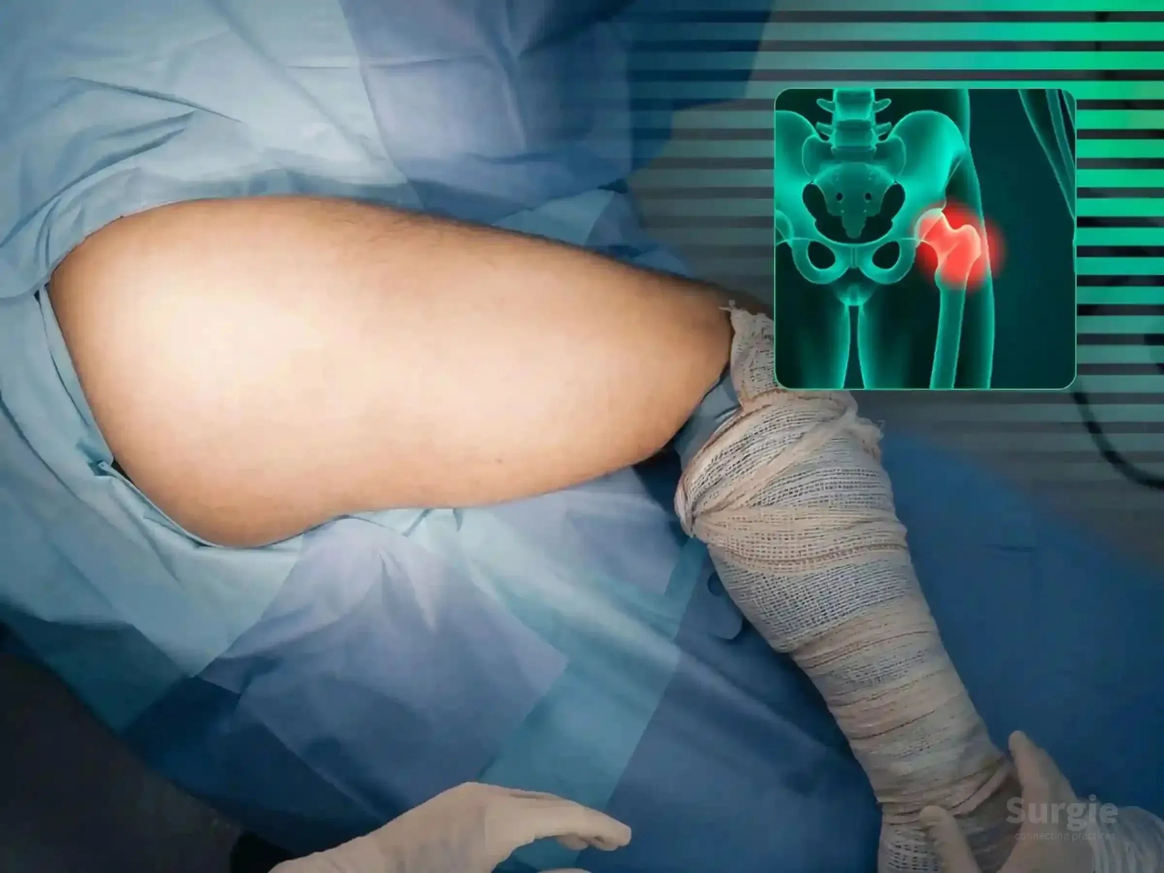
Articular disimpaction and posterior wall reconstruction
डॉ. सावीओ चामीऑर्थोपेडिस्ट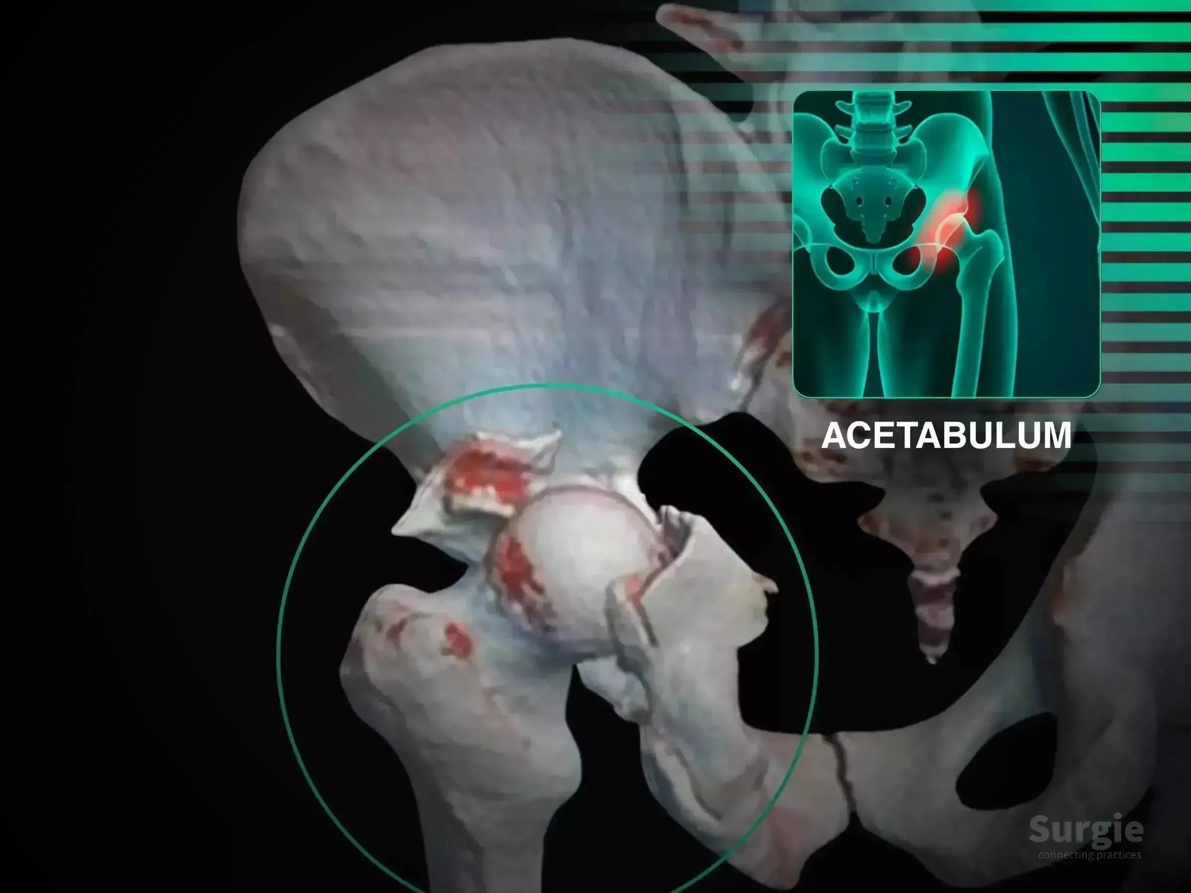
Kocher-Langenbeck exposure with posterior wall fixation and nerve protection
डॉ. सावीओ चामीऑर्थोपेडिस्ट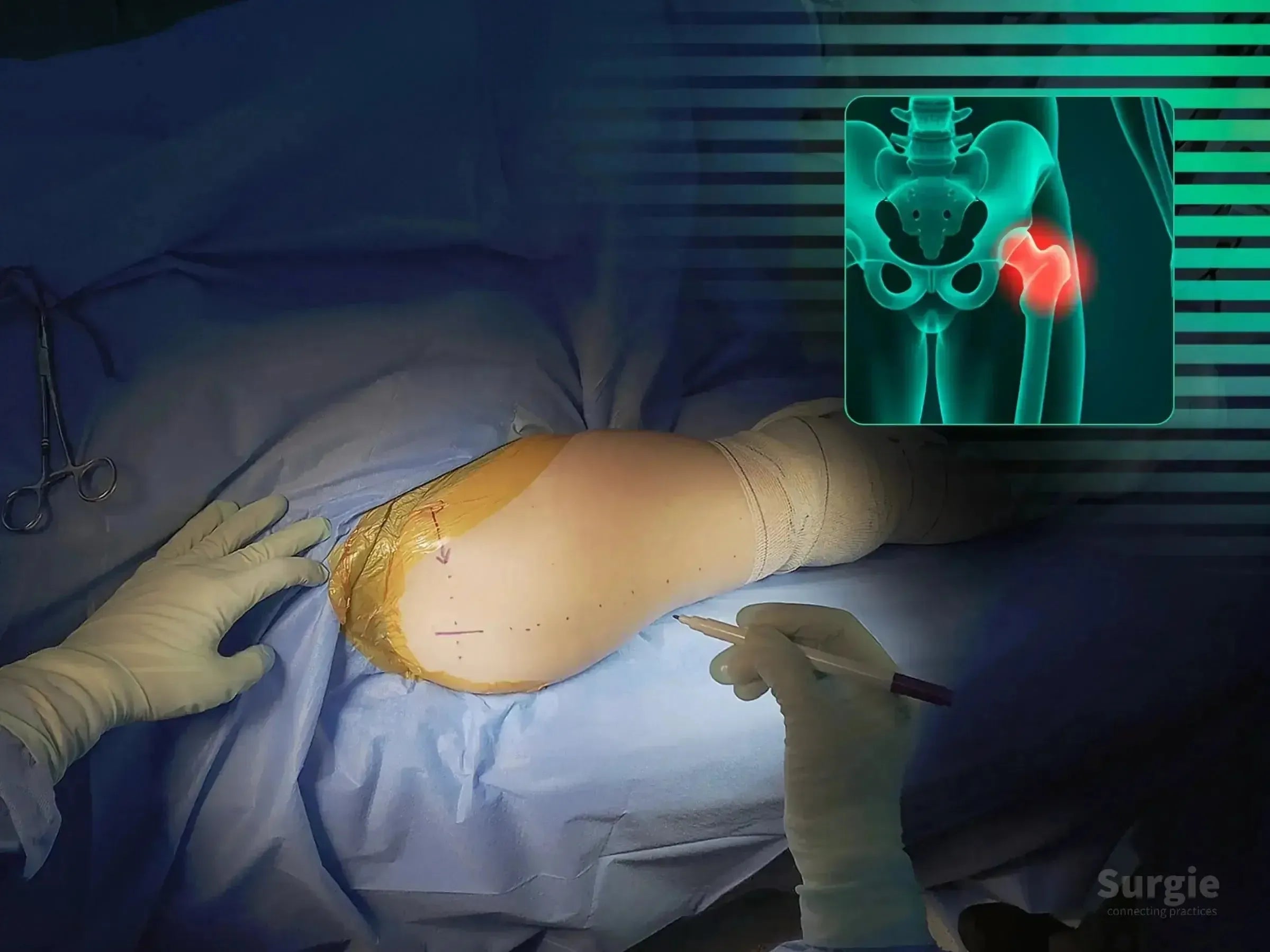
Rocker-assisted calcar reduction without a traction table
डॉ. सावीओ चामीऑर्थोपेडिस्ट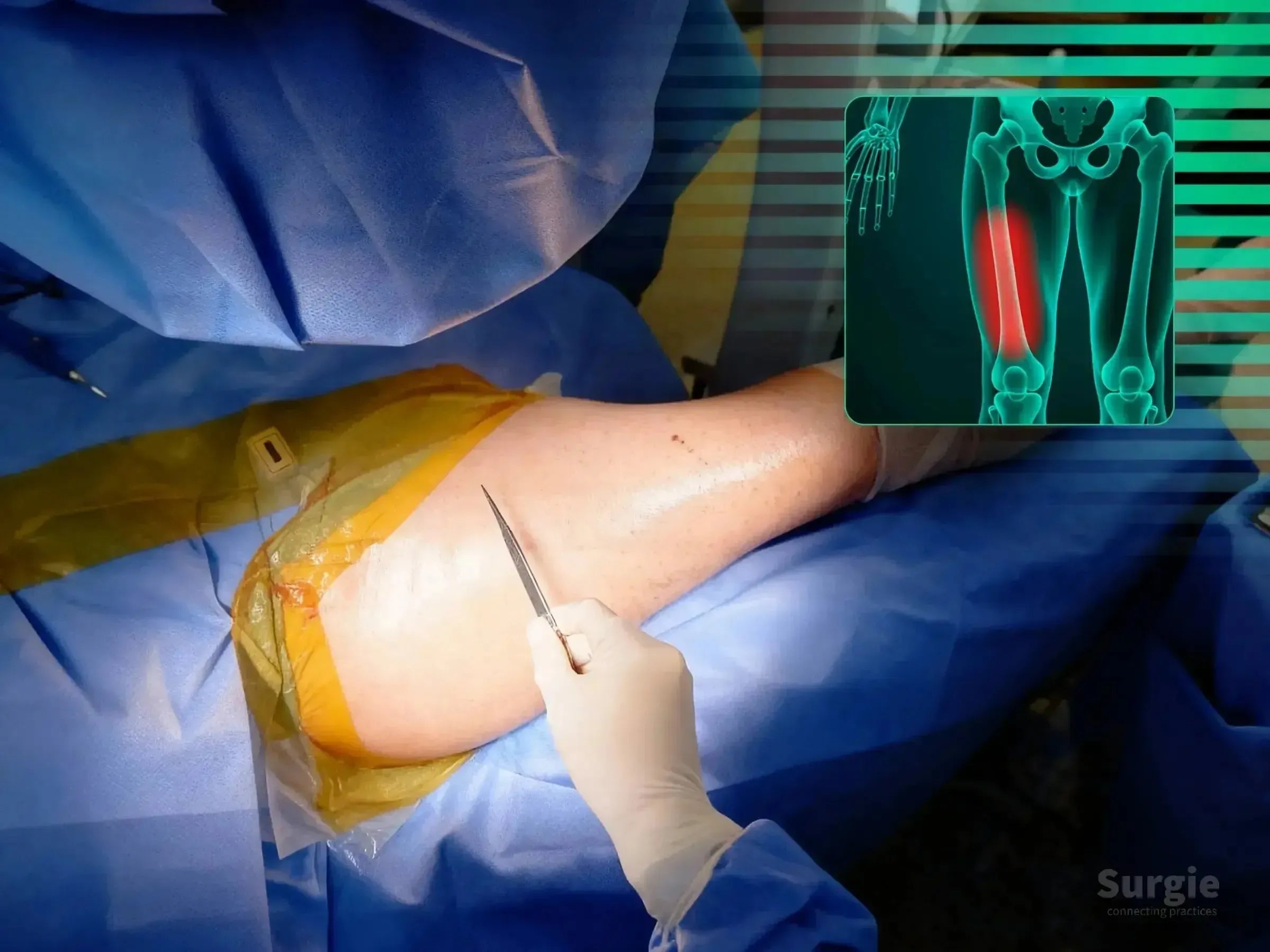
Double see-saw reduction for shaft and calcar realignment
डॉ. सावीओ चामीऑर्थोपेडिस्ट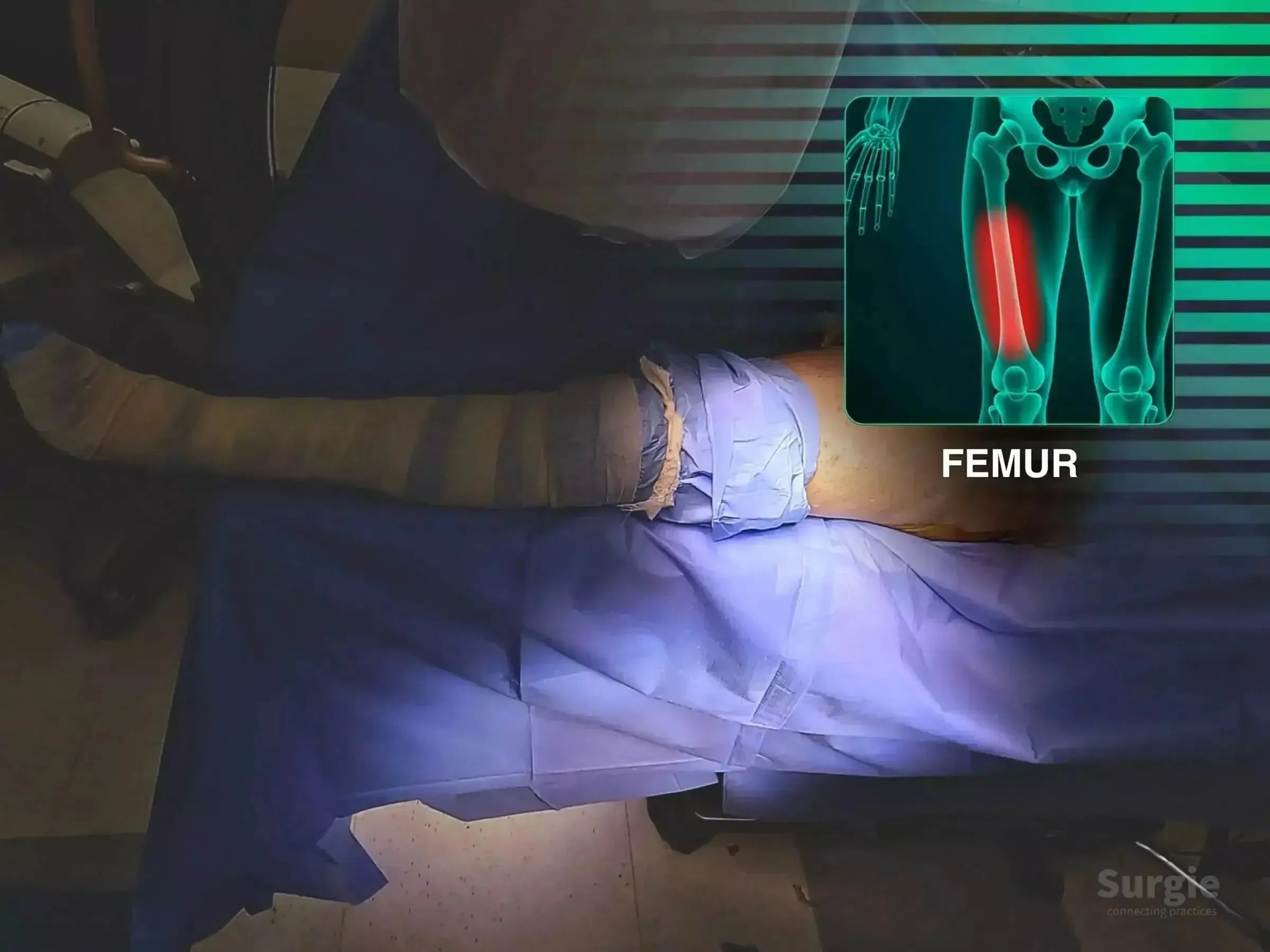
See-saw reduction of an unstable proximal femur fracture
डॉ. सावीओ चामीऑर्थोपेडिस्ट