
Filtri
50% preview
4K
Ankle
19:13
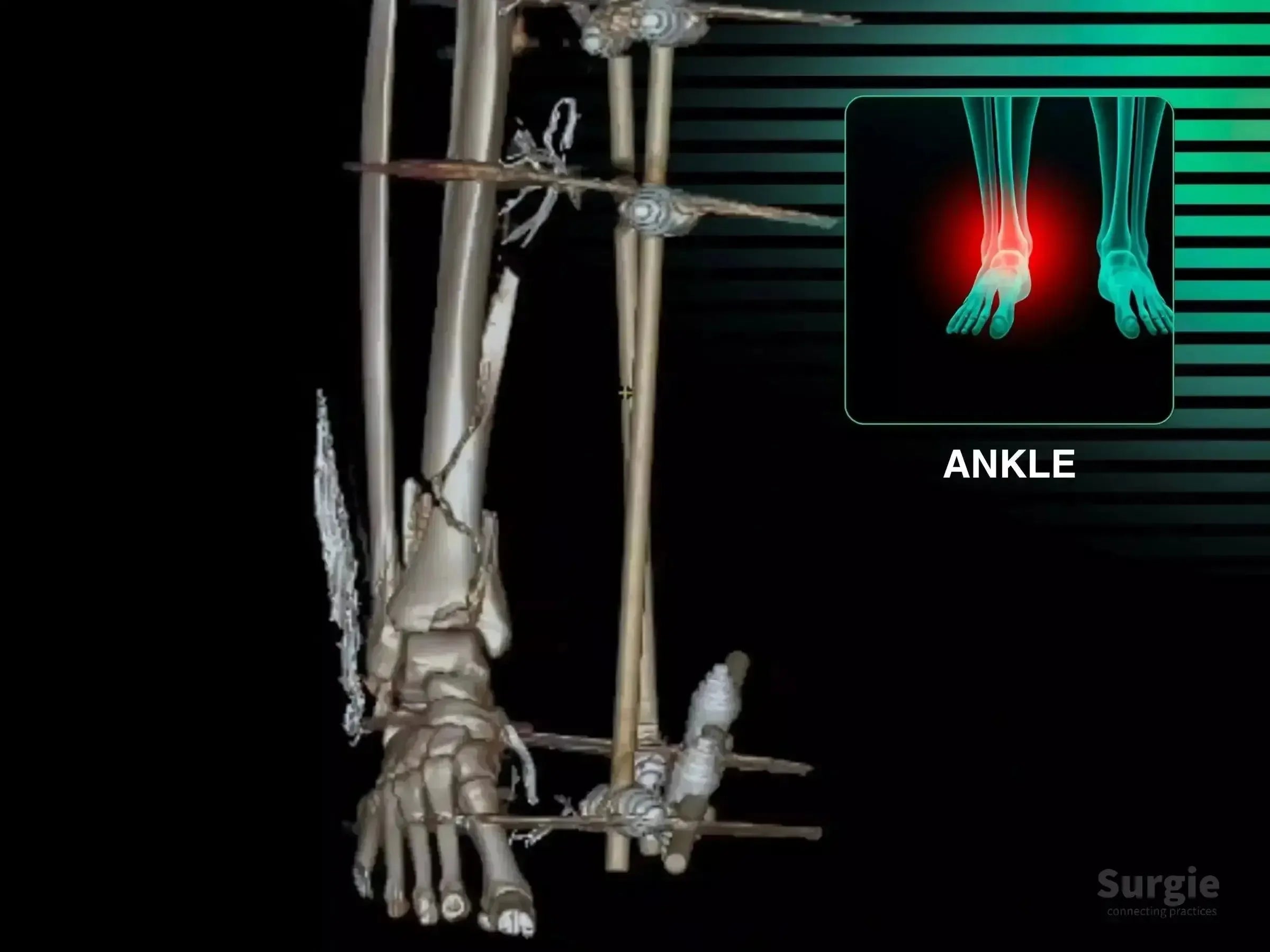
Comminuted Tibial Pilon Fracture: Staged Fixation
 Dr. Sávio ChamiOrthopedic Surgeon
Staged pilon reconstruction with articular reduction and multiplanar fixation
 Dr. Sávio ChamiOrthopedic Surgeon
9
languages

50% preview
4K
Ankle
08:17
Complex Ankle Injury: 360-Degree Reconstruction
Dr. Sávio ChamiOrthopedic Surgeon
Achilles, posterior tibial tendon, deltoid, and syndesmosis reconstruction
Dr. Sávio ChamiOrthopedic Surgeon
9
languages
50% preview
4K
Ankle
02:29
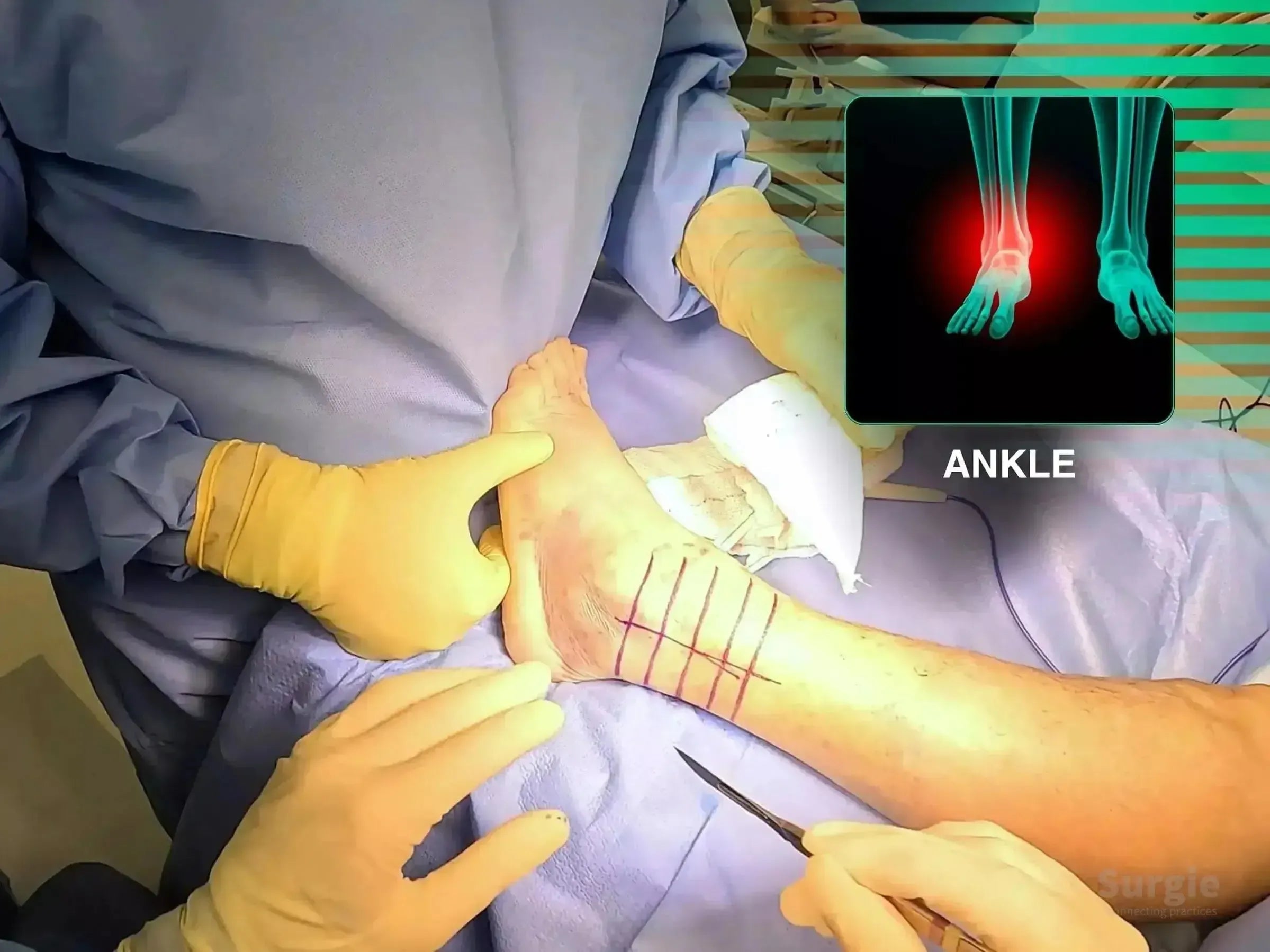
Lateral Malleolus Fracture with Wagstaffe Fragment
Dr. Sávio ChamiOrthopedic Surgeon
Wagstaffe fragment compression with locked lateral malleolus fixation
Dr. Sávio ChamiOrthopedic Surgeon
9
languages
Free
4K
Ankle
07:41
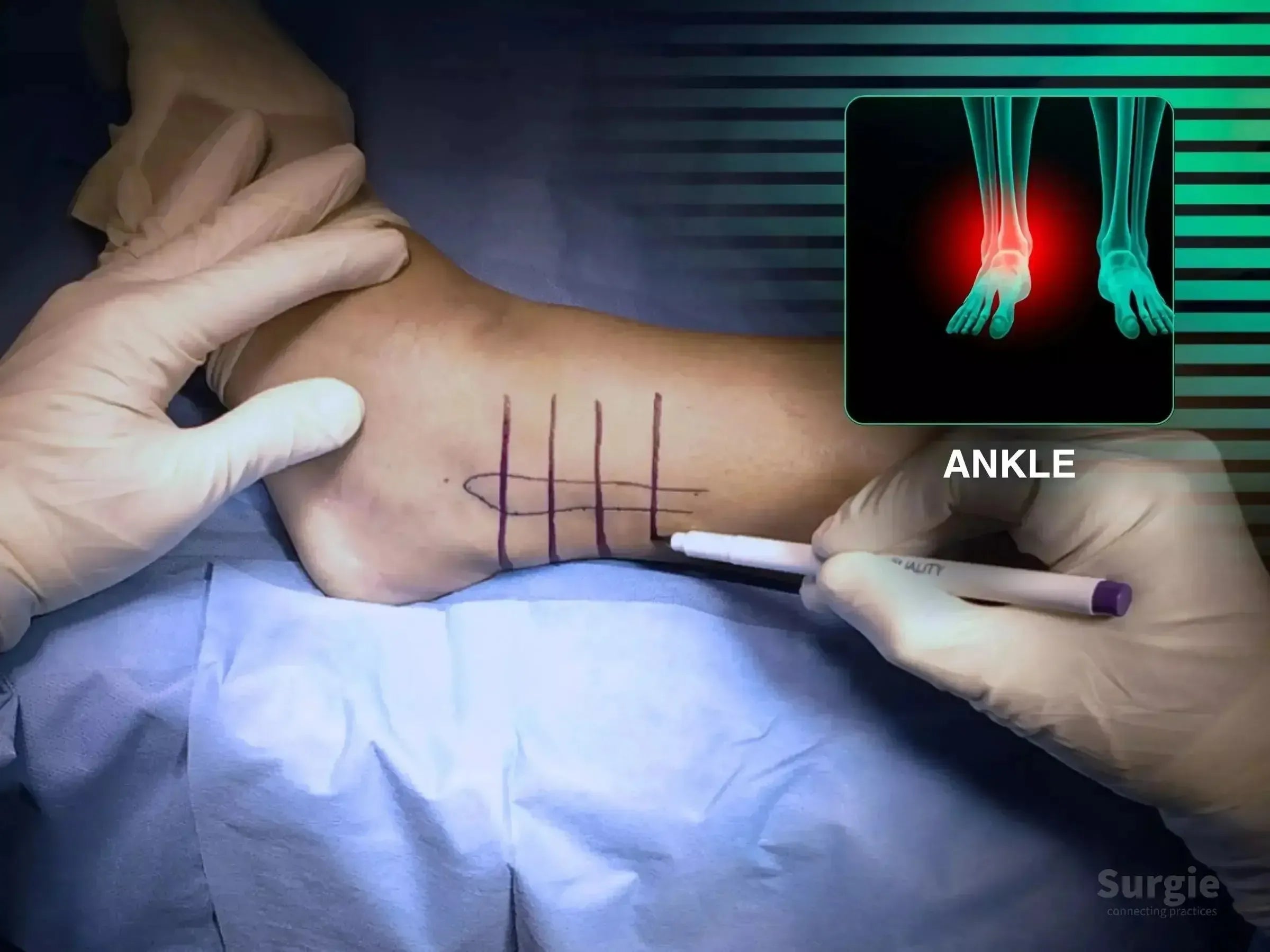
Lateral Malleolus Fracture: Posterior Plate Technique
Dr. Sávio ChamiOrthopedic Surgeon
Posterior antiglide plating for shear control and tissue preservation
Dr. Sávio ChamiOrthopedic Surgeon
9
languages
50% preview
4K
Ankle
04:14
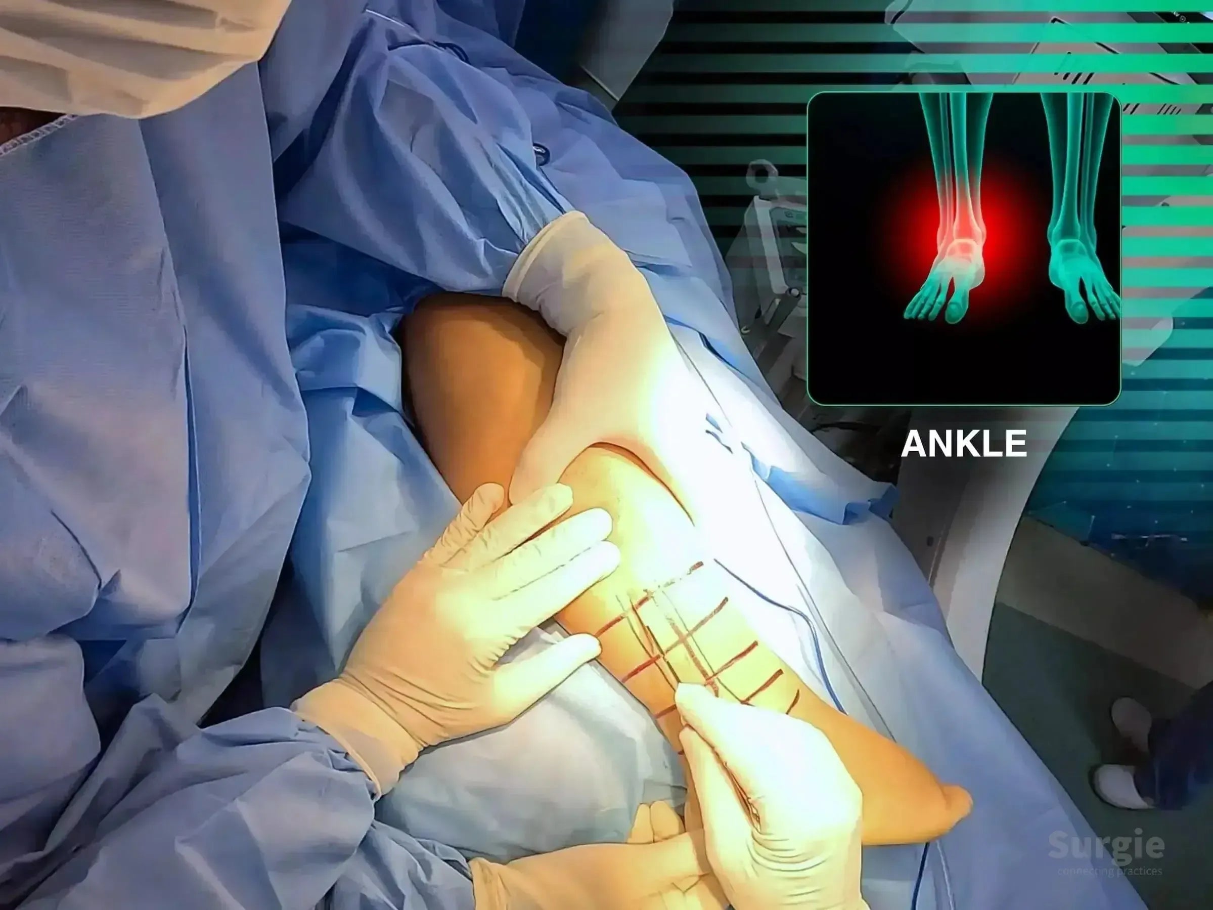
Posterior and Lateral Malleolus Fractures: Posterolateral Approach
Dr. Sávio ChamiOrthopedic Surgeon
Single posterolateral exposure for posterior and lateral malleolus fixation
Dr. Sávio ChamiOrthopedic Surgeon
9
languages
50% preview
4K
Ankle
22:30
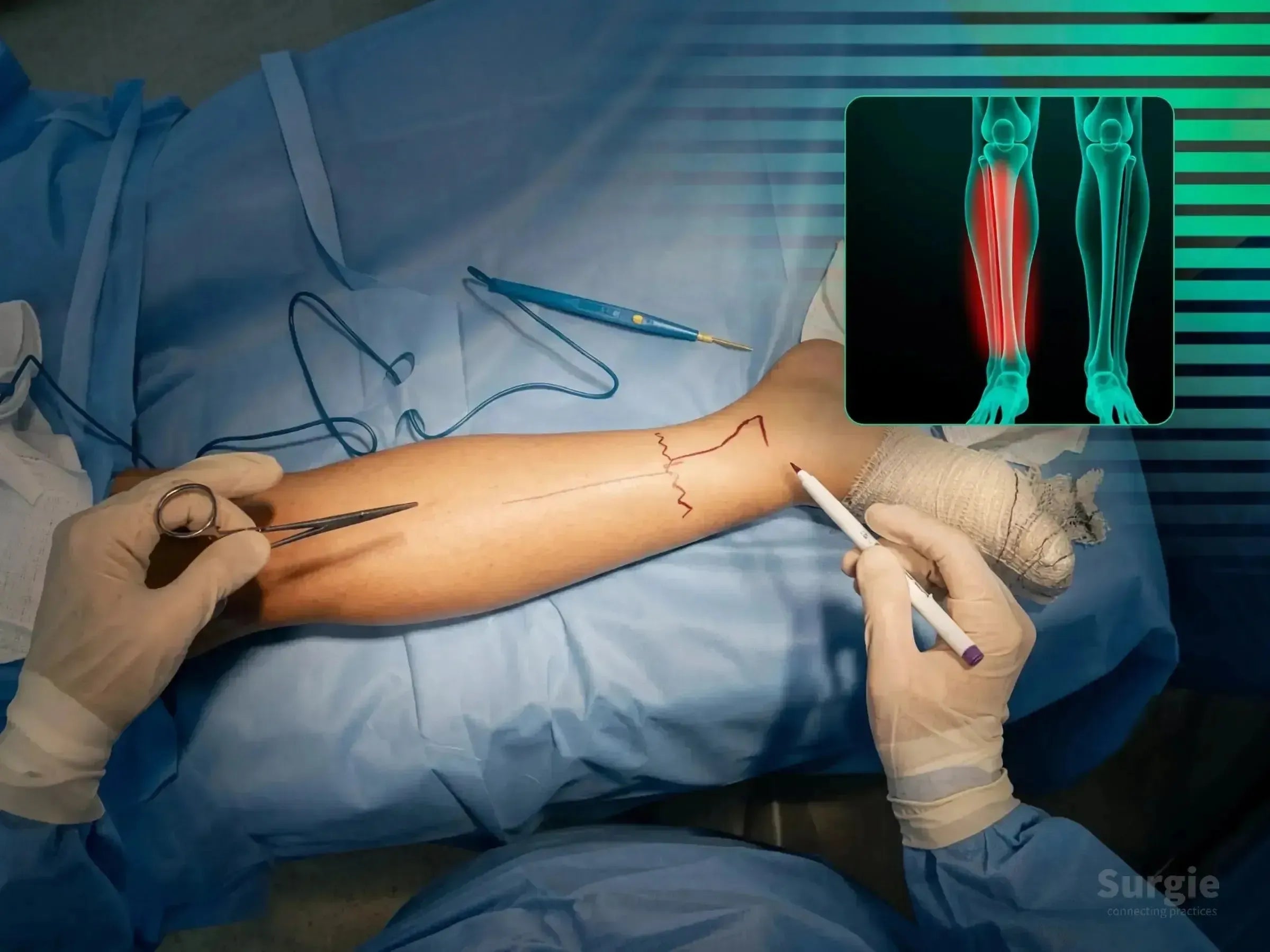
Tibial Pilon Fracture, Case 2: Dual-Approach Fixation
Dr. Sávio ChamiOrthopedic Surgeon
CT-guided dual-approach fixation of posteromedial pilon shear
Dr. Sávio ChamiOrthopedic Surgeon
9
languages
50% preview
4K
Ankle
28:05
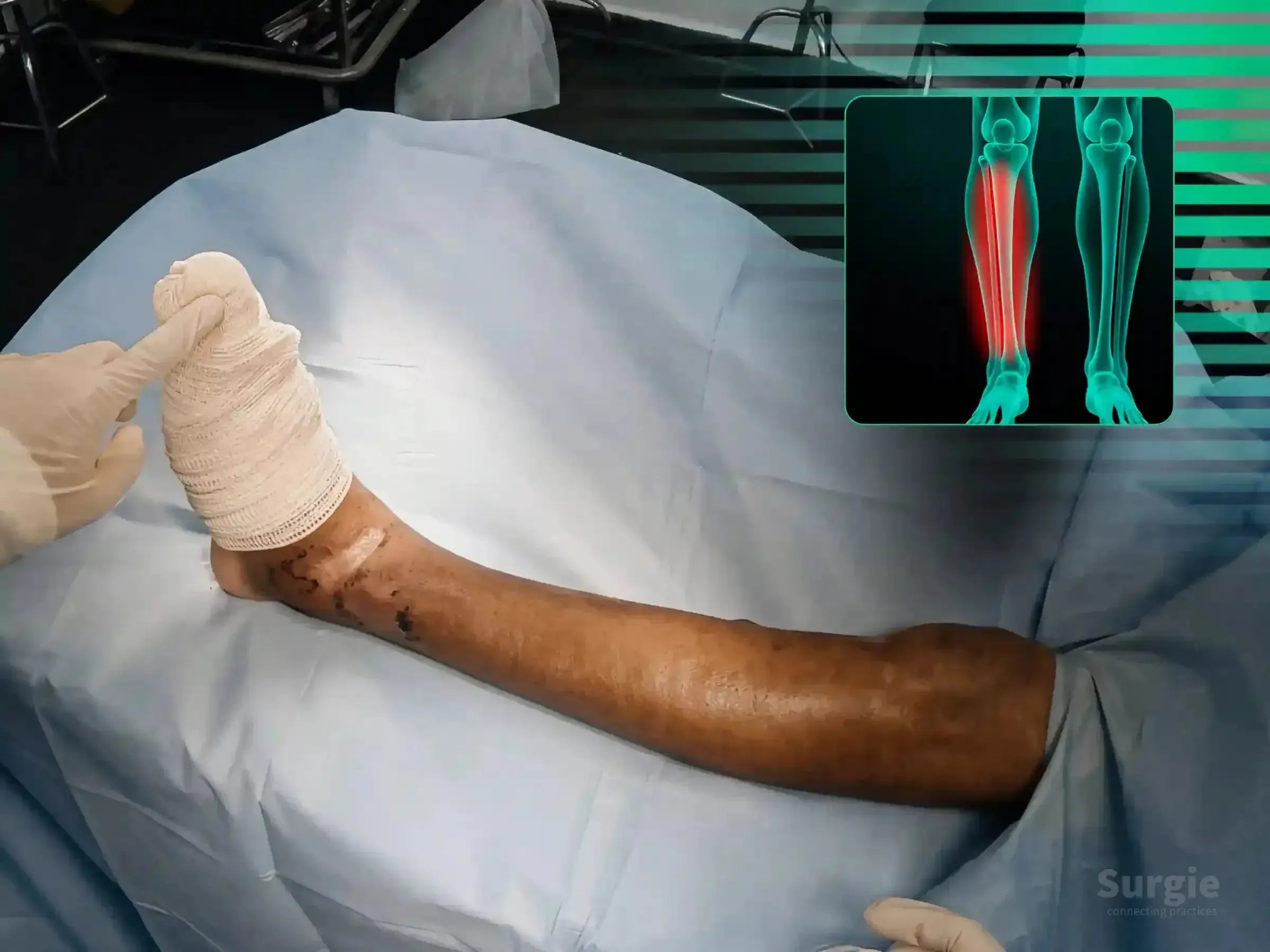
Tibial Pilon Fracture, Case 3: Anterolateral and Posteromedial Approaches
Dr. Sávio ChamiOrthopedic Surgeon
Staged dual-approach pilon reconstruction guided by CT morphology
Dr. Sávio ChamiOrthopedic Surgeon
9
languages
50% preview
4K
Ankle
23:37
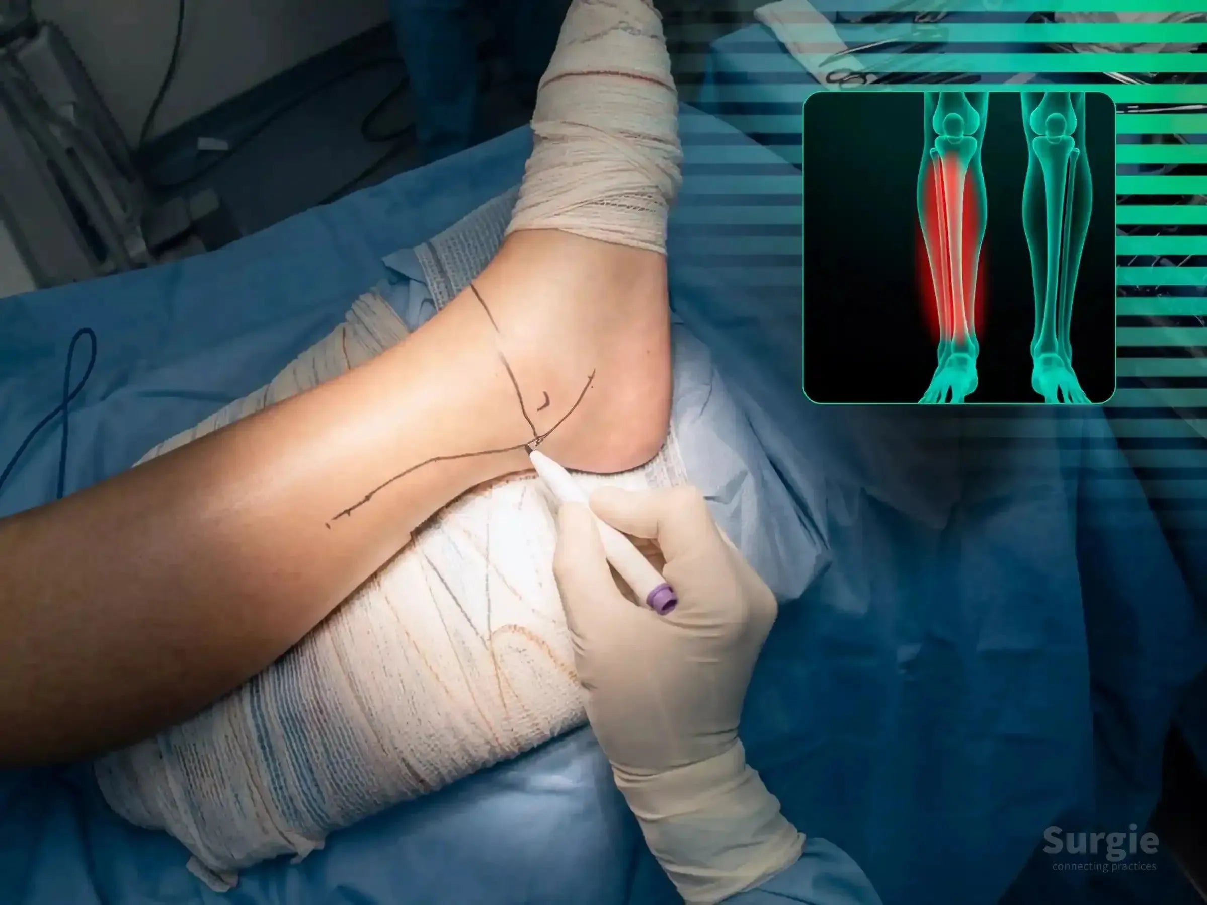
Trimalleolar Fracture, Case 3: Posterolateral Approach
Dr. Sávio ChamiOrthopedic Surgeon
Posterolateral ankle fixation with staged malleolar reconstruction
Dr. Sávio ChamiOrthopedic Surgeon
9
languages
50% preview
4K
Ankle
28:08
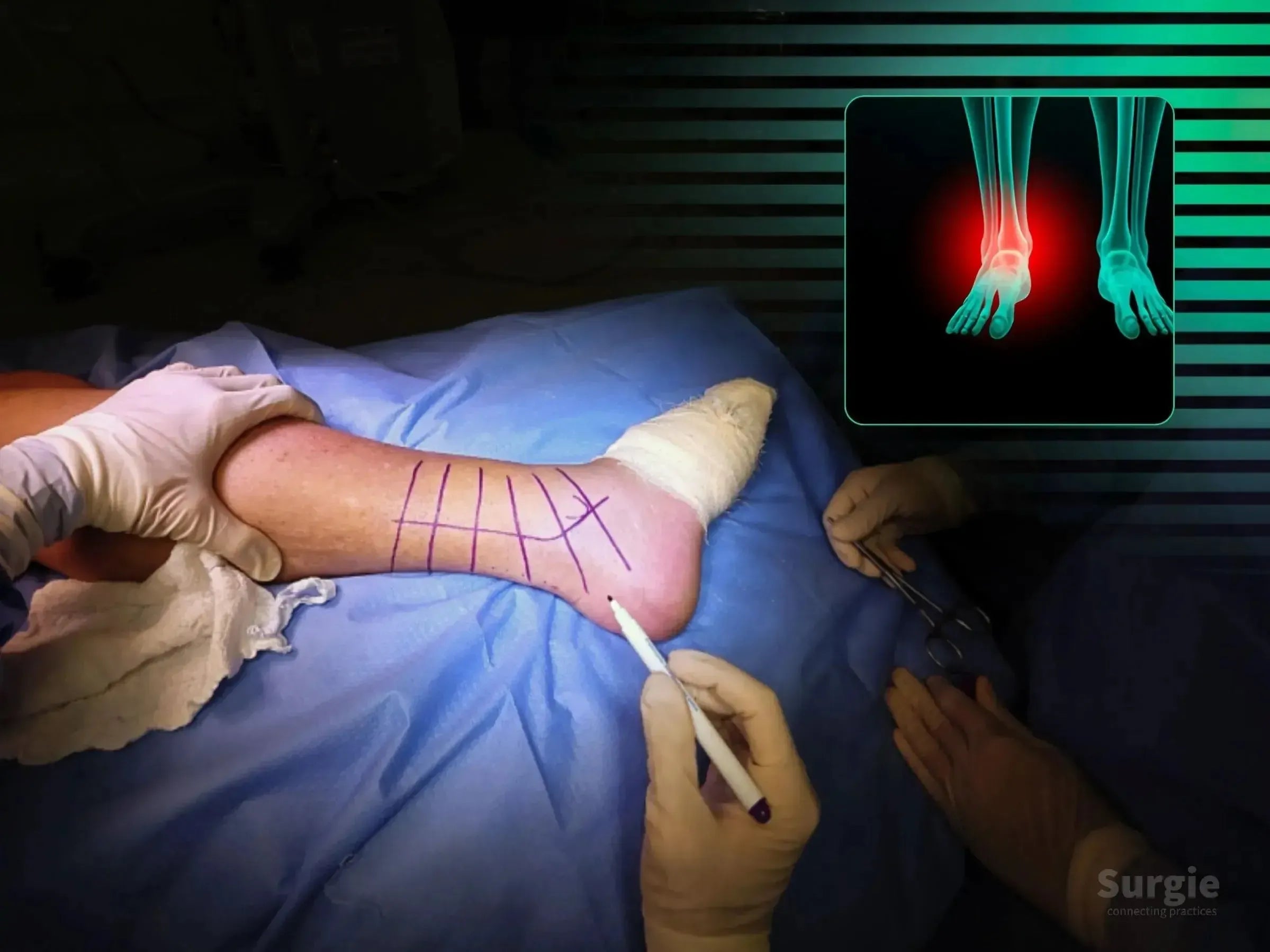
Trimalleolar Fracture: Lateral Decubitus Approach
Dr. Sávio ChamiOrthopedic Surgeon
Sequential posterior, lateral, and medial fixation in lateral decubitus
Dr. Sávio ChamiOrthopedic Surgeon
9
languages