
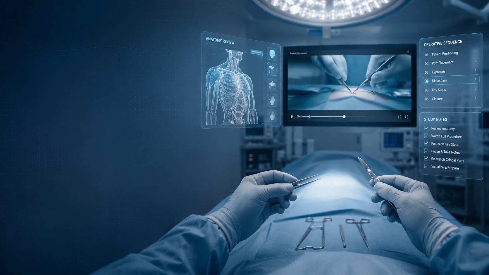
Watching a surgical procedure is easy.
Studying one is different.
A physician can watch an entire operation from beginning to end and still retain only a general impression of what happened. The procedure may look familiar, the sequence may seem logical and the final result may make sense, yet the learner may struggle to reconstruct the operation independently.
That distinction matters.
Effective surgical preparation is not simply about remembering that a procedure was seen. It is about developing a mental representation of the operation: the anatomy, sequence, exposure, instrument positioning, decisions, risks and technical transitions that connect one step to the next.
Surgical video becomes more valuable when the learner stops asking:
"What happened in this operation?"
and begins asking:
"Could I explain what should happen next, and why?"
That shift turns passive viewing into structured surgical study.
Why passive viewing is not enough
Surgery is not a sequence of isolated movements.
Each technical action depends on information that came before it.
The surgeon identifies an anatomical landmark, interprets the operative field, selects an instrument, determines a trajectory, performs a movement, evaluates the result and decides what should happen next.
Much of this process happens quickly.
When a surgical video is watched only once, the viewer's attention is divided among anatomy, instruments, movements, narration, imaging and the overall progression of the case.
It is difficult to study everything simultaneously.
A better approach is to revisit the same procedure with a different objective during each viewing.
The goal is not simply to watch the operation repeatedly.
The goal is to make each repetition answer a different surgical question.
Step 1: Understand the clinical problem before watching the technique
Before focusing on instruments or operative movements, understand why the operation is being performed.
Review the diagnosis, relevant imaging, indication, anatomical problem and objective of the procedure.
Ask what the surgeon is attempting to restore, remove, stabilize, reconstruct or expose.
This creates context for everything that follows.
Without understanding the indication, technical steps can become disconnected movements.
With clinical context, each step begins to have a purpose.
For example, the surgical approach is no longer simply an incision. It becomes a strategy for reaching a particular anatomical target while respecting structures that must be protected.
The implant is no longer simply hardware. Its position and orientation are connected to the biomechanical problem being treated.
Start with the problem.
Then study the solution.
Step 2: Watch the entire procedure once without constantly pausing
The first viewing should establish the overall flow of the operation.
Resist the temptation to analyze every detail immediately.
Instead, identify the major phases.
Where does exposure begin?
When does the operation transition from exposure to reconstruction or fixation?
Which moment appears to be technically decisive?
When does fluoroscopy, endoscopy or another form of imaging influence the next step?
How does the surgeon confirm the final result?
At this stage, you are building a map.
You may not yet understand every street, but you should begin to understand how the destination is reached.
When the procedure finishes, try to reconstruct its major phases without replaying the video.
If you cannot describe the sequence, watch the complete procedure again before moving into detailed analysis.
Step 3: Rewatch specifically for anatomy and exposure
The second detailed viewing should focus primarily on anatomy.
Ignore some of the technical execution temporarily and follow the exposure.
Identify visible landmarks.
Observe how the surgeon develops the operative plane.
Notice which structures become visible first and which appear only after further dissection or retraction.
Study the direction of exposure.
Pay particular attention to structures the surgeon protects, retracts or deliberately avoids.
In many operations, understanding the exposure is fundamental because every subsequent maneuver depends on the orientation created during these initial steps.
Surgeon point-of-view video can be especially useful here because the learner can examine the operative field from a perspective closer to the operator's line of sight.
Instead of only seeing where the surgeon is standing, the learner can study what is actually visible while the procedure is being performed.
Step 4: Study the hands, instruments and trajectories
The next viewing changes the focus again.
Now observe movement.
Look at the surgeon's right hand.
Then the left.
Study how both interact.
Which hand provides exposure?
Which stabilizes tissue or bone?
Which controls the working instrument?
What is the direction of traction?
At what angle does an instrument enter the field?
Where is its tip directed?
How does the trajectory change as anatomy becomes exposed?
Instrument trajectory is an important component of operative understanding that may be difficult to appreciate from an external camera.
A movement that appears simple from outside the surgical field can depend on a very specific combination of angle, depth, hand position and visual target.
Do not study only where an instrument finishes.
Study where it begins, how it travels and what visual information guides that trajectory.
Step 5: Identify decision points and risk zones
Not every moment in an operation has equal educational importance.
Some steps represent transitions where the surgeon must interpret what is being seen and decide what happens next.
These are decision points.
Pause when the surgeon evaluates reduction, changes an instrument, modifies exposure, selects an implant position, adjusts a trajectory or abandons one strategy for another.
Ask why.
Also identify risk zones.
Which anatomical structures require protection?
Which maneuver could cause a technical problem if performed in the wrong plane or direction?
Which step has limited tolerance for error?
What does the surgeon check before proceeding?
Expert narration becomes particularly valuable here.
The image may show what happened.
The surgeon's explanation can reveal why it happened.
Step 6: Close the video and mentally rehearse the procedure
One of the most useful tests of procedural understanding happens when the screen is no longer visible.
Close the video.
Then mentally reconstruct the operation.
Start with patient positioning and imagine moving through the procedure sequentially.
Visualize the exposure.
Identify the anatomical landmarks.
Imagine the instruments entering the field.
Recall the important trajectories.
Move through the major decision points.
Try to anticipate each next step before mentally progressing.
When you reach a point you cannot reconstruct clearly, you have identified a knowledge gap.
Return specifically to that portion of the video.
This creates a different learning process from simply pressing replay.
The video becomes a reference that helps build and test a mental model.
Step 7: Return to the video after supervised operative experience
Surgical video can also become valuable after the operating room.
A procedure often looks different after participating in the real case.
Movements that seemed obvious on video may suddenly have new meaning.
An exposure may appear more complex.
An instrument trajectory may make more sense.
A particular technical difficulty may become easier to recognize.
Return to the procedure after supervised clinical participation and compare what you experienced with what you observe in the expert recording.
This creates a learning loop:
Observe → Understand → Repeat → Mentally rehearse → Perform under supervision → Review → Refine
The operation is no longer an isolated educational event.
Preparation happens before it.
Reflection continues after it.
Six questions to ask while studying any surgical video
A useful surgical video study session should eventually allow you to answer:
- What is the clinical objective of this procedure?
- Which anatomical landmarks organize the approach?
- What are the major operative steps, in sequence?
- How are the surgeon's hands and instruments positioned during critical maneuvers?
- Where are the principal decision points and structures at risk?
- Can I mentally reconstruct what should happen next without watching the screen?
If one of these questions remains unclear, that becomes the objective of the next viewing.
Why surgeon POV changes what can be studied
Camera perspective matters.
Traditional external surgical video can provide valuable information about team positioning, general workflow and the procedure as a whole.
But the surgeon operates according to what is visible from the operator's own position.
First-person surgical recording brings the viewer closer to that perspective.
This can make it easier to study relationships that are particularly relevant to technical execution: anatomical depth, hand coordination, instrument entry angles, traction vectors and the relationship between the surgeon's visual target and subsequent movement.
The educational difference is subtle but important.
Instead of only asking:
"What is the surgeon doing?"
the learner can begin asking:
"What is the surgeon seeing that leads to this movement?"
That question moves surgical study closer to operative reasoning.
Why narration matters as much as visualization
High-resolution surgical footage alone does not automatically reveal expertise.
A learner can see an instrument being repositioned without knowing why its original trajectory was changed.
A surgeon may alter traction because a structure has become visible, change an approach because exposure is inadequate or reposition an implant based on information that is not obvious to the viewer.
Expert narration helps expose this reasoning.
The most useful explanations do more than describe movement.
They explain intention.
They connect anatomy with action.
They identify risks.
They clarify alternatives.
They help the learner understand not only what the surgeon does, but how the surgeon interprets the operative field.
From surgical video to the SNIPER learning model
This relationship between visualization, explanation, repetition and mental preparation is central to the SNIPER Method used in Surgie.
SNIPER stands for Surgical Narrative & Immersive Perspective with Enhanced Recording.
The methodology combines first-person surgical visualization with structured technical narration, real procedures, pedagogical organization and repeated access to the operative experience.
The objective is not to make surgical video a substitute for clinical training.
It is to make observation more structured and preparation more deliberate.
A learner can revisit anatomy.
Repeat a difficult step.
Analyze instrument positioning.
Review decision points.
Mentally rehearse the sequence.
Then connect that preparation with appropriate supervised clinical experience.
What surgical video cannot replace
No surgical video can reproduce the complete responsibility, variability and complexity of treating a real patient.
Video cannot replace formal medical training.
It cannot replace qualified mentorship.
It cannot replace supervised operative participation, simulation, cadaveric training or institutional protocols when those are required.
And watching a procedure does not establish competence to perform it independently.
The appropriate role of digital surgical education is preparation and reinforcement.
It can make surgical expertise more observable and repeatable.
Clinical training transforms that knowledge into supervised experience.
The two should work together.
A more useful way to watch surgery
The next time you open a surgical video, do not begin by asking how many times you need to watch it.
Ask what you are trying to learn during that specific viewing.
First understand the case.
Then learn the sequence.
Then study the anatomy.
Then follow the hands and instruments.
Identify decisions and risks.
Close the screen and reconstruct the operation mentally.
Return after supervised experience and look again.
The procedure will rarely look exactly the same on the second, third or fourth viewing.
That is the point.
You are no longer simply watching surgery.
You are learning how to study it.
Study real procedures on Surgie
Surgie is built around real surgical procedures, experienced physicians and practical clinical knowledge.
Explore complete procedures across multiple specialties, study surgeon POV cases, follow expert explanations and return to the same operative sequence whenever you need to review it.
Start with a free procedure, explore a specialty or study a case relevant to your next supervised surgical experience.
Watch. Understand. Repeat. Prepare.
Surgie — real surgical education built around real procedures.

Leave a comment
This site is protected by hCaptcha and the hCaptcha Privacy Policy and Terms of Service apply.