

Distal Humerus Osteotomy – Cubitus Varus Deformity
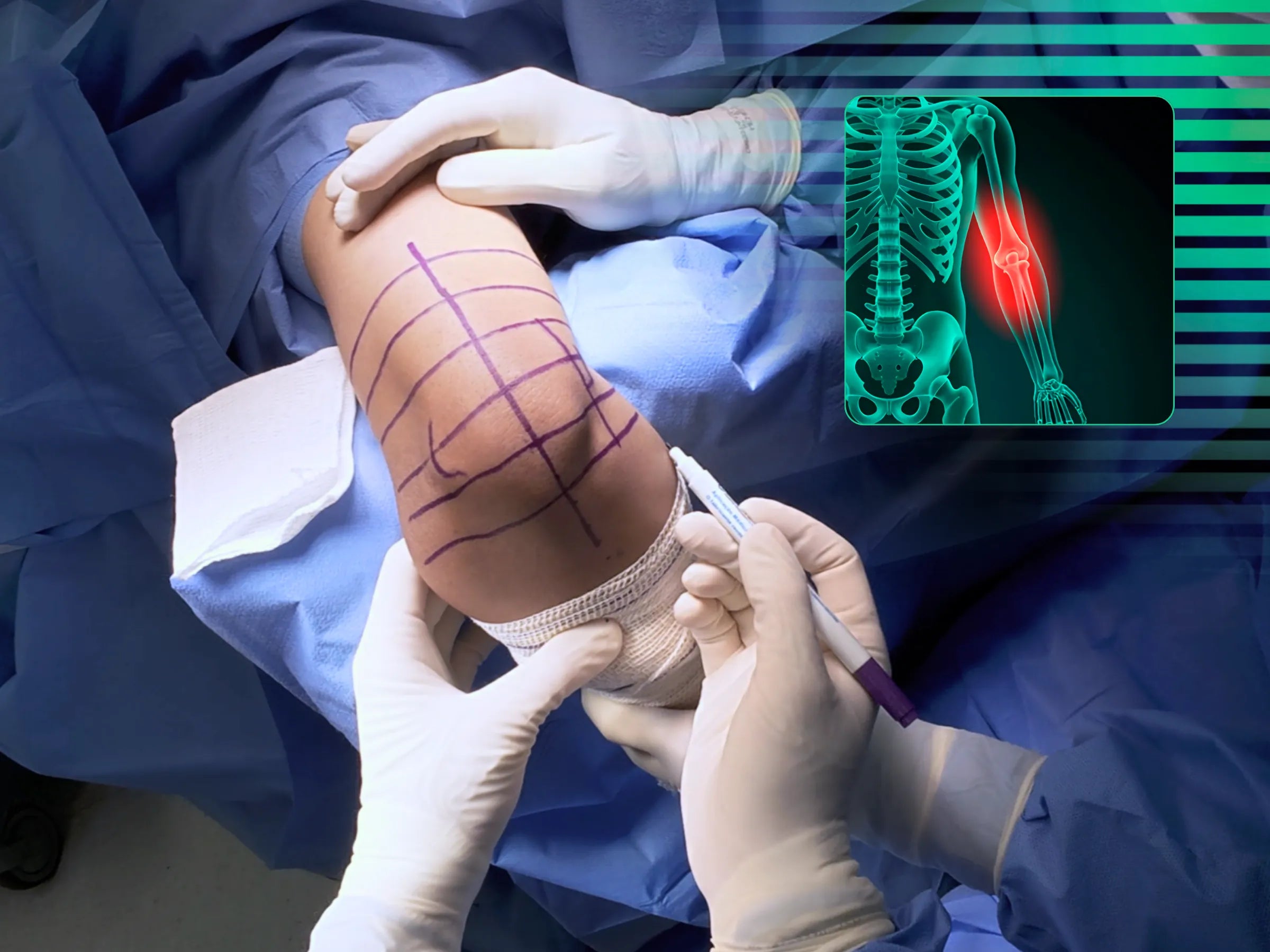
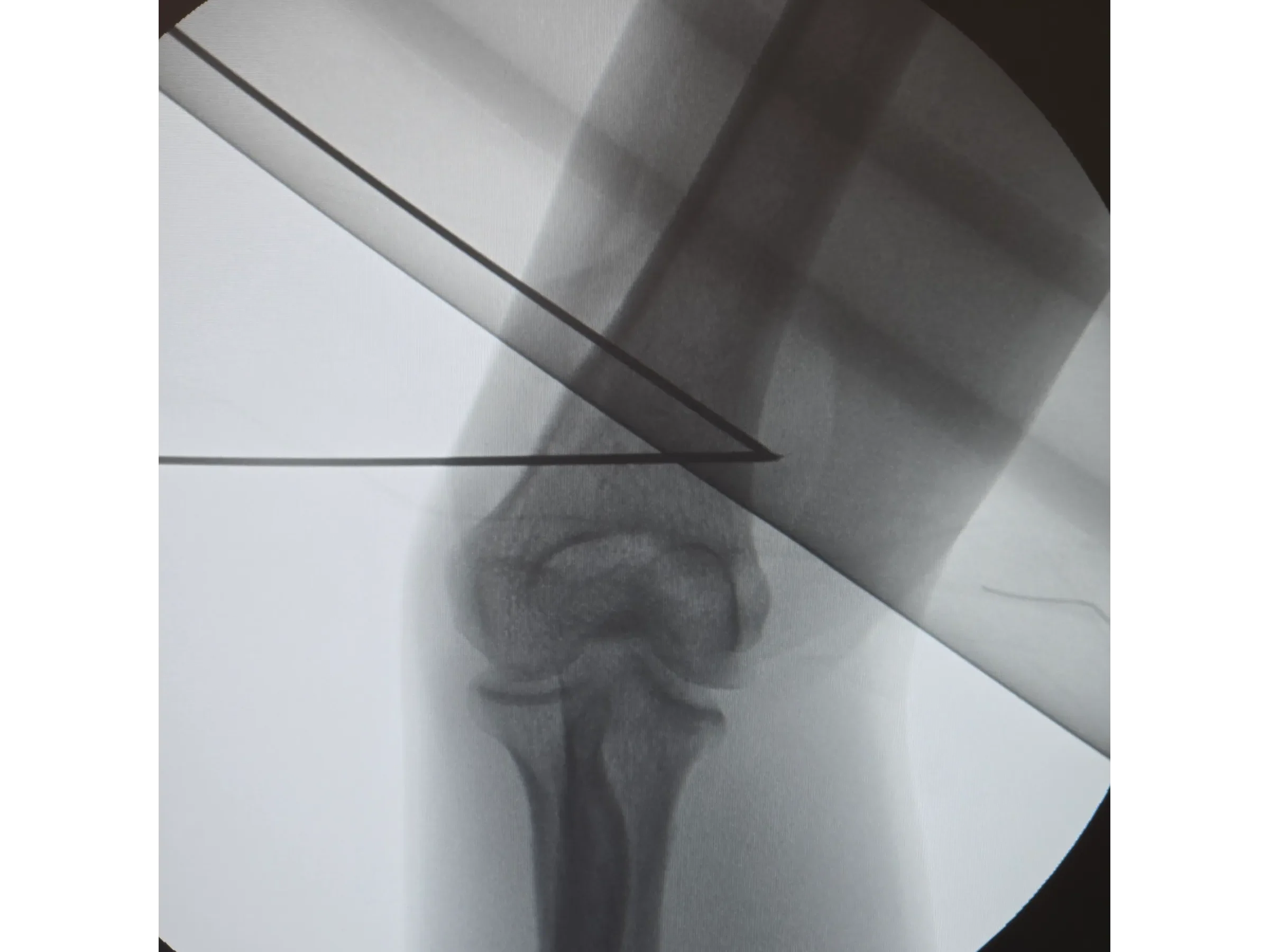
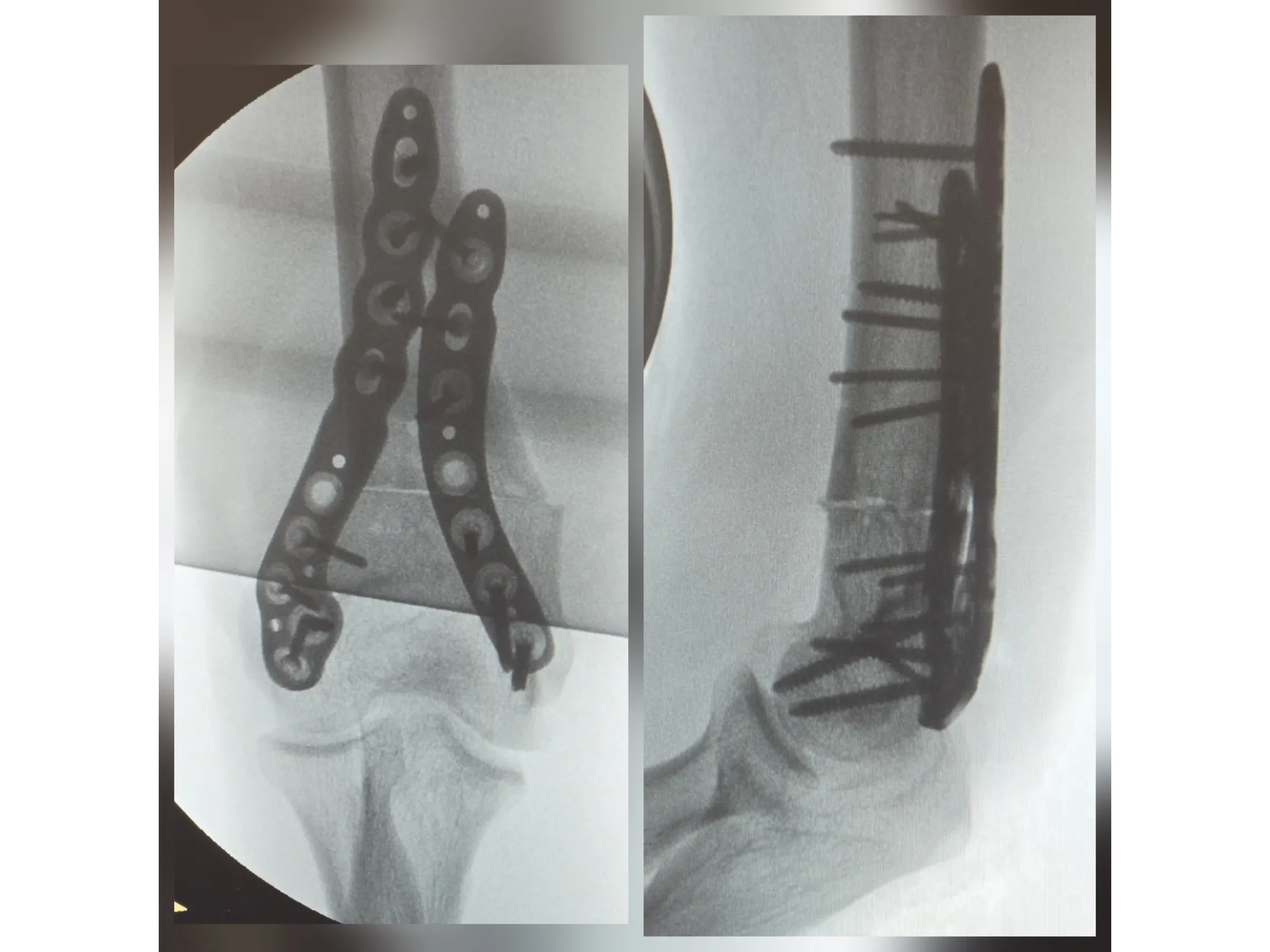
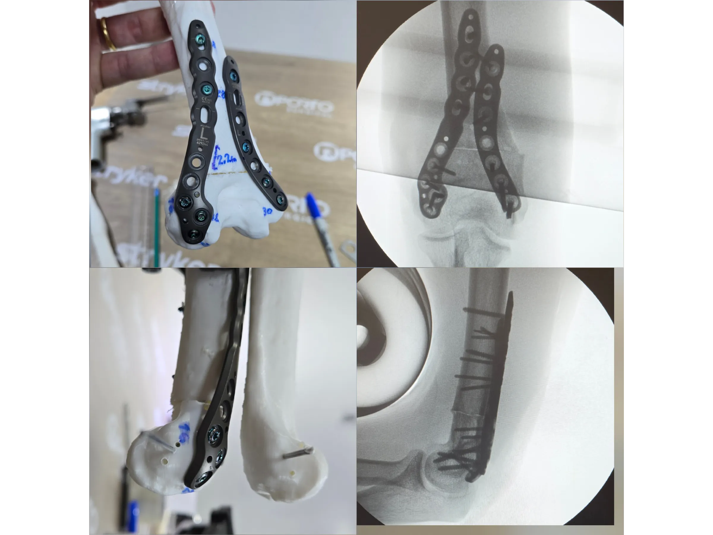
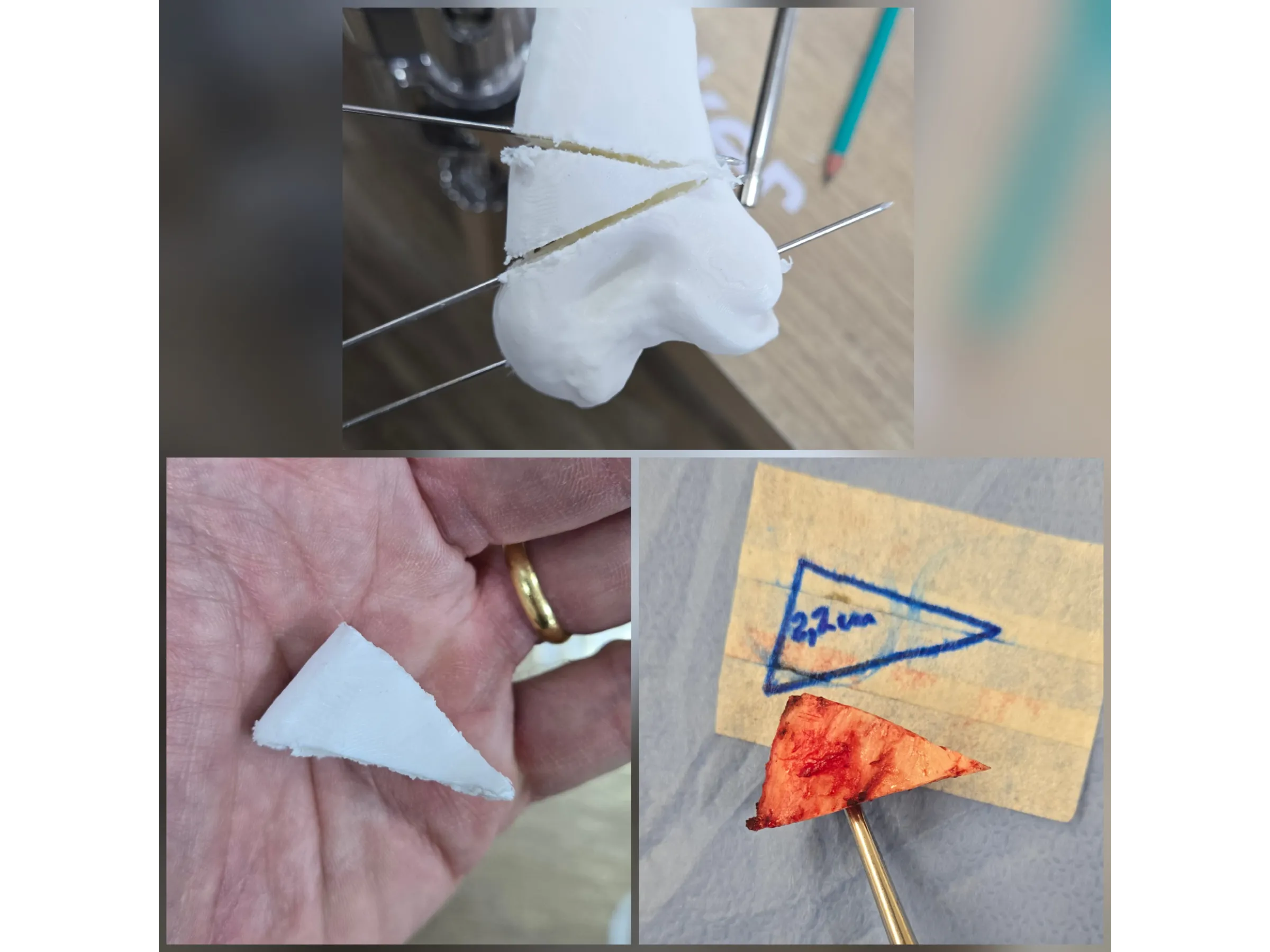
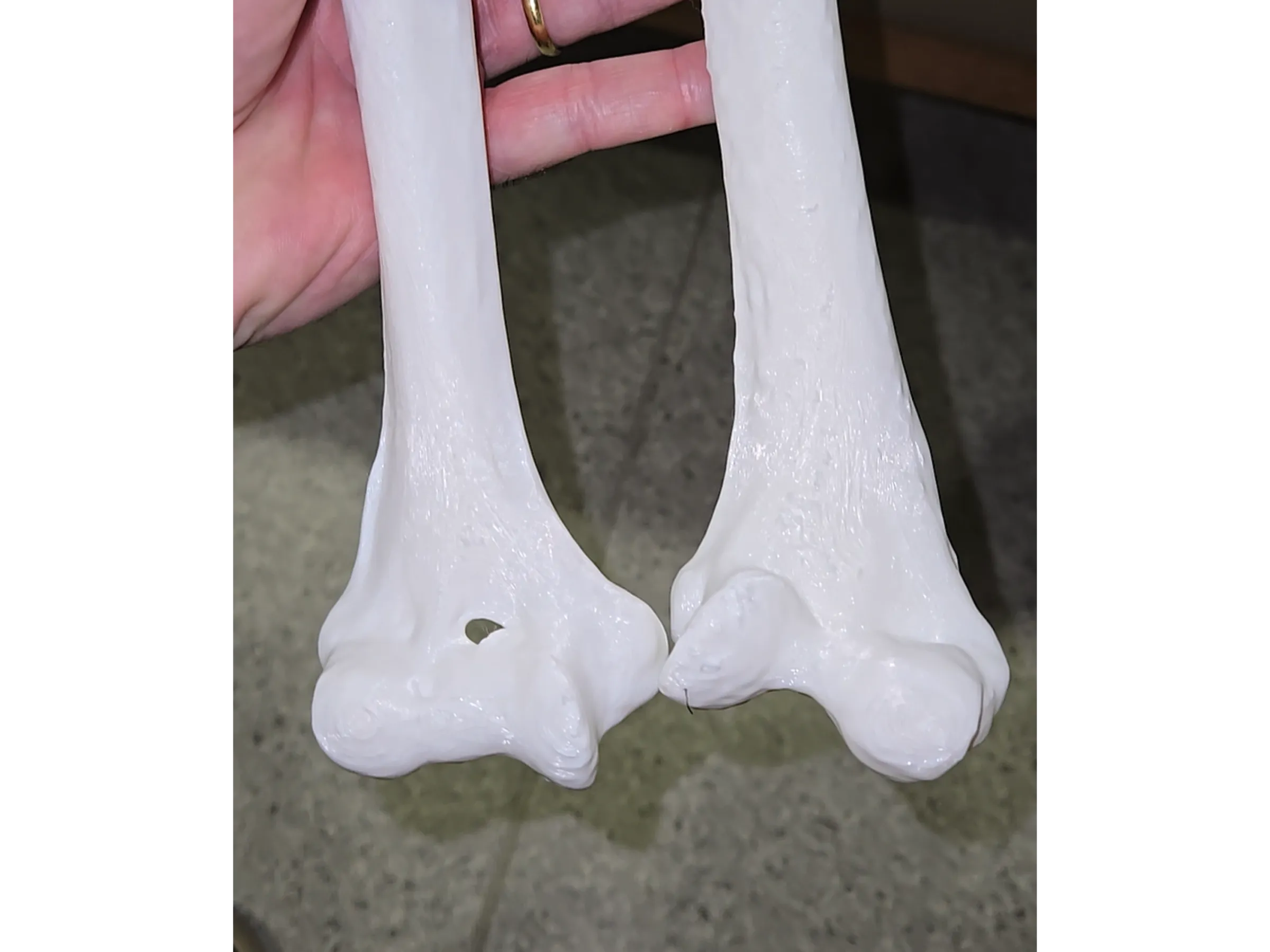
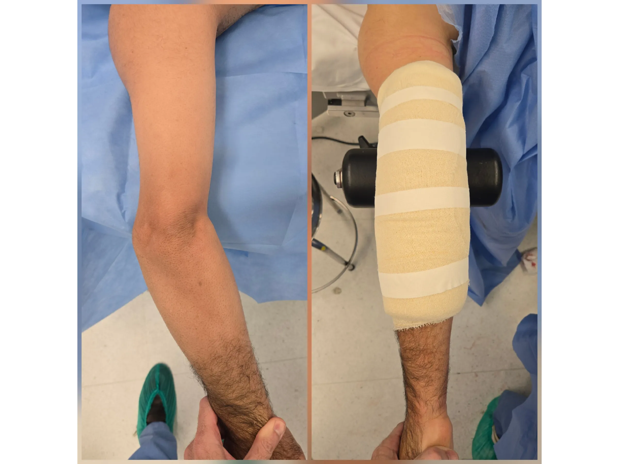
Correction of cubitus varus after supracondylar fracture represents a technical challenge in orthopedics. This training offers a practical and scientific approach to mastering distal humerus osteotomy, using three-dimensional planning with printed models, surgical guide, and fixation with locking plates, presented in an objective and highly visual format.
Training Focus:
- Preoperative planning with 3D printing and surgical guide;
- Strategic positioning for distal humerus exposure;
- Identification, release, and anterior transposition of the ulnar nerve;
- Guided osteotomy and fixation with locking plates;
- Postoperative evaluation focusing on stability and alignment.
Detailed Content:
- Precise Three-Dimensional Planning: reduction of intraoperative risks and anatomical fidelity;
- Safe Distal Humerus Exposure: muscle preservation and optimization of surgical access;
- Advanced Neurological Protection: release and anterior transposition of the ulnar nerve to prevent complications;
- Guided Osteotomy: angular correction with axial alignment and biomechanical stability;
- Stable Locked Fixation: systematic sequence of plates and screws for maximum safety;
- Functional Anatomical Closure: aesthetic preservation and accelerated recovery;
- Objective Postoperative Evaluation: effective correction with minimal morbidity.
Included Material:
Detailed PDF: the complementary material presents preoperative planning protocols with 3D printing, techniques for exposure and ulnar nerve protection, step-by-step guided osteotomy using a surgical guide, strategies for locked fixation, and recommendations for postoperative evaluation.
Enhance your practice in orthopedic trauma surgery. Enroll now and master the distal humerus osteotomy technique with 3D planning and locked fixation.
Elige opciones
Distal Humerus Osteotomy – Cubitus Varus Deformity

HD Surgical Videos
High-quality procedure-based training.