

Trimalleolar Fracture – Lateral Decubitus Approach
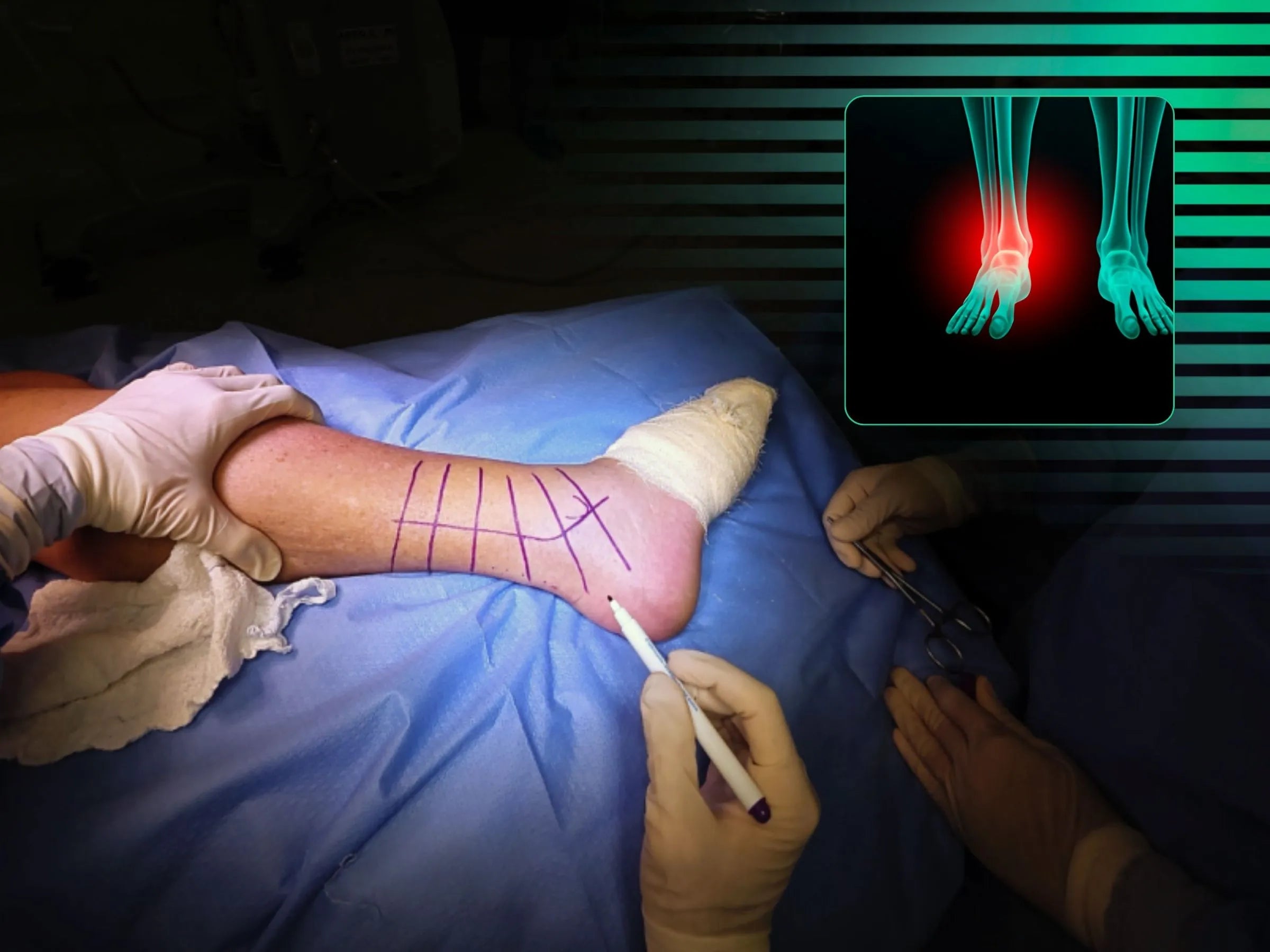
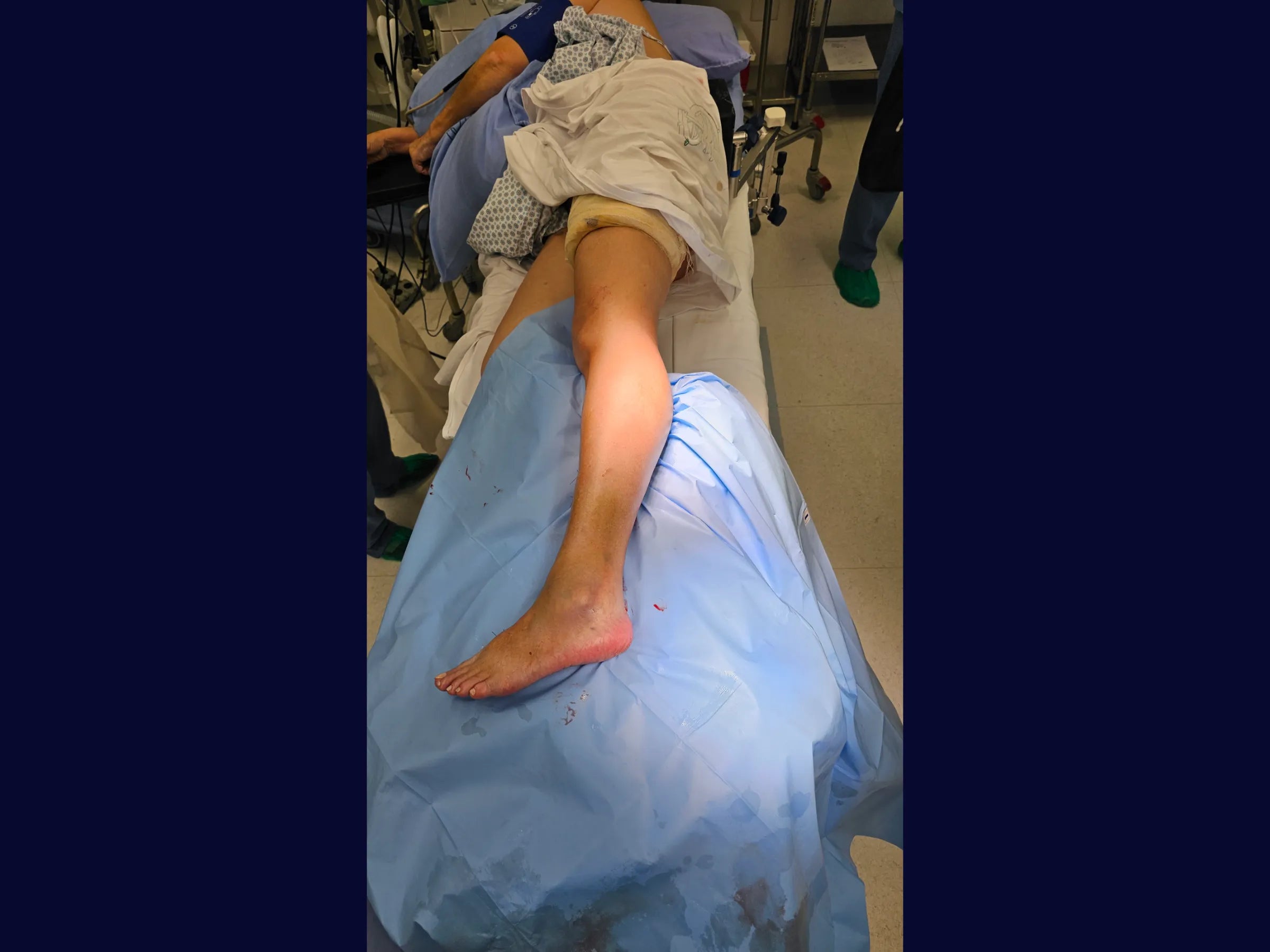
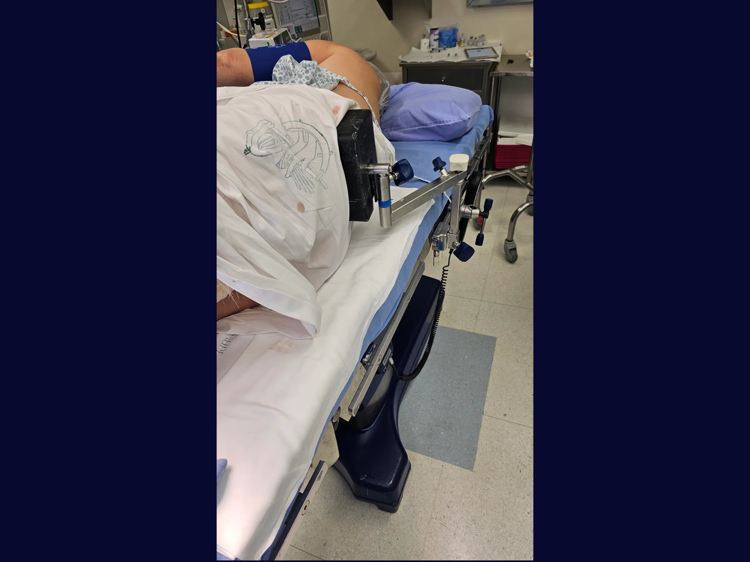
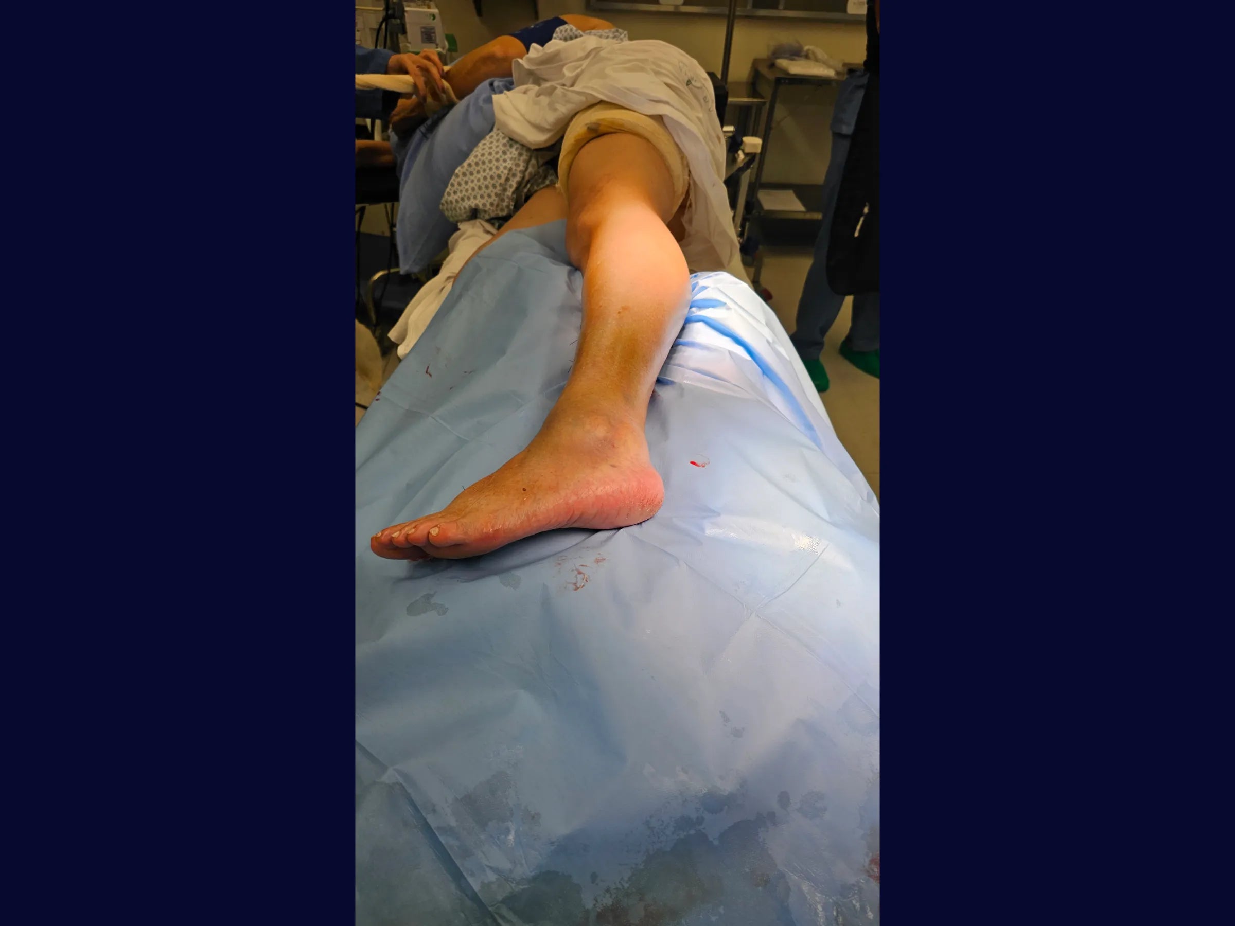
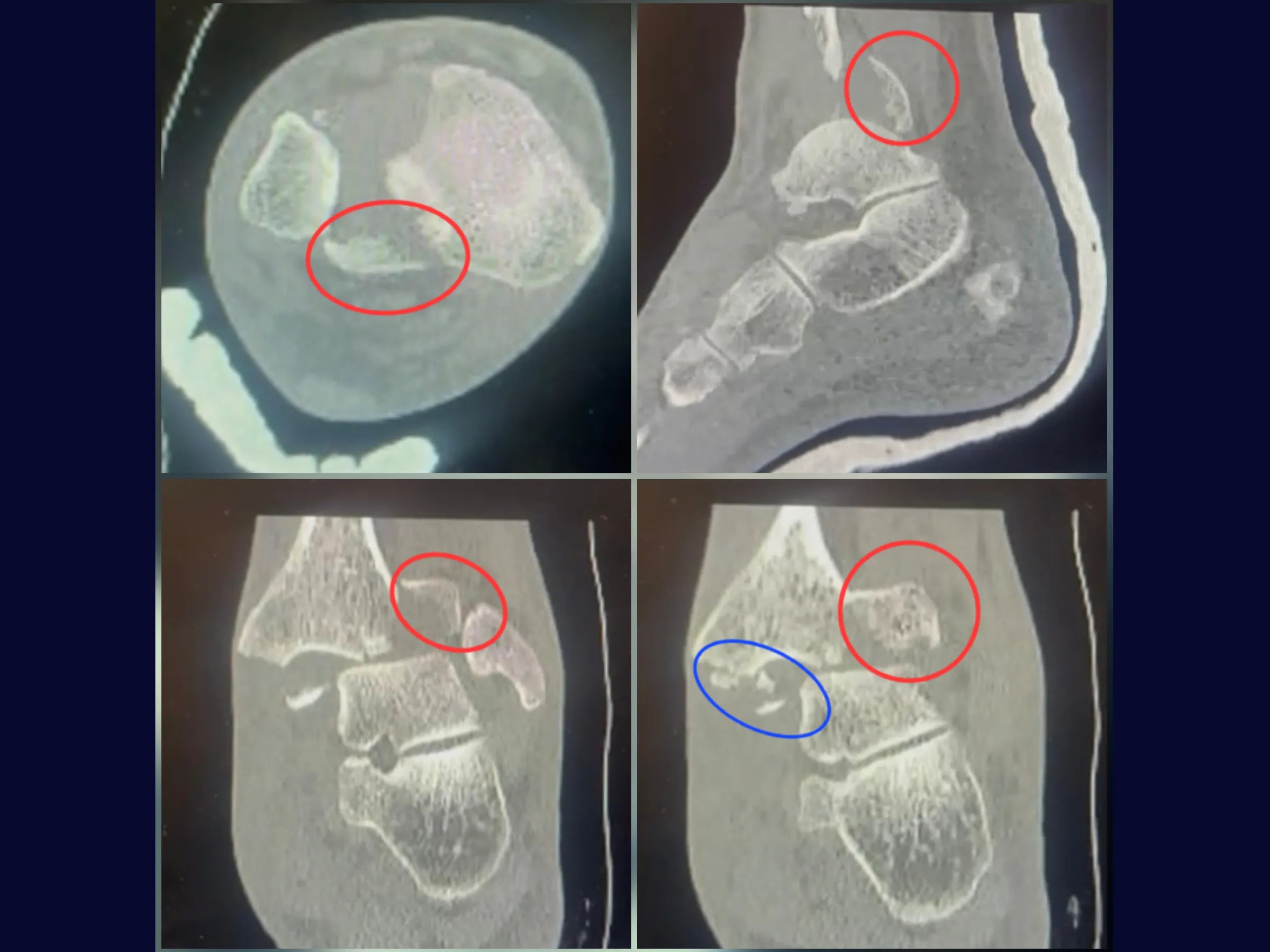
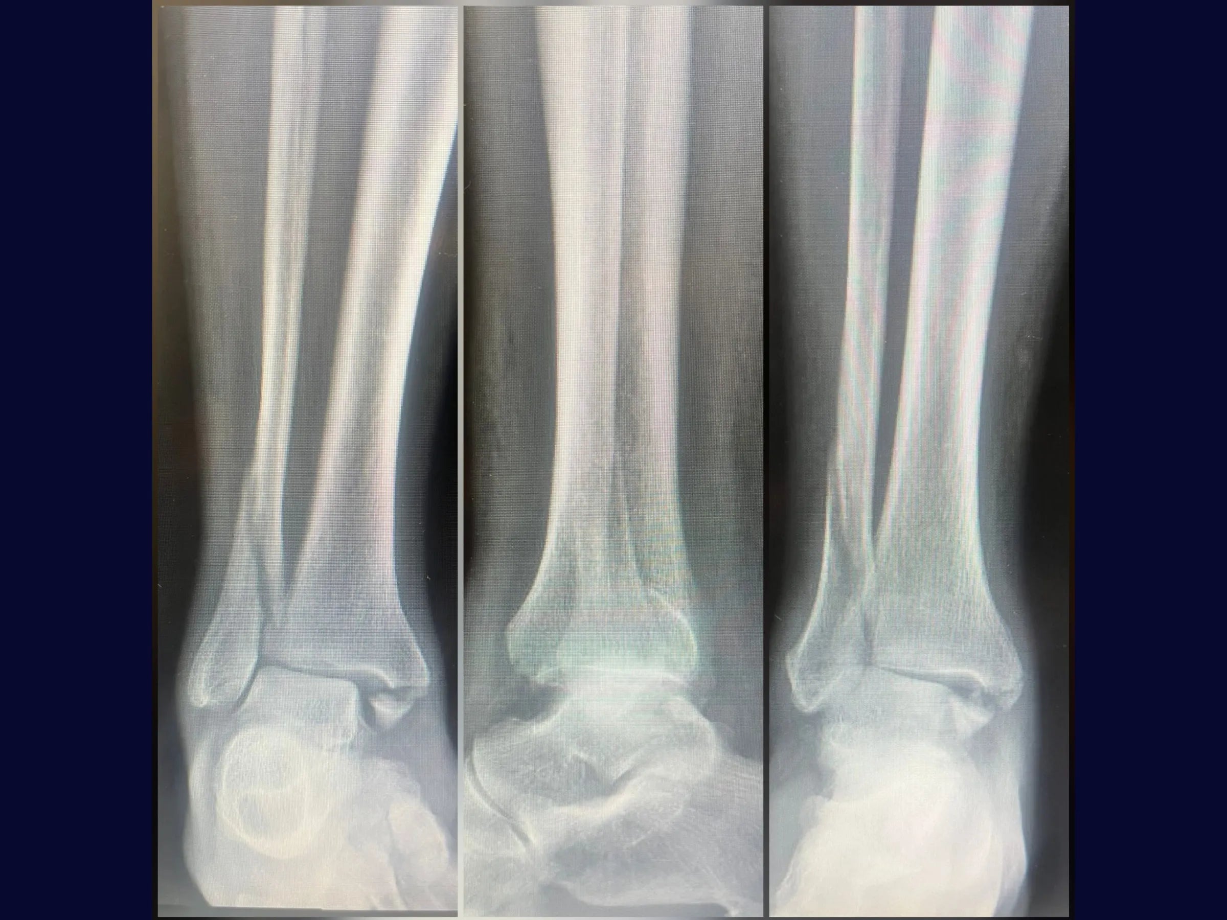
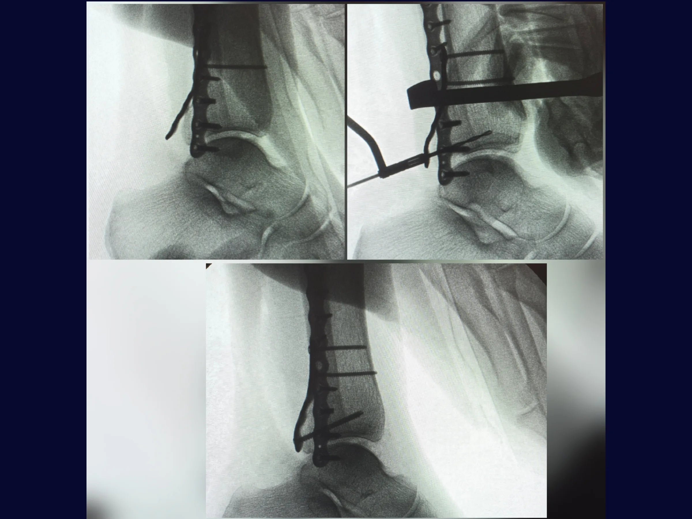
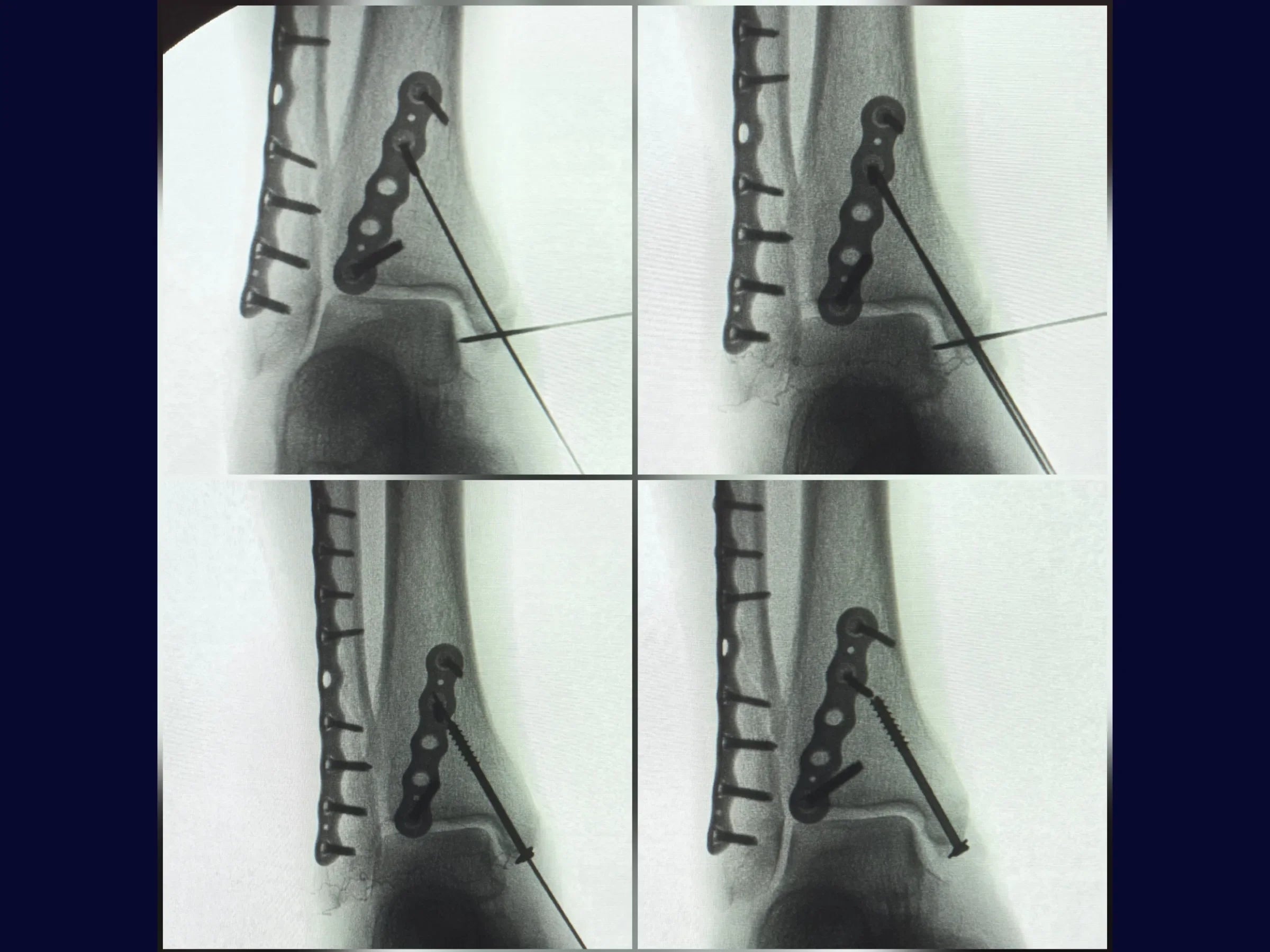
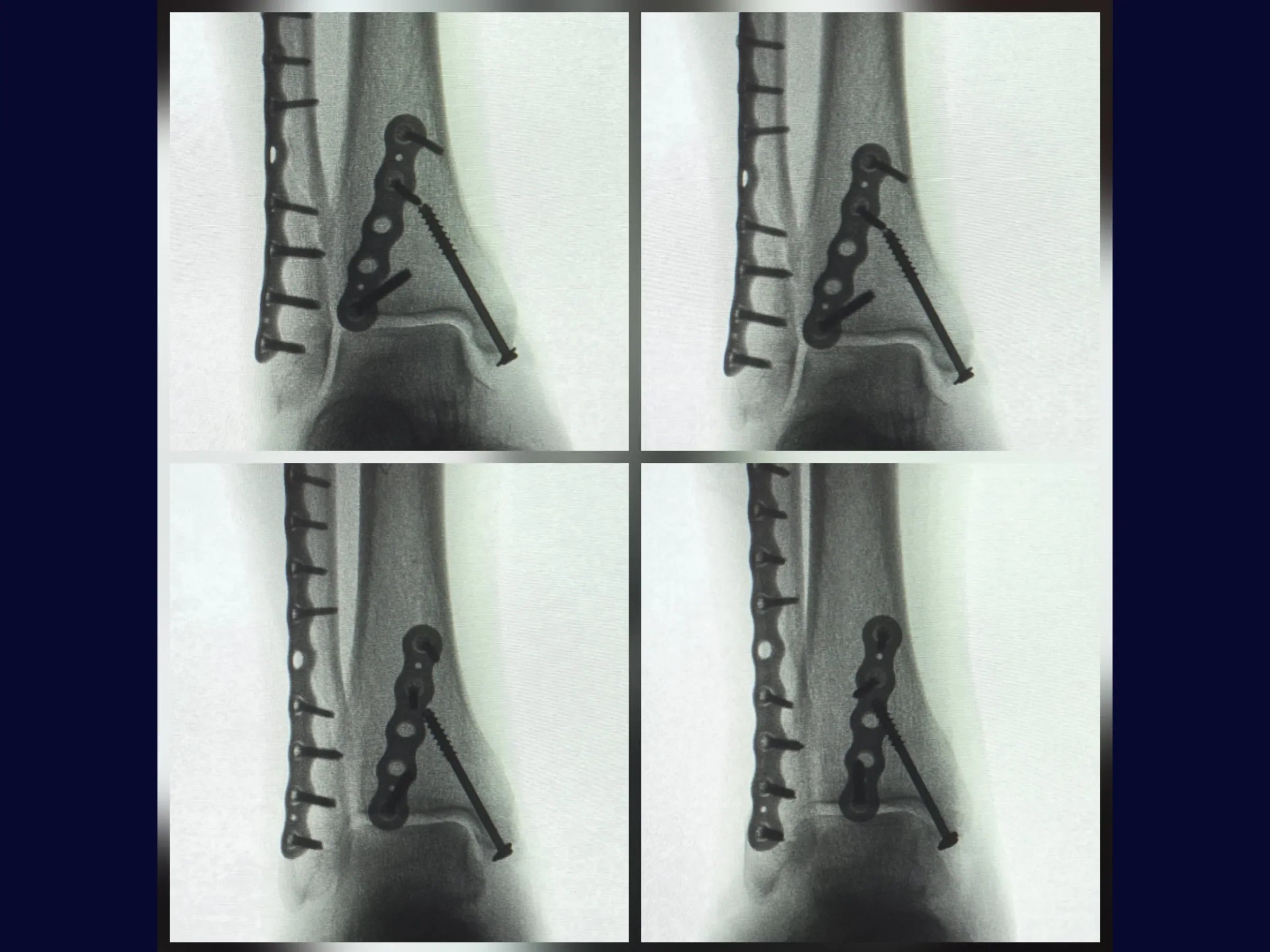
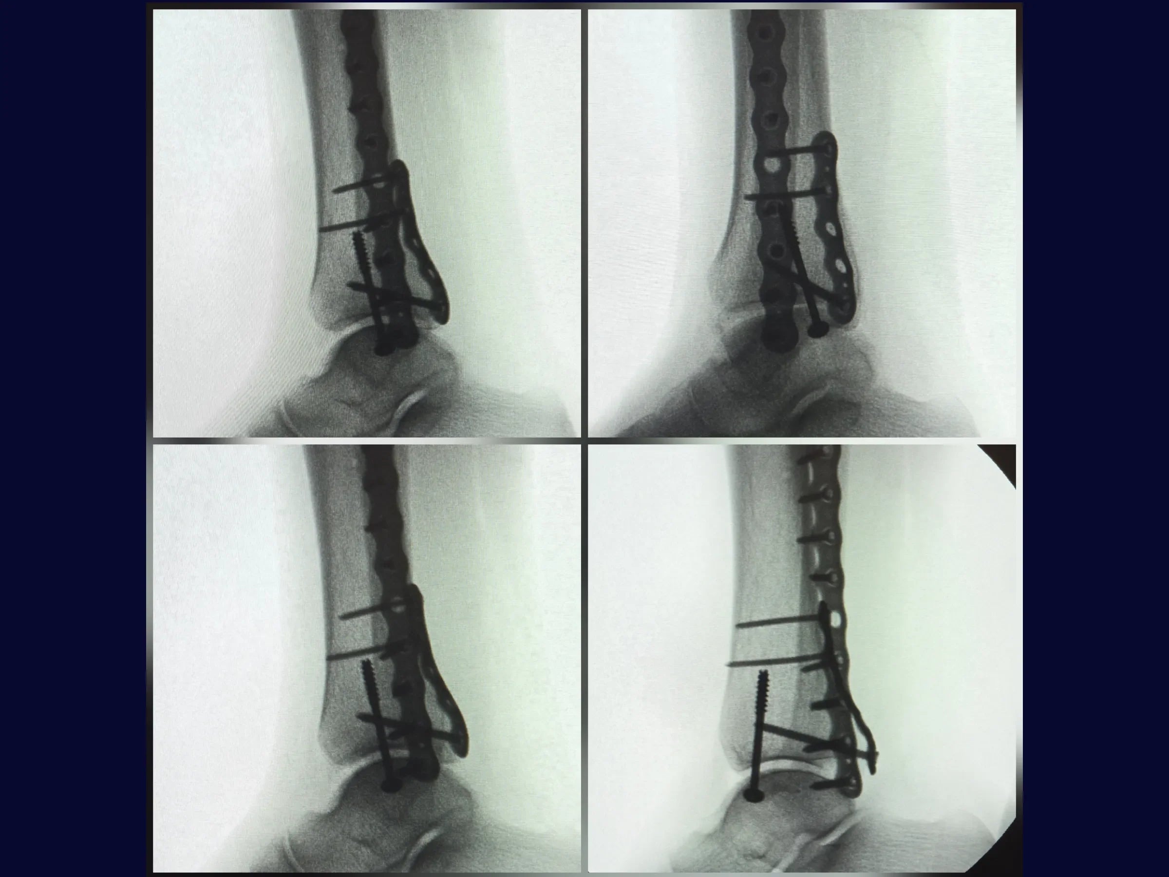
Trimalleolar fractures represent a challenge due to the anatomical complexity and the need for complete joint stability. This training shows, in 4K video from the surgeon’s perspective, the systematic approach to reduction and fixation of the three malleoli, ensuring 360° stability and functional recovery.
Training Focus:
· Strategic positioning in lateral and dorsal decubitus.
· Postero-lateral approach to the fibula and posterior malleolus.
· Fixation of the lateral malleolus with a low-profile locking plate.
· Indirect reduction and fixation of the posterior malleolus with a contoured plate.
· Medial approach with joystick wire and cannulated screw with washer.
Detailed Content:
· Anatomical Reduction of the Fibula: Use of fragmentary vertices and pointed reduction clamp to restore length.
· Secure Fibula Fixation: Plate aligned with the shear vector, ensuring compression and stability.
· Posterior Anti-Shear Stabilization: Technique with key screw and controlled dorsiflexion.
· Low-Profile Implant: Minimizes soft tissue irritation and preserves vascularization.
· Biological Preservation: Minimal periosteal stripping and careful dissection.
· Precise Medial Fixation: Use of joystick wire for rotational control and cannulated screw with washer for final compression.
· Radiographic Control: Intraoperative check with image intensifier.
· Safe Decubitus Sequence: Lateral-to-dorsal conversion without contamination risk.
Included Material:
Detailed PDF: Contains objective description of the technique, positioning sequence, approaches and fixations, as well as practical protocols for tissue preservation, use of low-profile implants, and strategies for 360° joint stability.
Enhance your precision in trimalleolar fracture fixation. Enroll now and master the combined approach techniques for the lateral, posterior, and medial malleoli.
Elige opciones
Trimalleolar Fracture – Lateral Decubitus Approach

HD Surgical Videos
High-quality procedure-based training.