

Dr. Sávio Chami
Orthopedist
Diaphyseal Fractures of the Radius and Ulna
⏳ 15s grátis
🌐 You can change subtitles and audio in the video settings ⚙️ 🇬🇧 🇧🇷 🇫🇷 🇪🇸 🇵🇹 🇺🇸
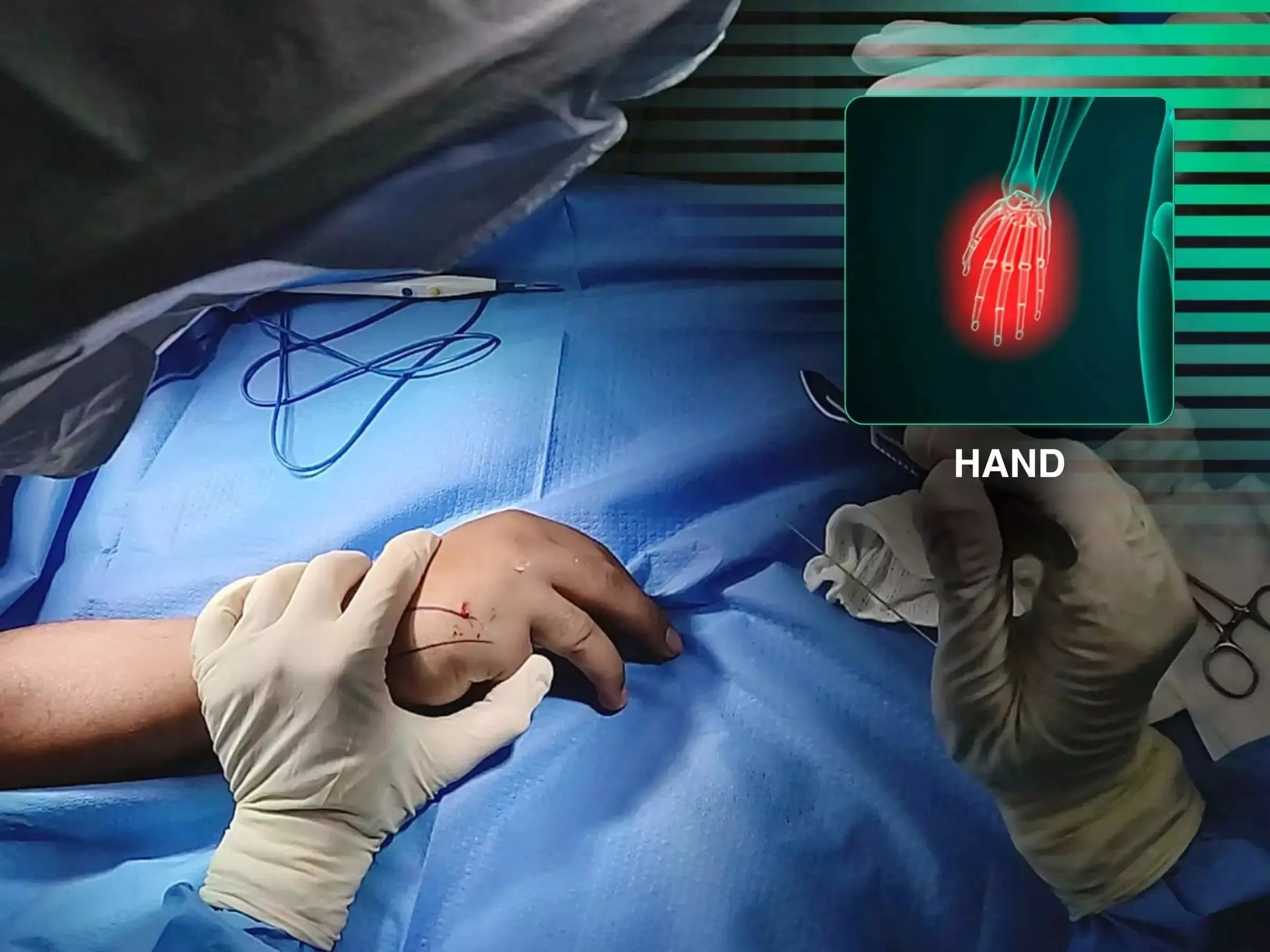
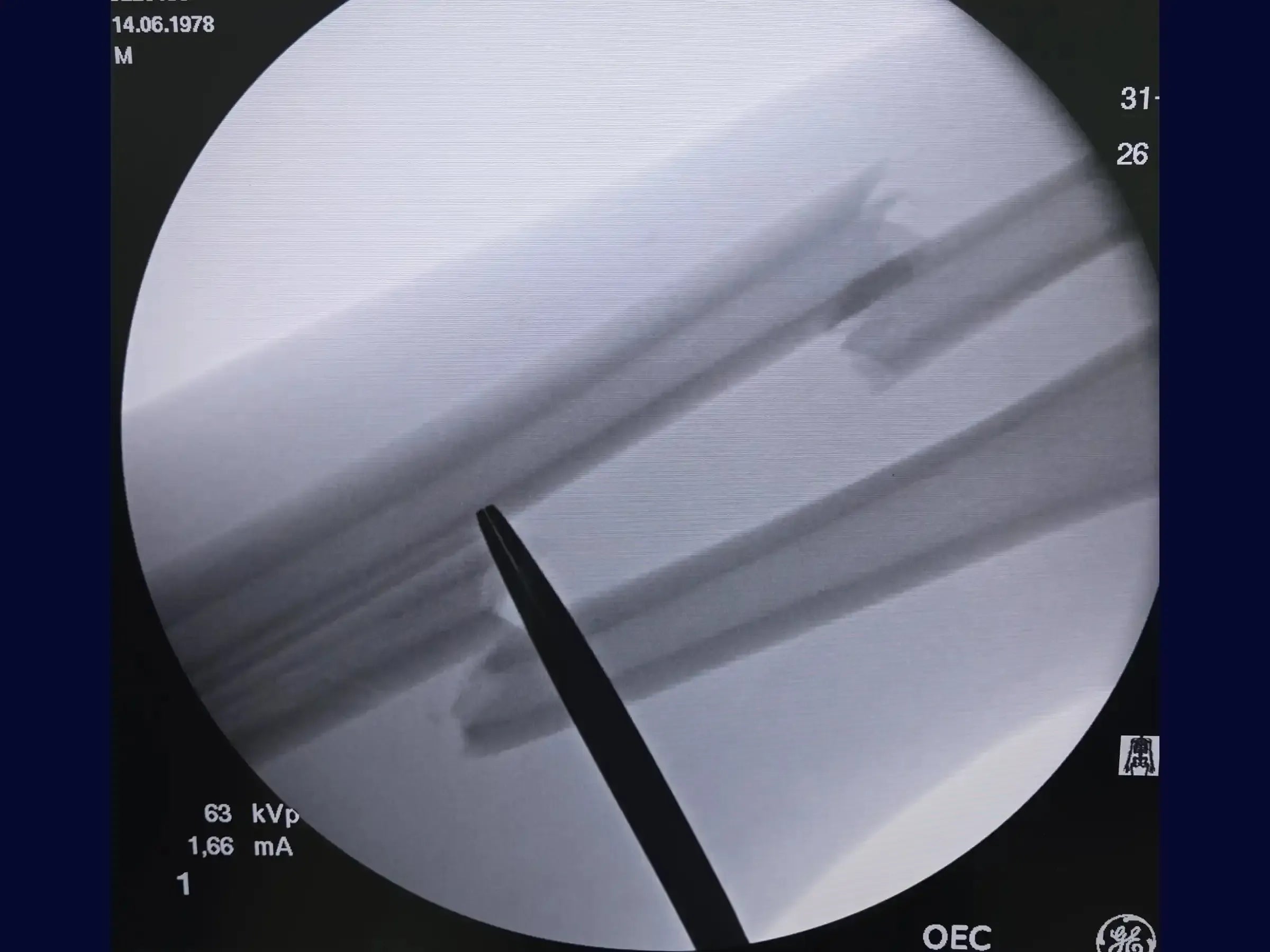
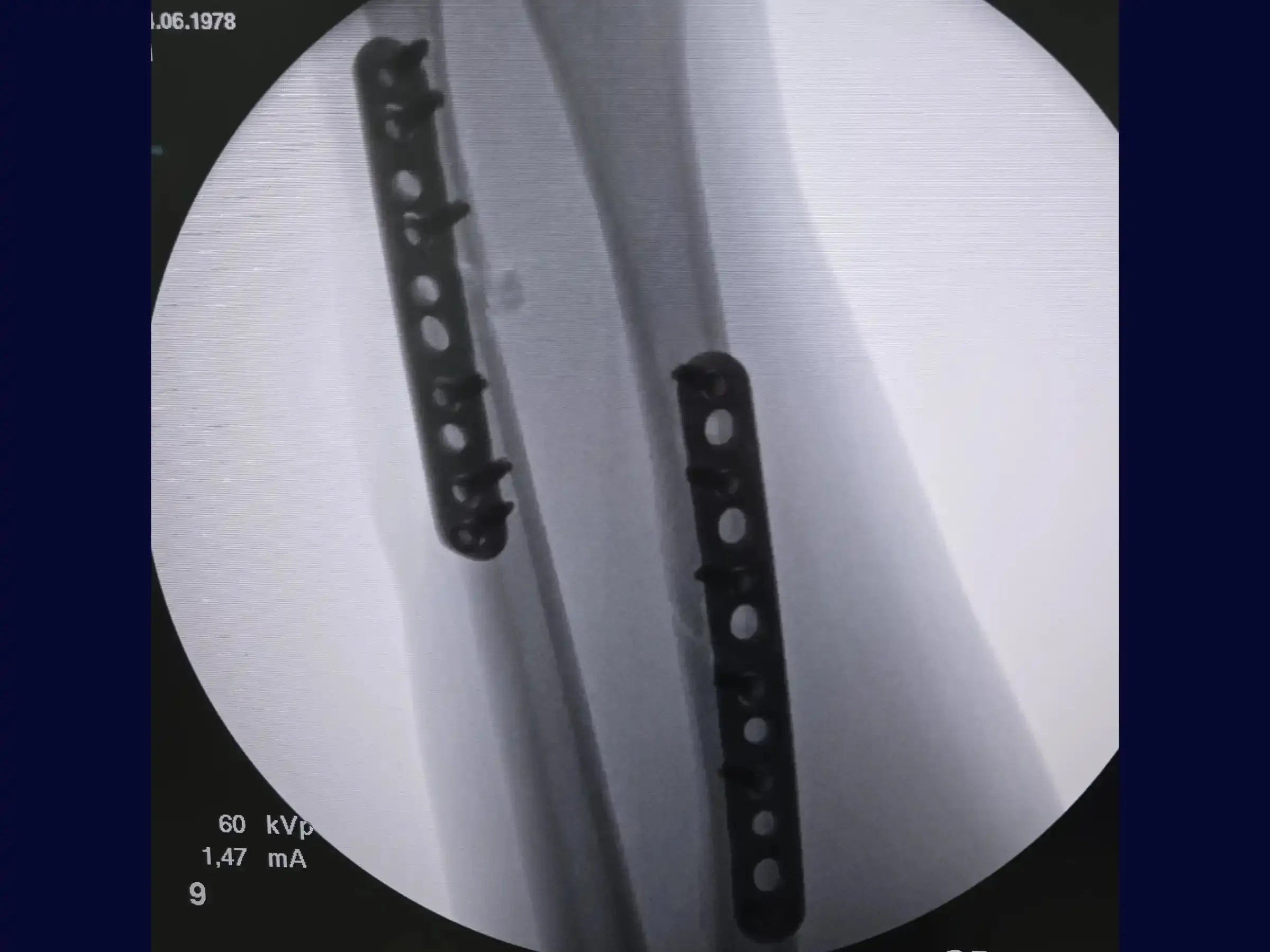
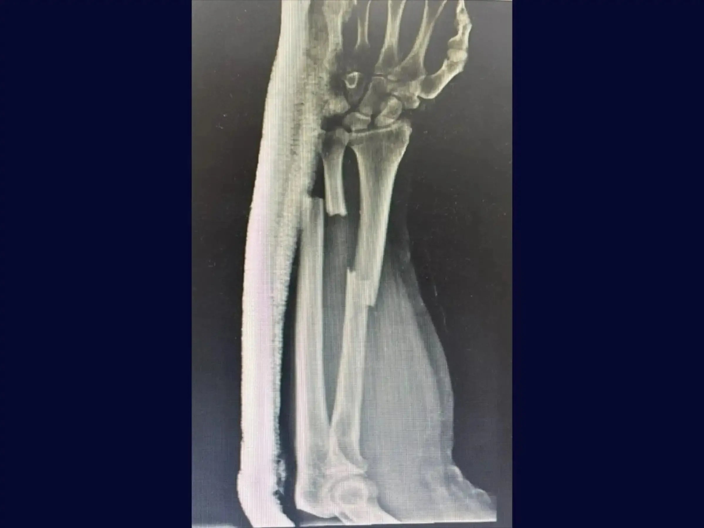
Master the surgical treatment of radial and ulnar shaft fractures with this comprehensive training. Explore surgical approaches that emphasize anatomic preservation and clean technique, from planning and access to reduction and fixation, aiming to optimize patient recovery.
Training Focus:
- Surgical treatment of radial and ulnar shaft fractures.
- Dorsal approach (Thompson) for radial fractures.
- Traditional approach for ulnar fractures.
- Anatomic reduction and fracture stabilization.
- Tissue preservation and minimization of bleeding.
Detailed Content:
Treatment of Radial Shaft Fracture (Thompson Approach):
- Planning and Marking: Learn to mark and palpate Lister's tubercle and the lateral epicondyle, drawing a line between them. Utilize muscle palpation to define the exact location of the skin incision and the image intensifier to locate the fracture site.
- Incision and Dissection: Master the skin incision with electrocautery and hemostasis. Deepen the dissection to identify muscle intervals, using gauze for blunt dissection and visualizing the fascia for the ideal access plane, preserving the natural planes of the forearm.
- Fascia Opening and Exposure: Make a small incision through the fascia, creating the interval between the extensor digitorum communis and extensor carpi radialis brevis muscles, with careful progression to preserve underlying structures and prevent muscle damage.
- Fracture Identification and Cleaning: Palpate the proximal radial fragment and define the trajectory towards the bone with the index finger. Make gentle contact with the scalpel on the bone surface to find the apex and the proximal fragment, maintaining dissection along the anatomic planes and exposing the fracture ends. Clean the fracture edges to find a reference point for reduction.
Treatment of Ulnar Shaft Fracture (Distal Third):
- Positioning and Access: Position the patient in supine decubitus with the arm flexed. Perform dissection between the extensor carpi ulnaris and flexor carpi ulnaris muscles, opening the fascia carefully to avoid muscle damage.
- Fracture Identification and Periosteum Preservation: Identify the fracture, even with distorted anatomy. Elevate the periosteum anatomically to minimize soft tissue trauma and facilitate closure over the plate.
- Bone Bed Preparation and Reduction: Create the bone bed precisely on the dorsal (tension) cortex of the ulna. Clean the fracture edges in search of reference points, using a "bone crest" as a guide for anatomic reduction and avoiding the creation of a segmental fracture.
- Stabilization and Plating: Reposition the reduction clamp distally to optimize manipulation. With the radius already stabilized, achieve ulnar reduction for excellent stability and alignment. Perform plate pre-tensioning to avoid a gap in the opposite cortex and use a 3.5 mm locked DCP (Dynamic Compression Plate).
- Closure and Recovery: Close the periosteum over the plate, preserving all muscular structures. Respect anatomic dissection with total muscular preservation and perform layered closure for significantly improved postoperative recovery.
Included Material:
- Complete Technical PDF: A detailed didactic document covering surgical techniques for treating radial (Thompson approach) and ulnar (traditional approach) shaft fractures. Includes everything from initial planning and access to reduction and fragment fixation.
Enhance your forearm surgery skills. Enroll now and master advanced osteosynthesis techniques for radial and ulnar shaft fractures, ensuring anatomic reduction and optimized recovery for your patients.
オプションを選択
Dr. Sávio Chami
Orthopedist
Diaphyseal Fractures of the Radius and Ulna
⏳ 15s grátis
🌐 You can change subtitles and audio in the video settings ⚙️ 🇬🇧 🇧🇷 🇫🇷 🇪🇸 🇵🇹 🇺🇸
Diaphyseal Fractures of the Radius and Ulna
セール価格R$ 20,00
Dr. Sávio Chami
Orthopedist
R$ 20,00
Members pay R$ 16,00 — save 20%

HD Surgical Videos
High-quality procedure-based training.