

Multifragmentary Distal Radius Fracture (Volar Approach)
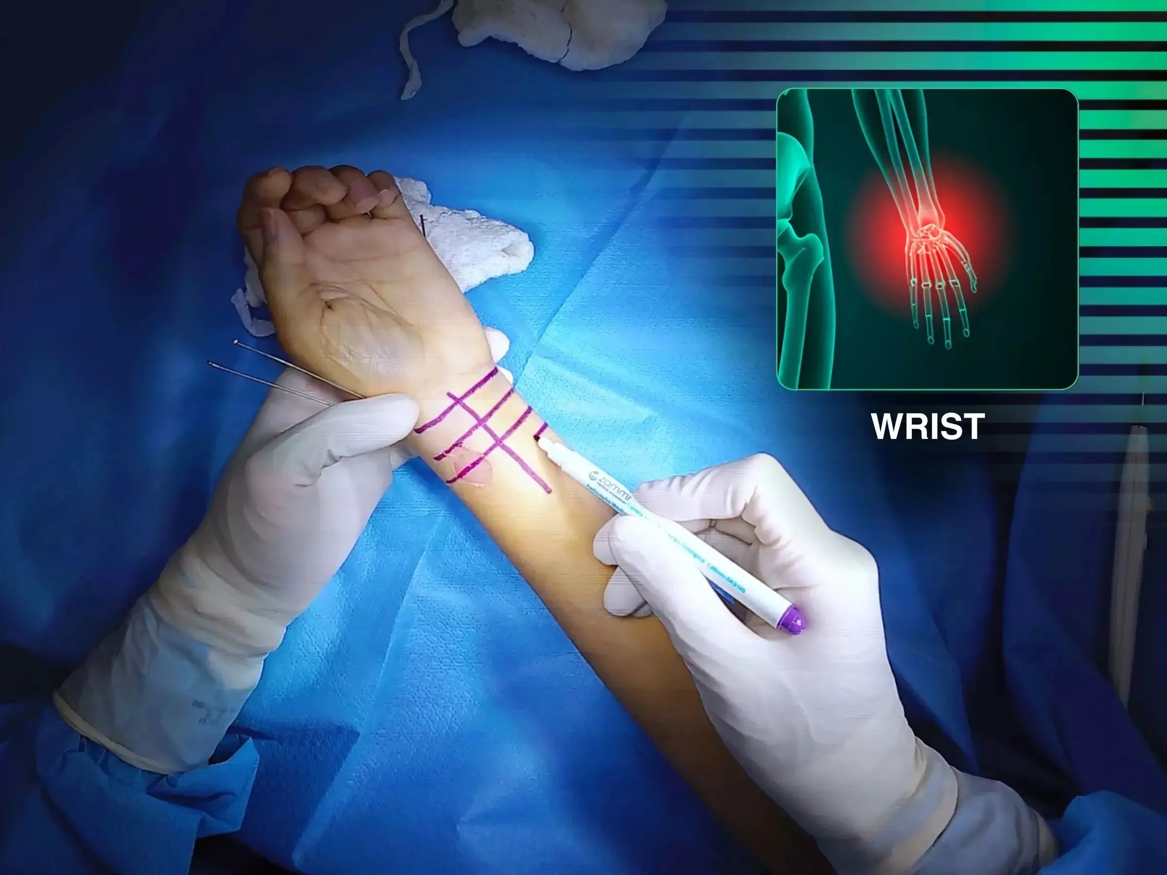
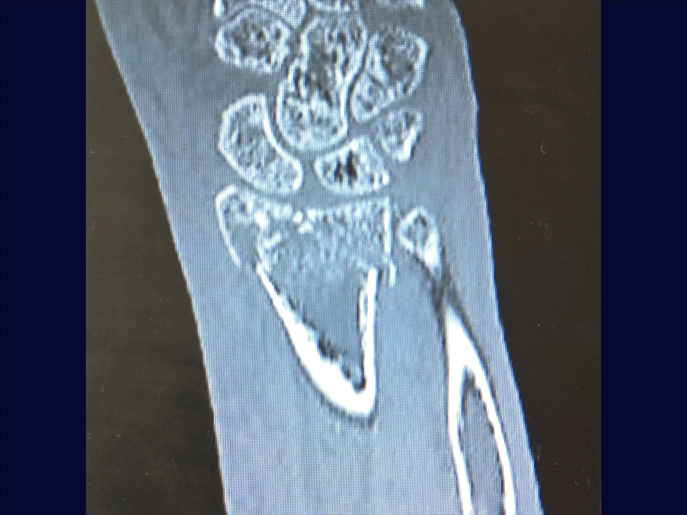
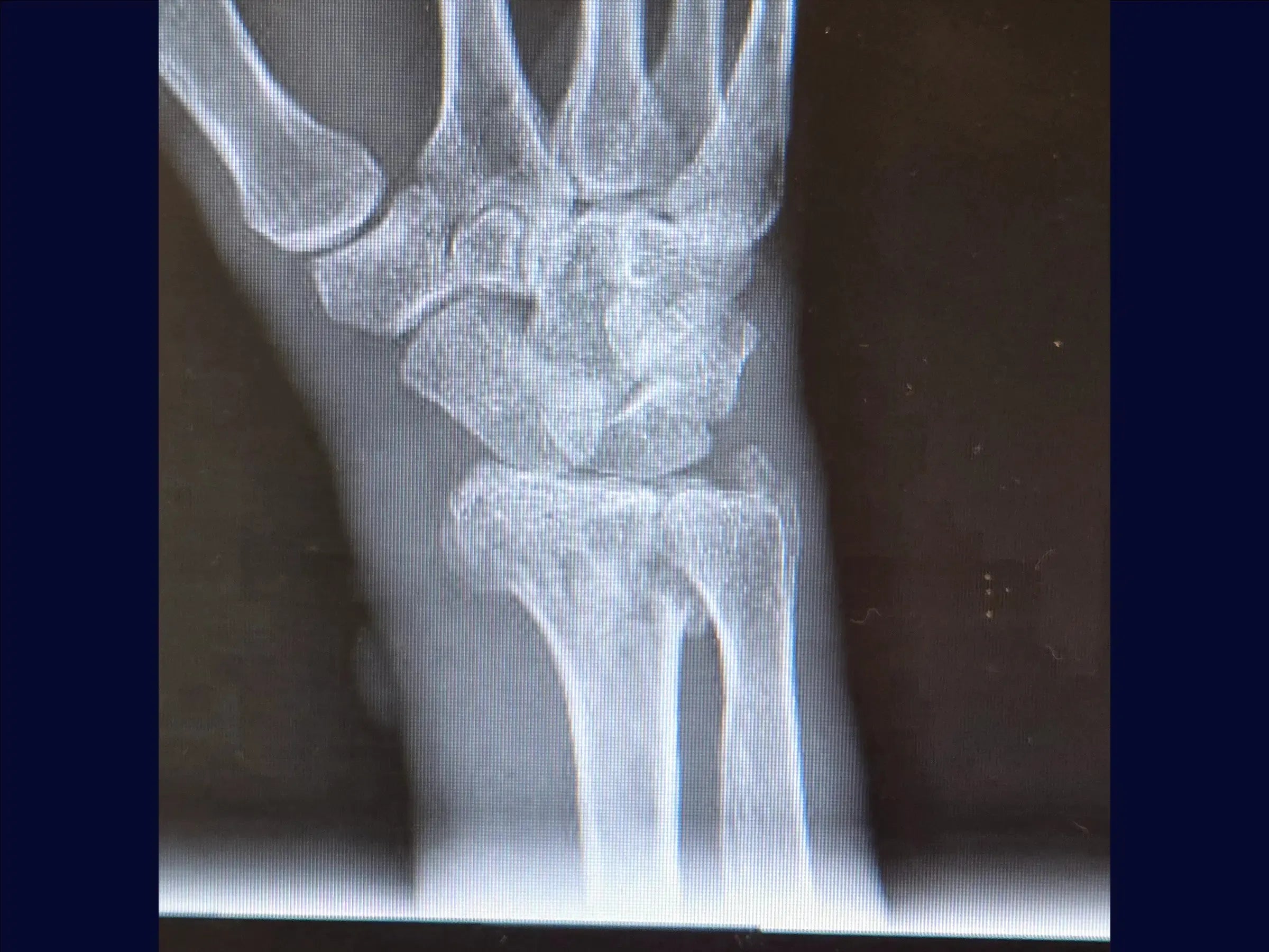
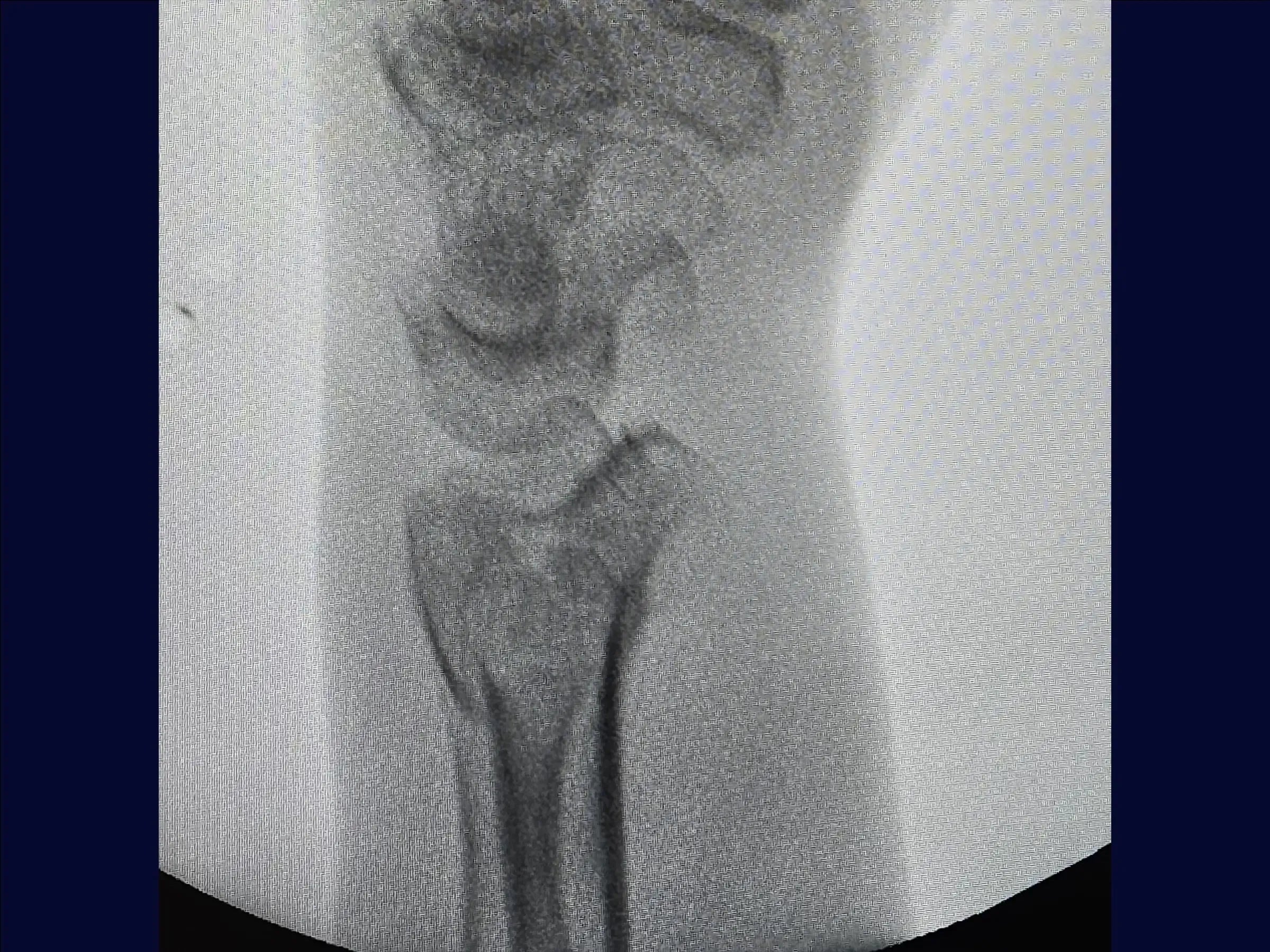
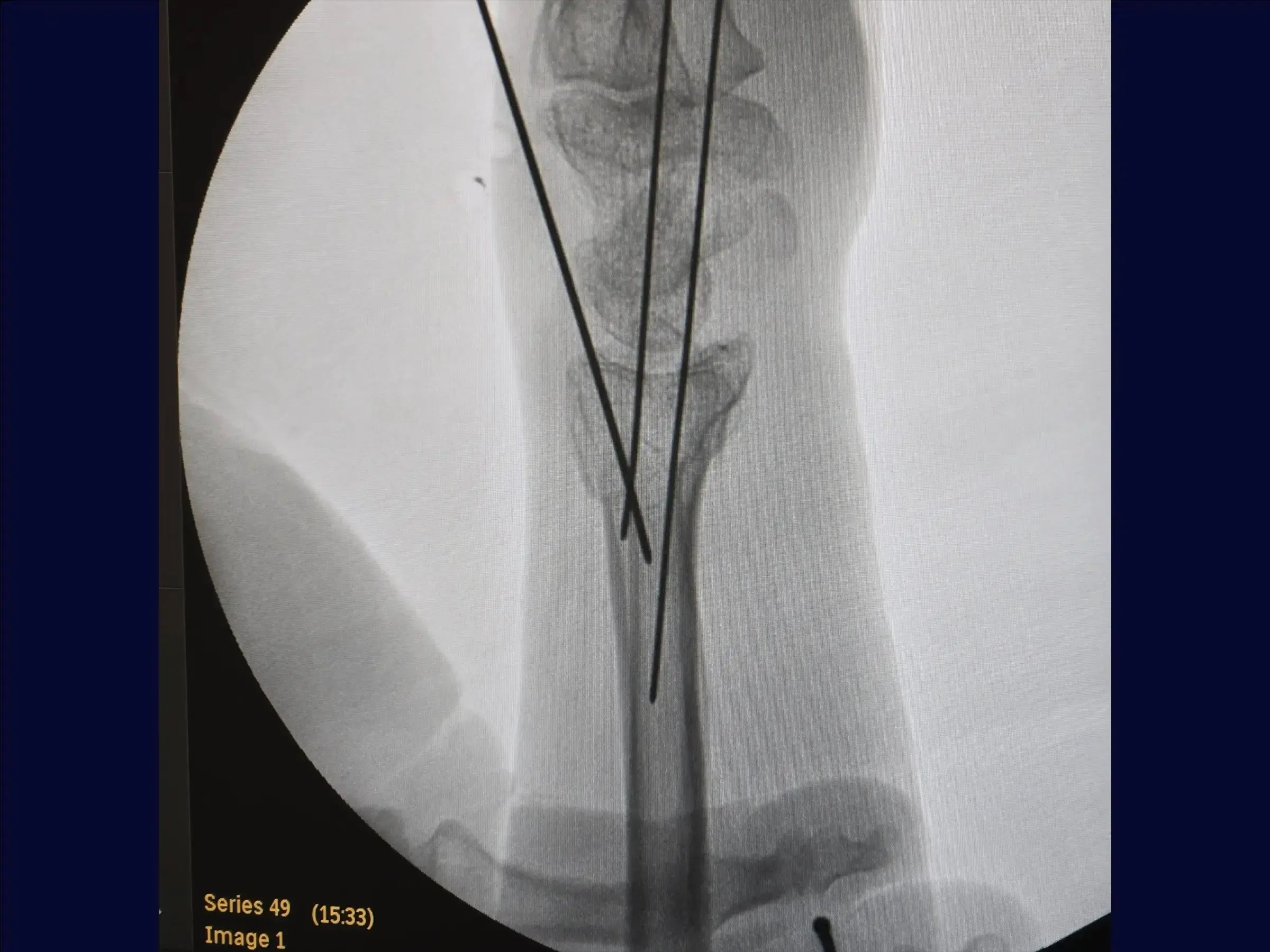
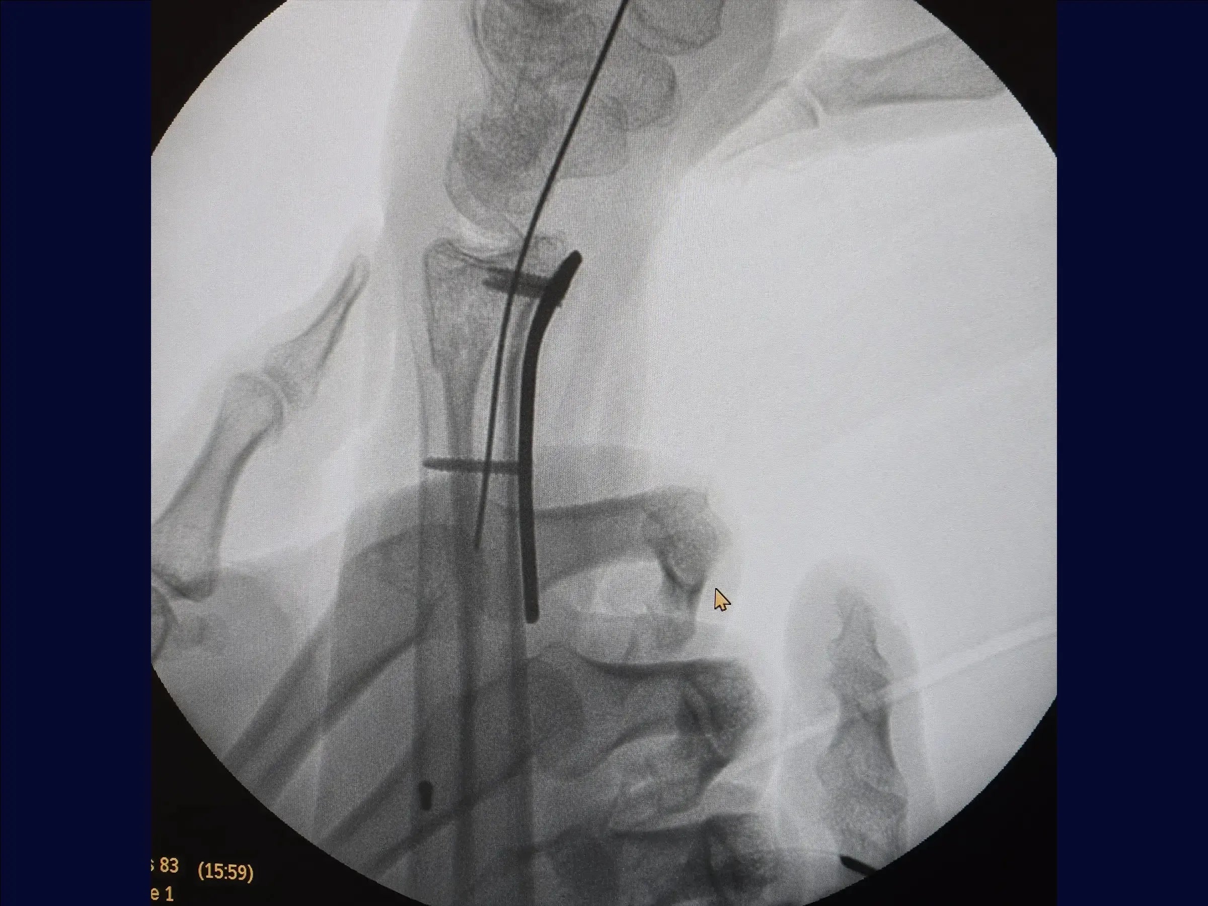
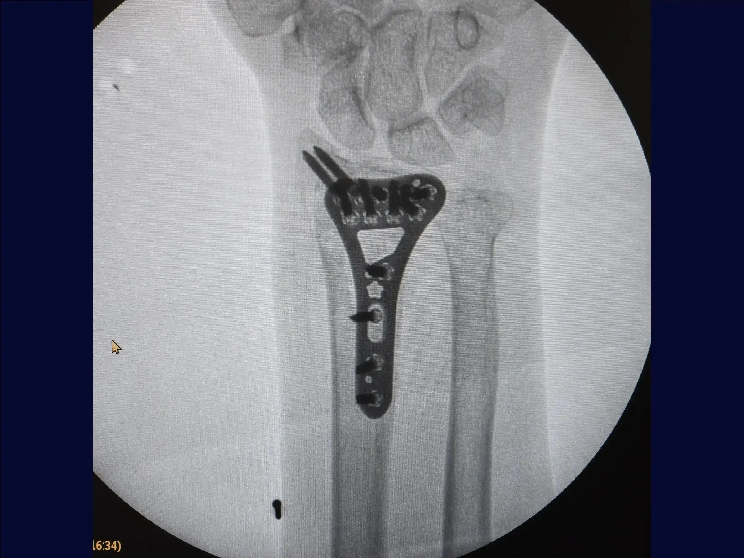
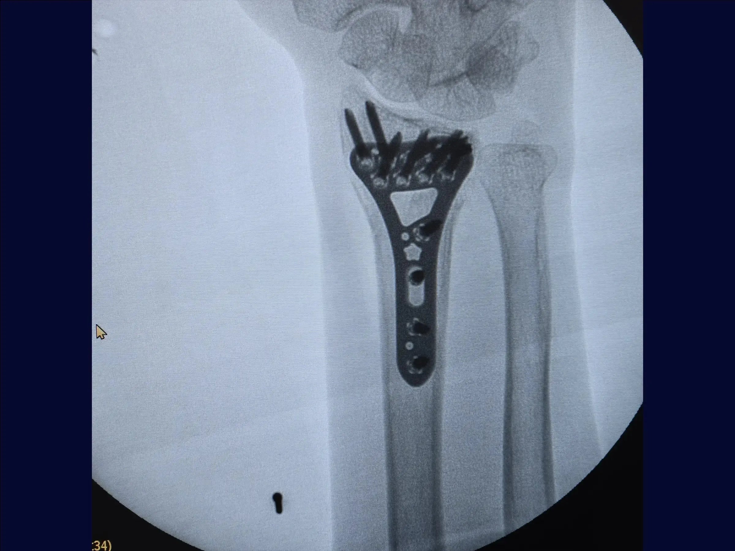
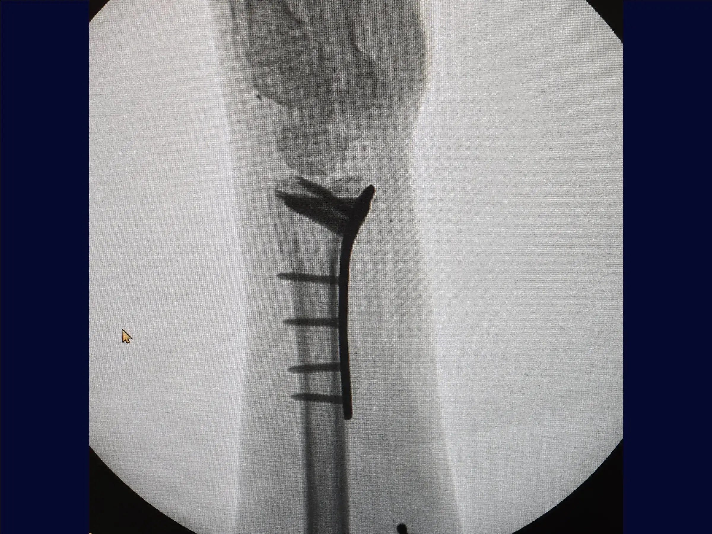
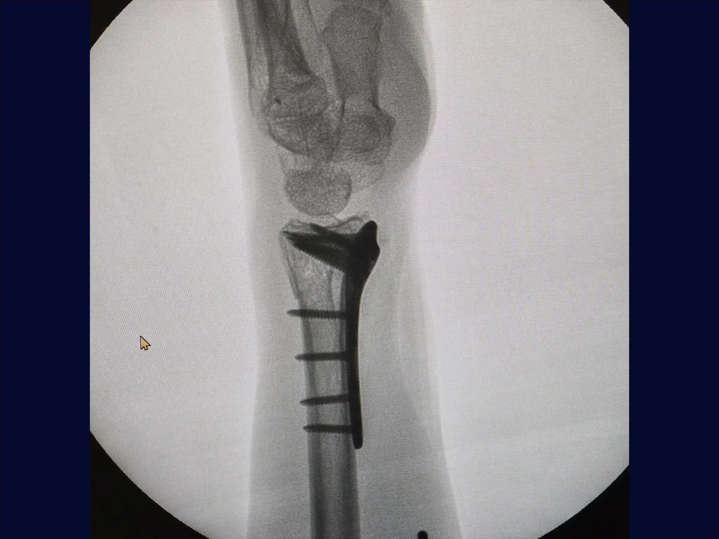
Master the surgical treatment of multifragmentary distal radius fractures with this training focused on the volar approach. The procedure aims to restore radial length and articular congruity, using a combination of manual traction, provisional percutaneous fixation, and a fixation strategy with a volar plate and screws of different lengths to stabilize the multiple fragments, including the dorsal component, without the need for an additional dorsal approach.
Training Focus:
- Treatment of multifragmentary distal radius fractures.
- Volar approach for reduction and fixation.
- Strategies to restore radial length and articular congruity.
- Fixation with a volar plate and screws of different lengths.
- Stabilization of the dorsal component without additional access.
Detailed Content:
- Assessment and Provisional Reduction: The training covers the analysis of images of multifragmentary distal radius fractures and reduction by manual traction (ligamentotaxis). It includes provisional percutaneous fixation with K-wires in the radial styloid and Lister's tubercle for fragment stabilization.
- Volar Surgical Approach: Details of the incision, coagulation, and dissection of tissue planes. It teaches how to retract tendons (flexor pollicis longus and flexor carpi radialis ulnarly) and protect the radial artery radially for a wide approach. The fascia is incised linearly.
- Exposure of the Pronator Quadratus: The course covers the identification of the pronator quadratus and how to position retractors. A needle delineates the articular margin. Access to the pronator quadratus is made in an L-shape, with subperiosteal elevation to expose the fracture lines, observing the already reduced volar surface.
- Preparation and Volar Fixation: Retractors are exchanged, fracture vertices are cleaned, and the plate is introduced respecting the articular margin. It begins with a cortical screw in the oval hole for compression. Short screws are used to fix the volar fragment, and with digital maneuvers, long screws stabilize the dorsal surface without the need for another approach. The short screws are exchanged for long ones (18–20 mm), including the styloid, completing the osteosynthesis in the remaining holes.
- Closure and Final Results: The pronator quadratus is sutured in a straight fashion. The reduction is satisfactory, with radial anatomy close to normal. Skin closure is performed for a satisfactory outcome.
Included Material:
- Detailed PDF: Covers provisional fixation, volar surgical approach, radius exposure and plate implantation, and closure and final results. It also includes a practical summary of the procedure with key points.
Enhance your wrist surgery skills. Enroll now and master the techniques for treating multifragmentary distal radius fractures, ensuring functional restoration and stability for your patients.
Choose options
Multifragmentary Distal Radius Fracture (Volar Approach)

HD Surgical Videos
High-quality procedure-based training.