Skip to content

Cart

Your cart is empty

Bone Reconstruction

Sort by

76 products

Filters

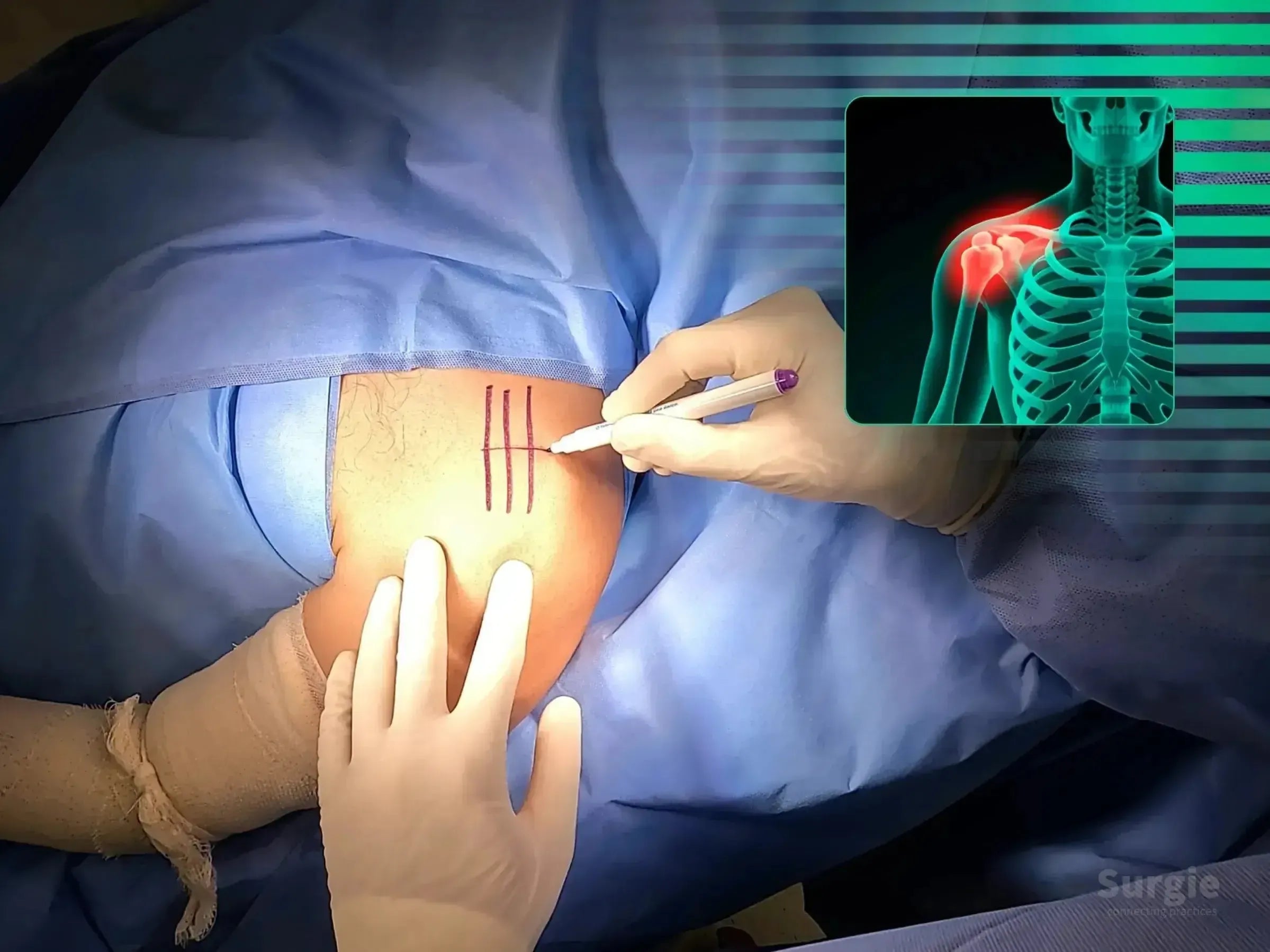
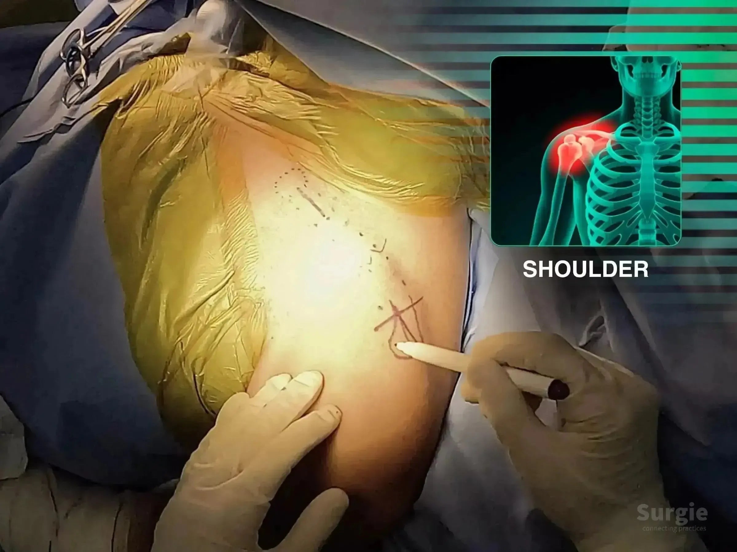
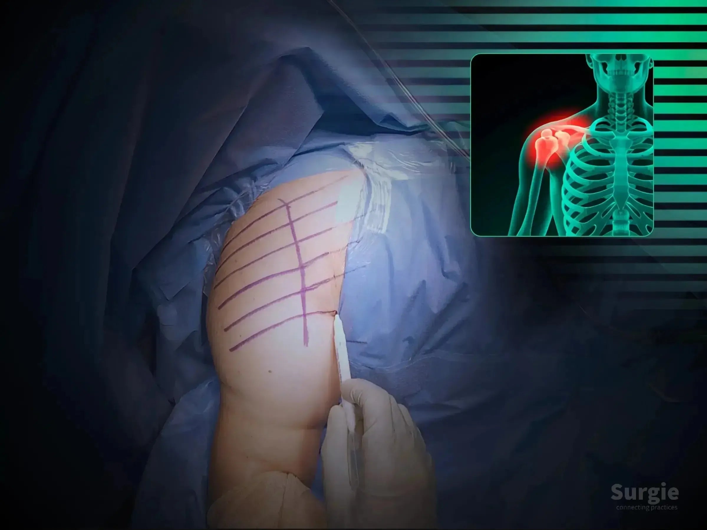
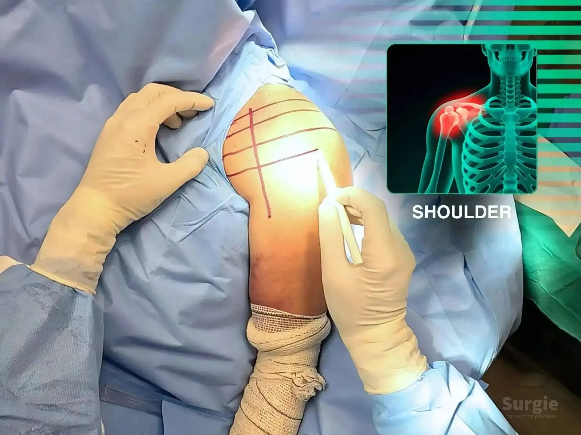
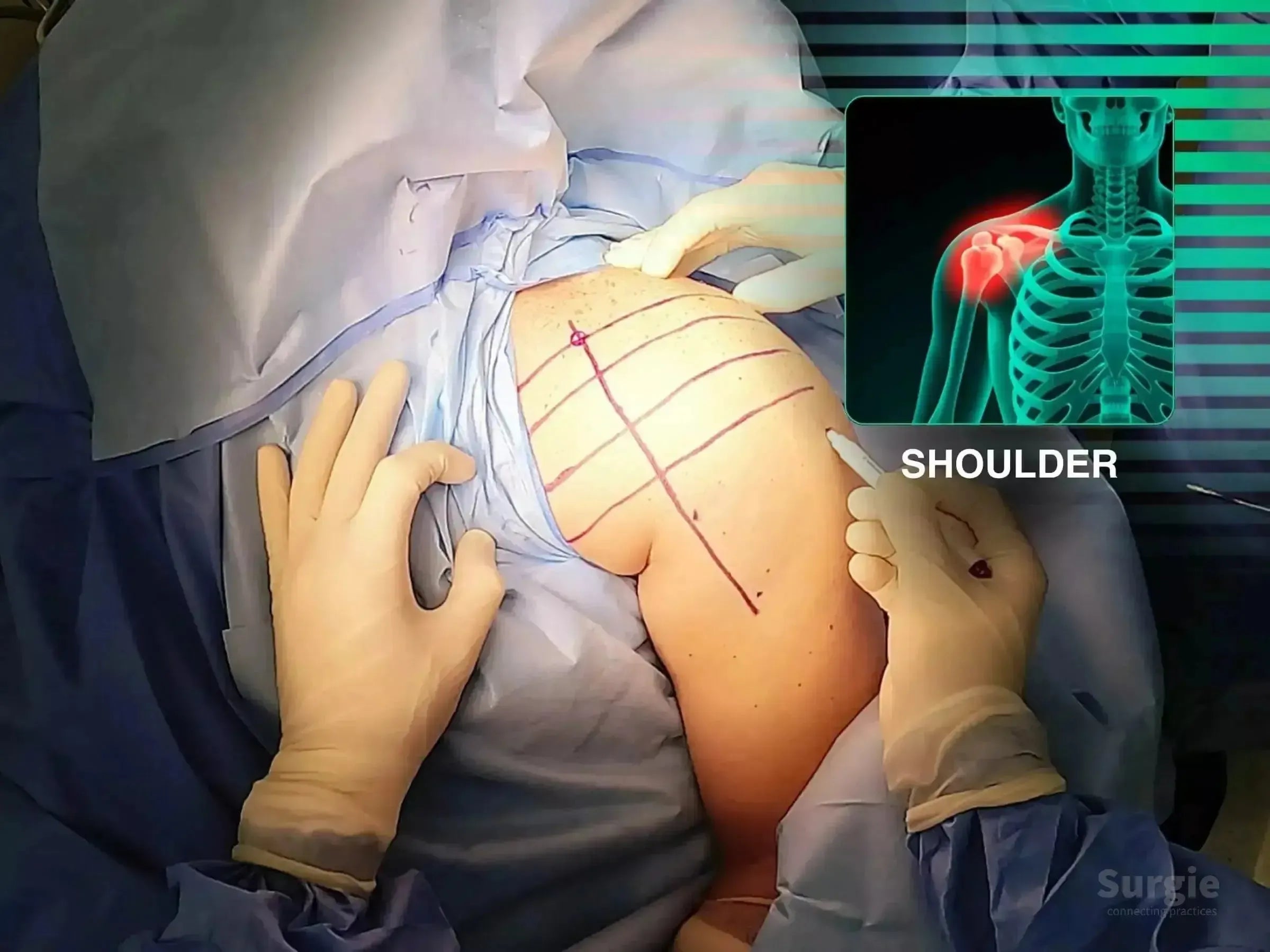
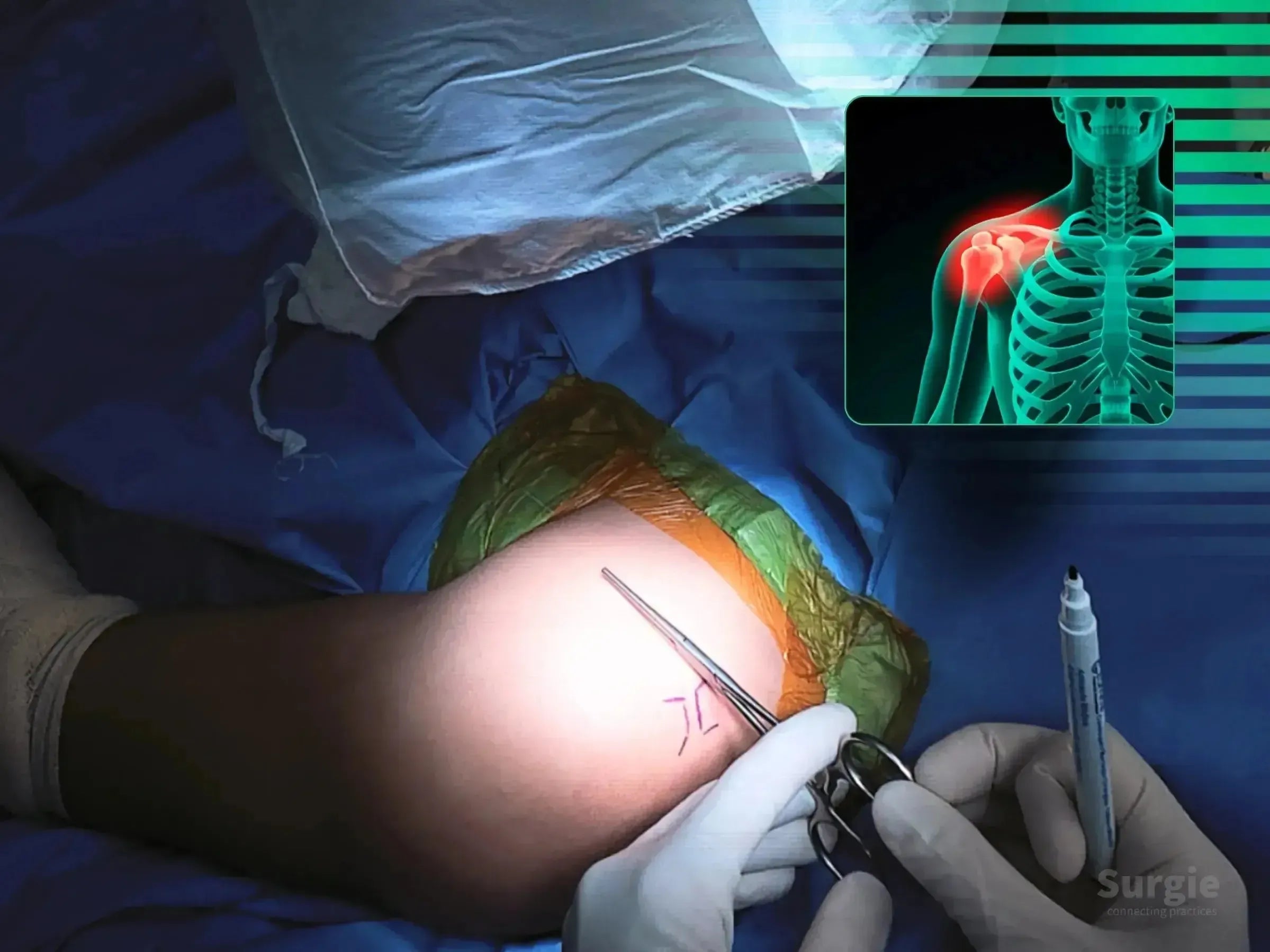
Surgeon marking shoulder skin for TightRope AC joint reconstruction with an anatomical overlay showing dislocation.
50% preview 4K Shoulder
07:08
AC Joint Dislocation, Case 2: TightRope Tunnel Technique

Precise clavicle and coracoid tunneling for TightRope fixation

Dr. Sávio Chami Dr. Sávio ChamiOrthopedic Surgeon
9 languages
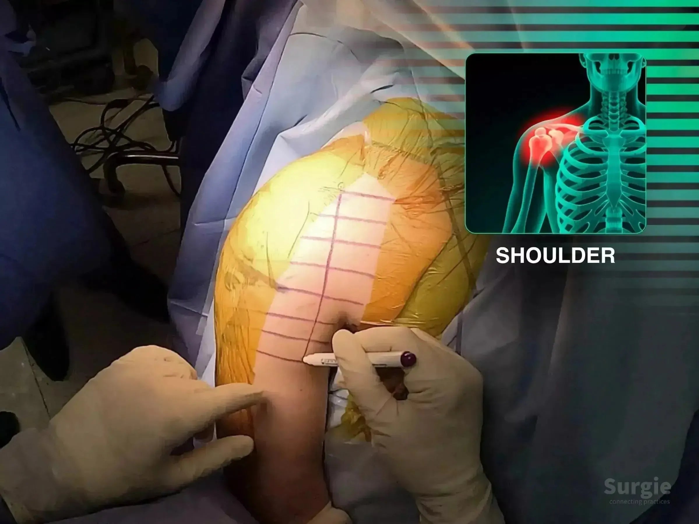
Surgeon marking a patient's shoulder for AC joint surgery with a TightRope device, featuring a skeletal X-ray overlay.
50% preview 4K Shoulder
06:42
AC Joint Dislocation: Open TightRope Fixation

Open coracoclavicular tunnel creation and TightRope stabilization

Dr. Sávio Chami Dr. Sávio ChamiOrthopedic Surgeon
9 languages
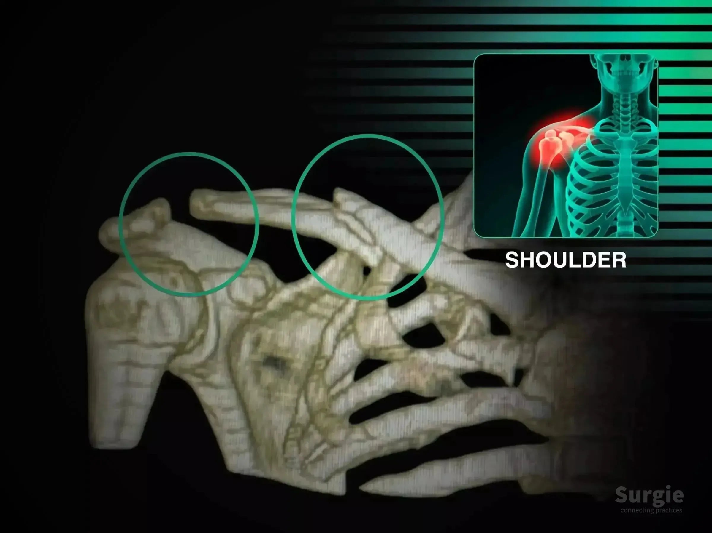
3D skeletal model showing clavicle fracture and acromioclavicular dislocation with highlighted surgical zones.
50% preview 4K Shoulder
05:26
Clavicle Fracture with Posterior AC Joint Dislocation

Combined clavicle fixation and multidirectional AC joint stabilization

Dr. Sávio Chami Dr. Sávio ChamiOrthopedic Surgeon
9 languages
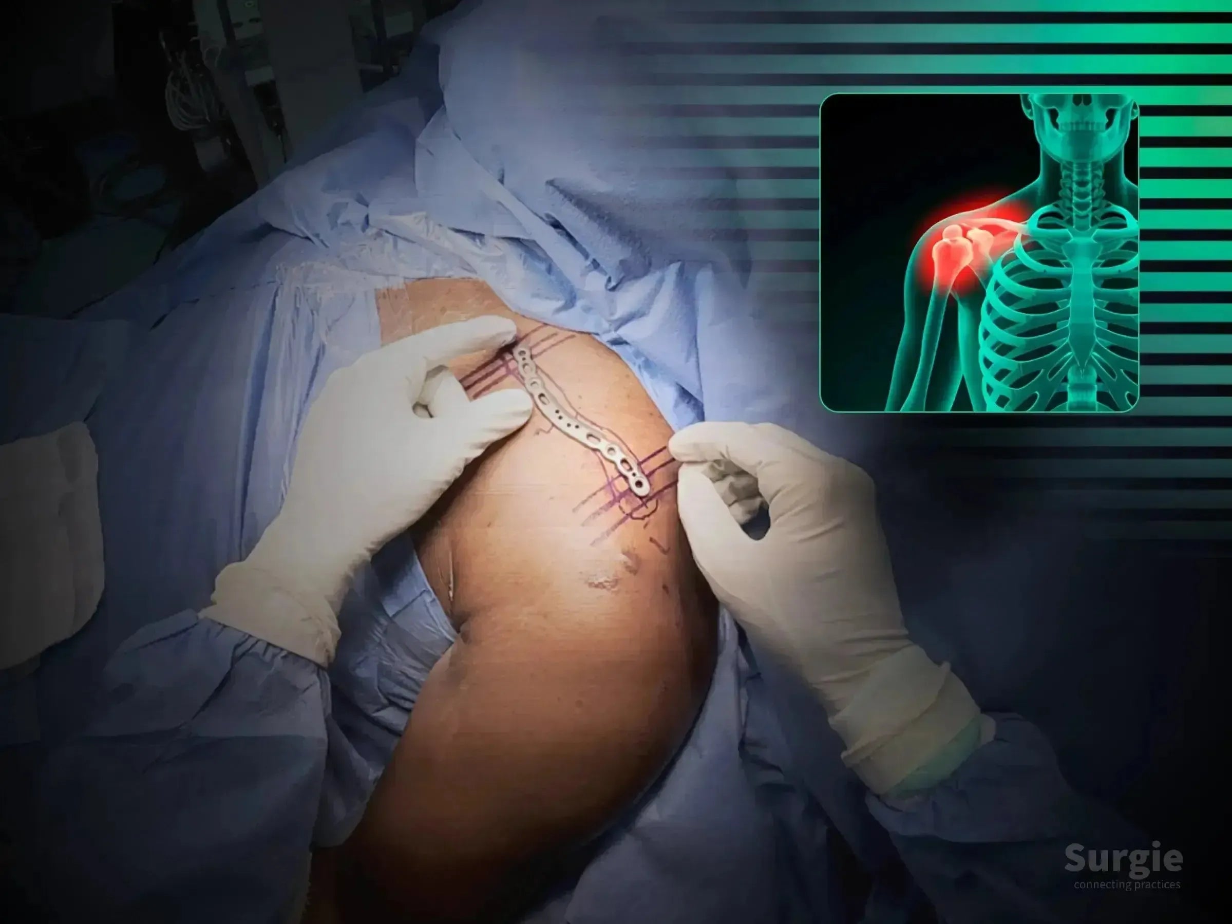
Surgeon placing a metallic anatomic clavicle plate on a patient with a skeletal diagram showing a shoulder fracture.
50% preview 4K Shoulder
15:50
Clavicle Fracture, Case 2: Minimally Invasive Fixation

Two-portal clavicle fixation with fluoroscopic reduction

Dr. Sávio Chami Dr. Sávio ChamiOrthopedic Surgeon
9 languages
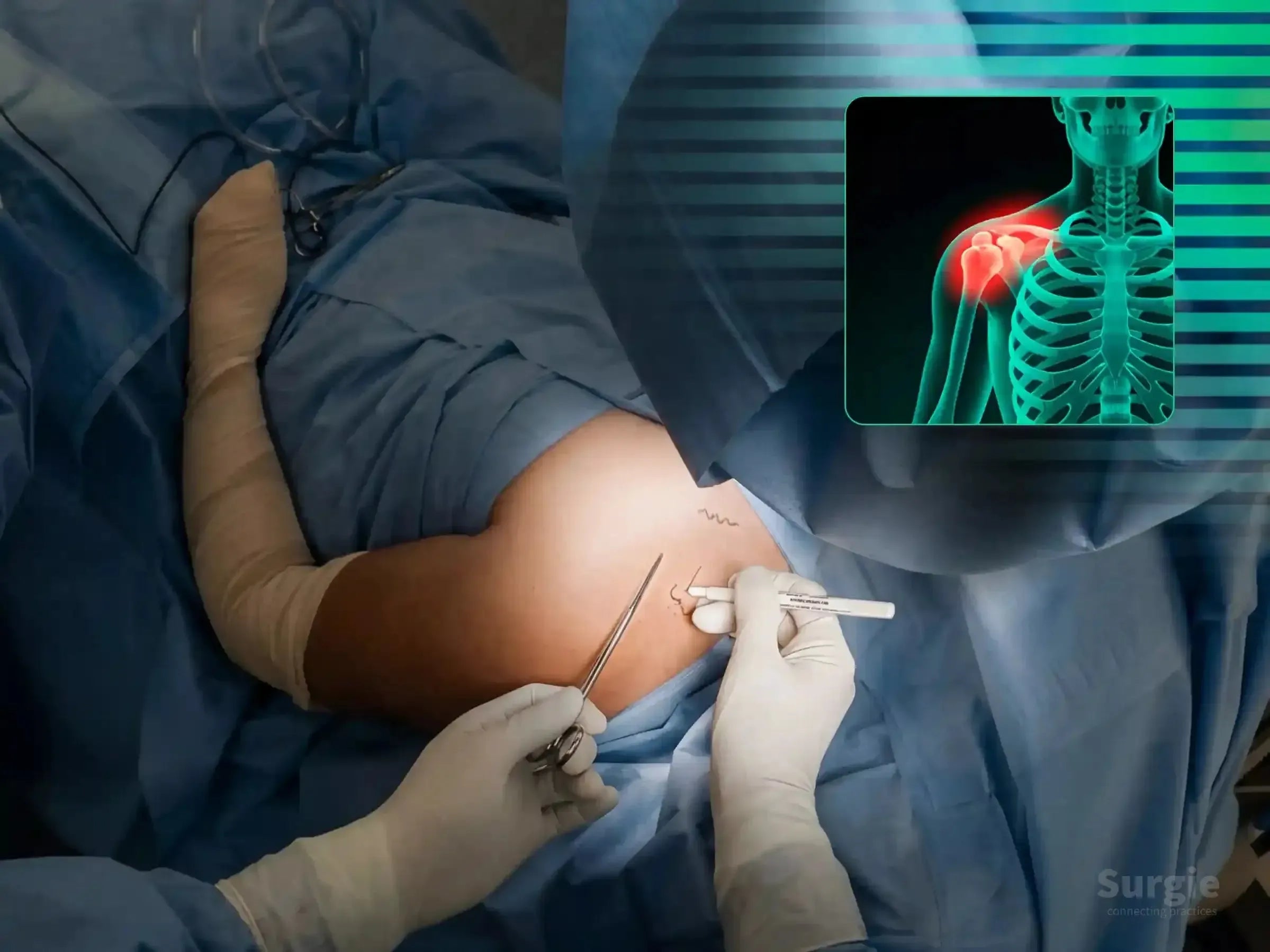
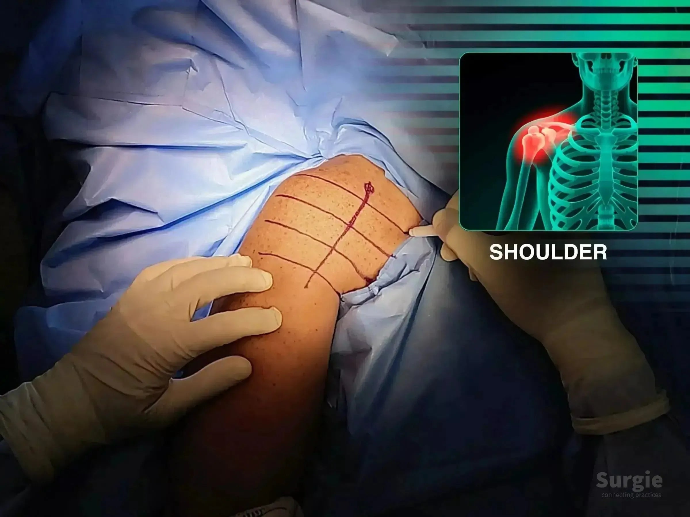
Surgeon marking a clavicle fracture site with an anatomical overlay showing a highlighted shoulder injury.
50% preview 4K Shoulder
12:34
Clavicle Fracture, Case 3: Percutaneous Osteosynthesis

Two-portal percutaneous plating with fluoroscopic guidance

Dr. Sávio Chami Dr. Sávio ChamiOrthopedic Surgeon
9 languages
Surgeon marking skin for minimally invasive clavicle bridge plating with anatomical shoulder diagram overlay.
50% preview 4K Shoulder
02:22
Clavicle Fracture: Minimally Invasive Bridge Plating

Two-portal bridge plating with soft-tissue preservation

Dr. Sávio Chami Dr. Sávio ChamiOrthopedic Surgeon
9 languages
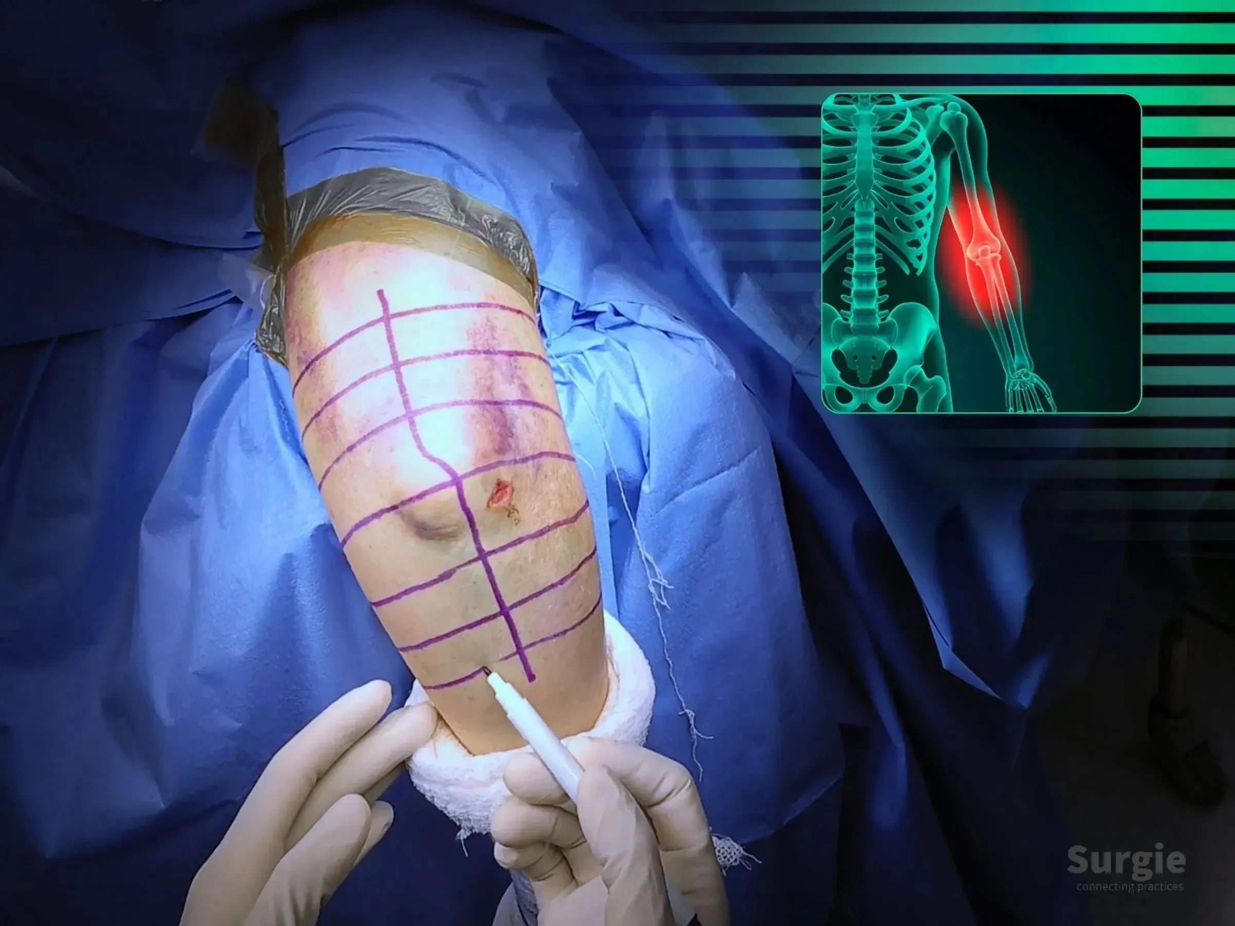
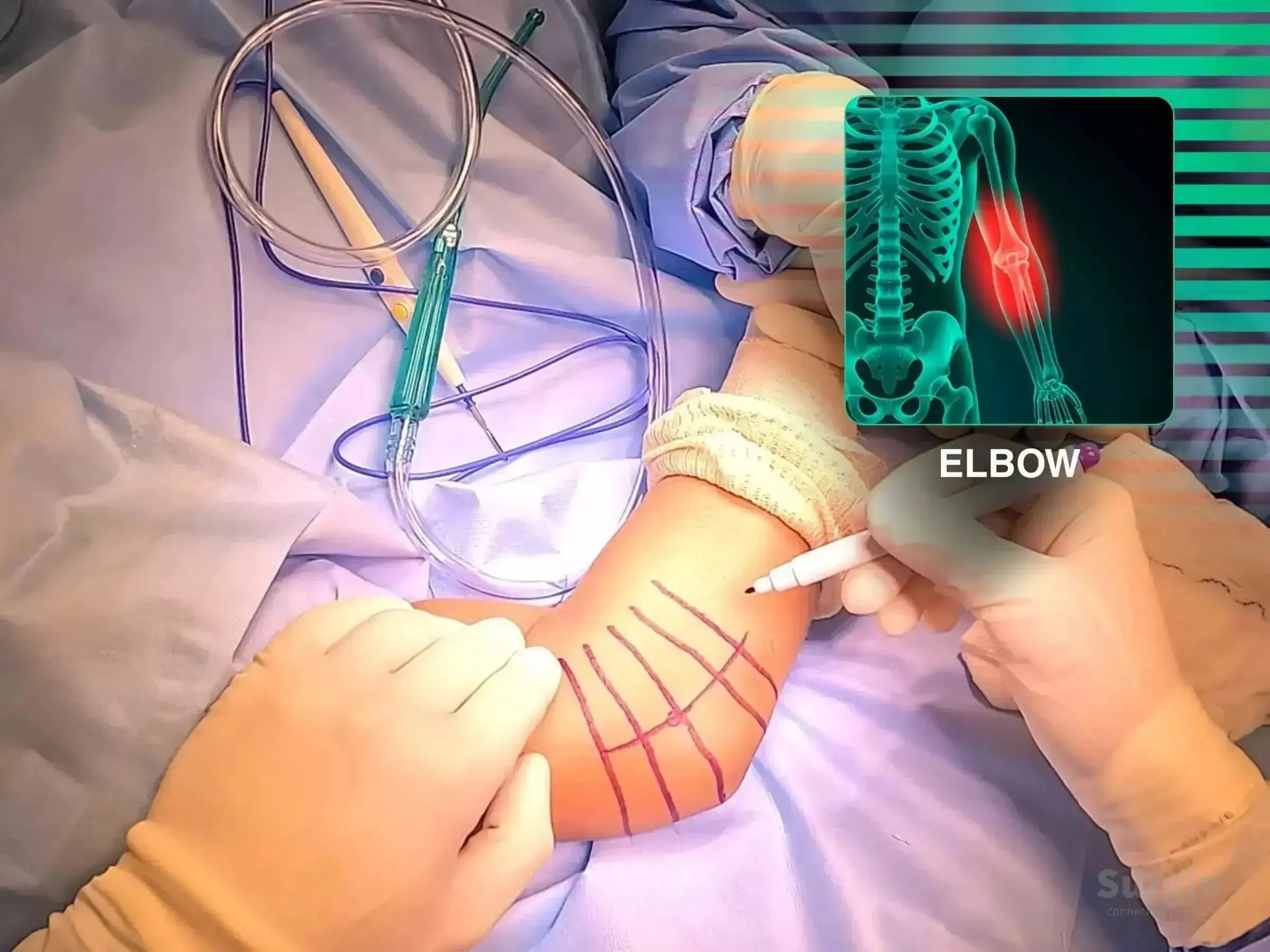
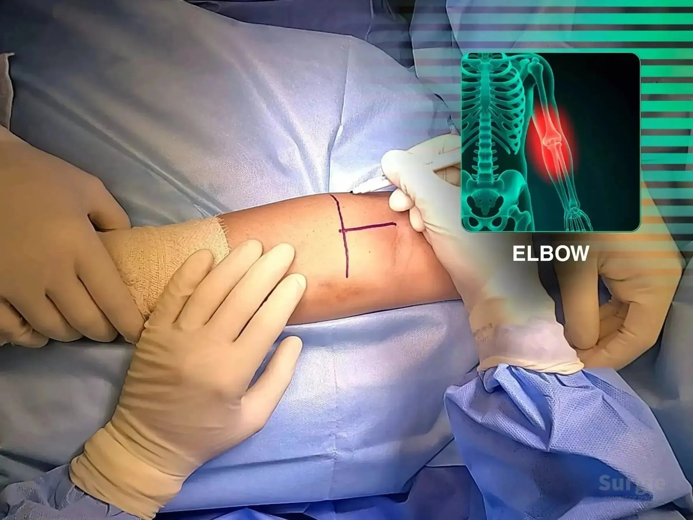
Surgical marking on a patient's elbow with a distal humerus fracture overlay showing the injury site in a clinical setting.
50% preview 4K Elbow
36:05
Comminuted Articular Distal Humerus Fracture: Fixation

Olecranon osteotomy, articular reduction, and dual-column fixation

Dr. Sávio Chami Dr. Sávio ChamiOrthopedic Surgeon
9 languages
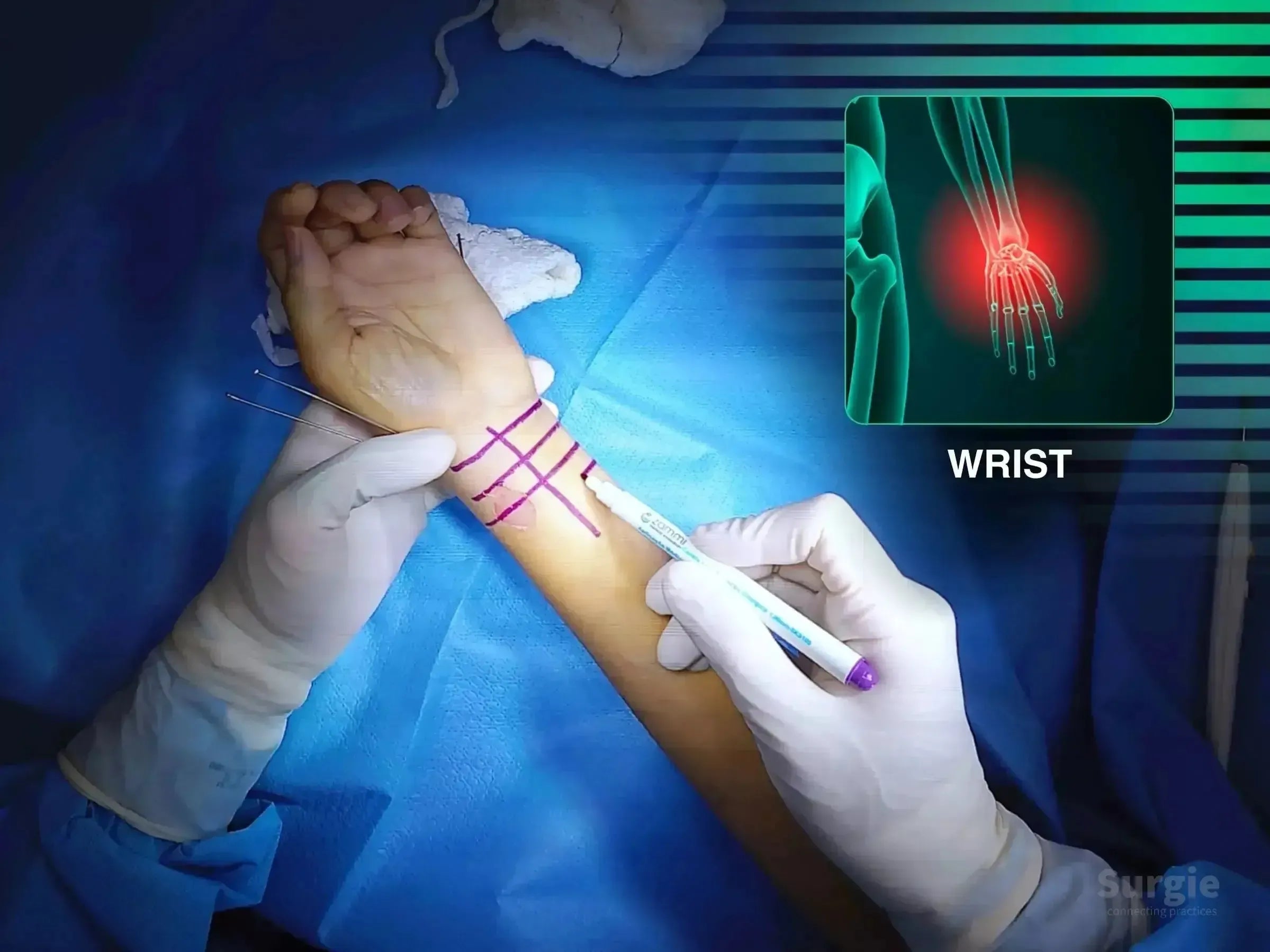
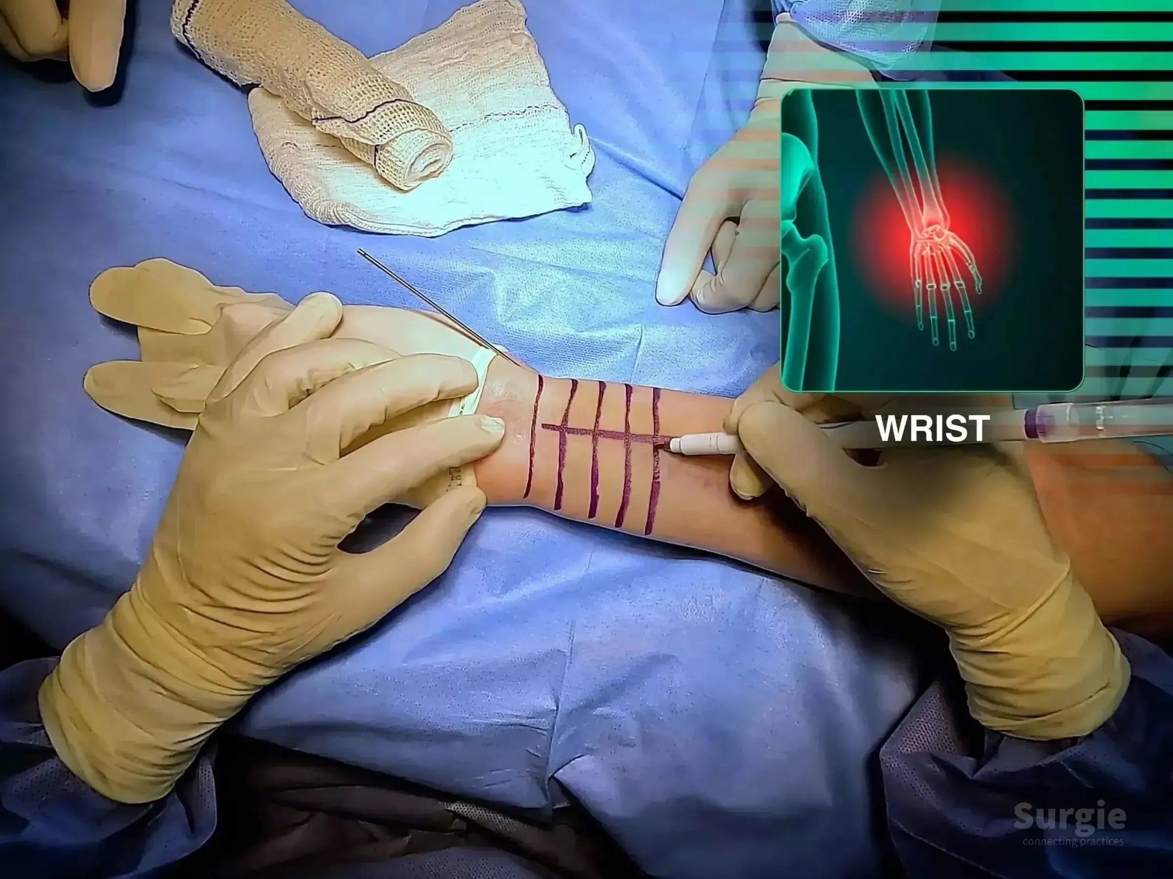
Surgeon marking a patient's wrist for volar approach surgery with a digital anatomical overlay of the distal radius.
50% preview 4K Wrist
11:08
Comminuted Distal Radius Fracture: Volar Plate Fixation

Restore radial length and articular congruity through volar plating

Dr. Sávio Chami Dr. Sávio ChamiOrthopedic Surgeon
9 languages
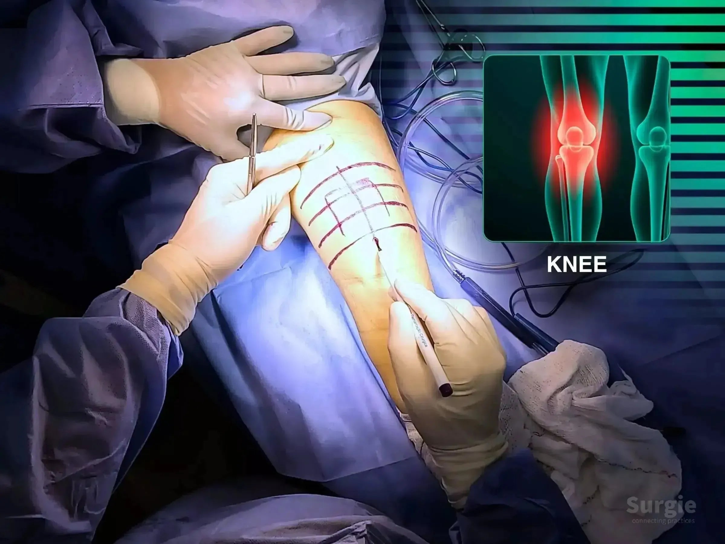
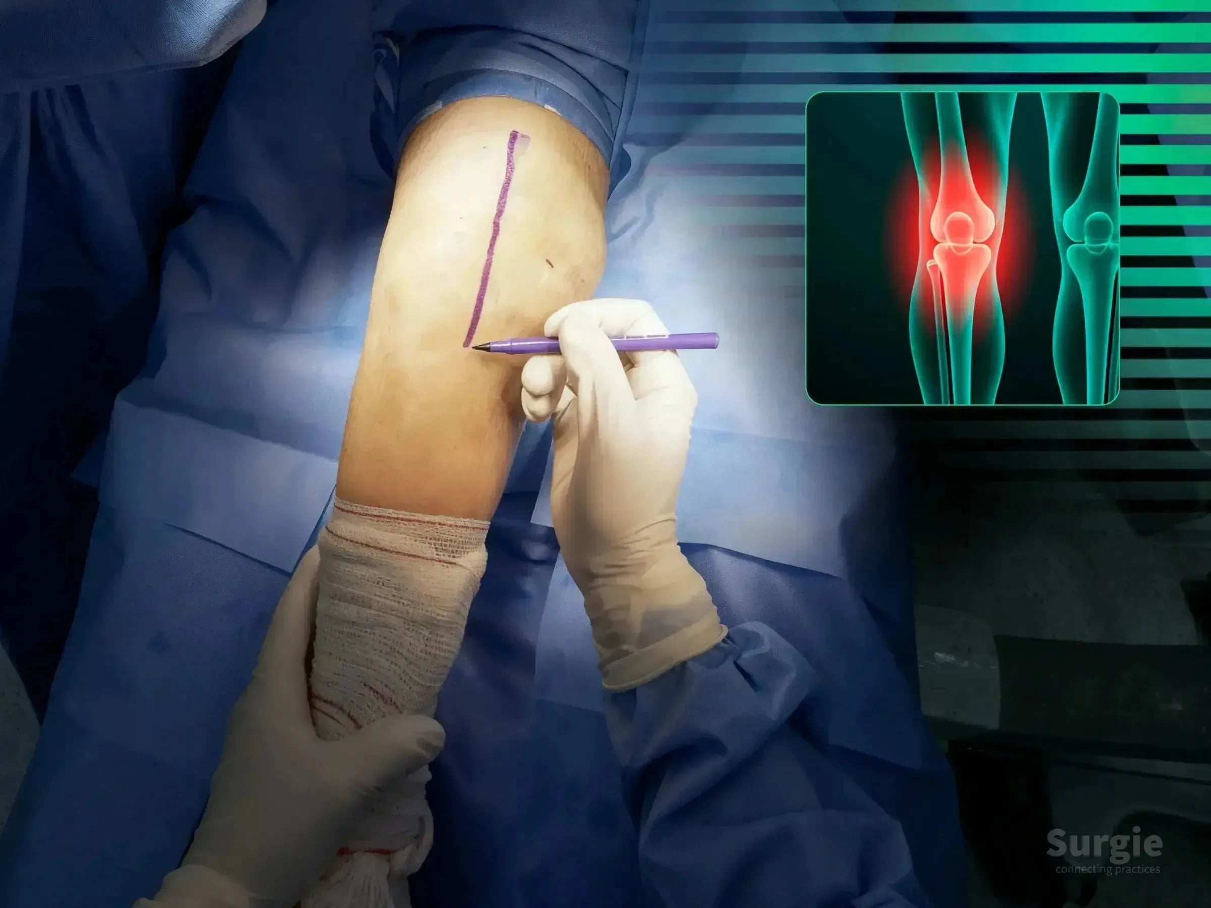
Surgeon marking a patient's knee for patella fracture surgery with an anatomical diagram overlay.
50% preview 4K Knee
13:28
Comminuted Patellar Fracture: Surgical Fixation

Cannulated screws and cerclage for extensor mechanism reconstruction

Dr. Sávio Chami Dr. Sávio ChamiOrthopedic Surgeon
9 languages
Surgeon marking a patient's elbow for radial head arthroplasty with an anatomical X-ray overlay highlighting the joint.
50% preview 4K Elbow
05:35
Comminuted Radial Head Fracture: Arthroplasty

Radial head replacement with ligament repair and stability testing

Dr. Sávio Chami Dr. Sávio ChamiOrthopedic Surgeon
9 languages
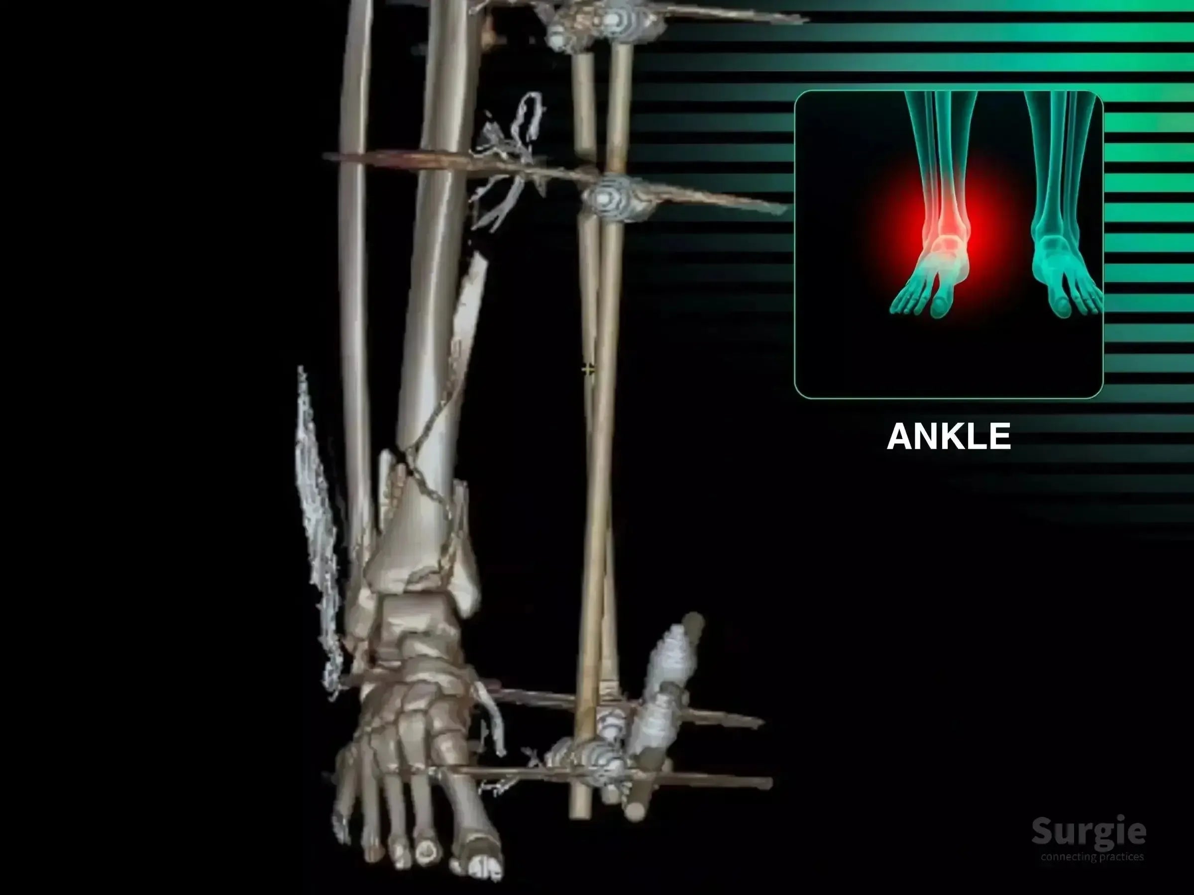
3D CT scan of a multi-fragmentary tibial pilon fracture with external fixation and an ankle pain highlight.
50% preview 4K Ankle
19:13
Comminuted Tibial Pilon Fracture: Staged Fixation

Staged pilon reconstruction with articular reduction and multiplanar fixation

Dr. Sávio Chami Dr. Sávio ChamiOrthopedic Surgeon
9 languages
Complex Ankle Injury: 360-Degree Reconstruction
50% preview 4K Ankle
08:17
Complex Ankle Injury: 360-Degree Reconstruction

Achilles, posterior tibial tendon, deltoid, and syndesmosis reconstruction

Dr. Sávio Chami Dr. Sávio ChamiOrthopedic Surgeon
9 languages
Surgeon marking a patient's shoulder for a deltopectoral approach with an anatomical diagram overlay.
50% preview 4K Shoulder
16:11
Complex Four-Part Proximal Humerus Fracture Reconstruction

Four-part reconstruction with valgus correction, plate, and suture fixation

Dr. Sávio Chami Dr. Sávio ChamiOrthopedic Surgeon
9 languages
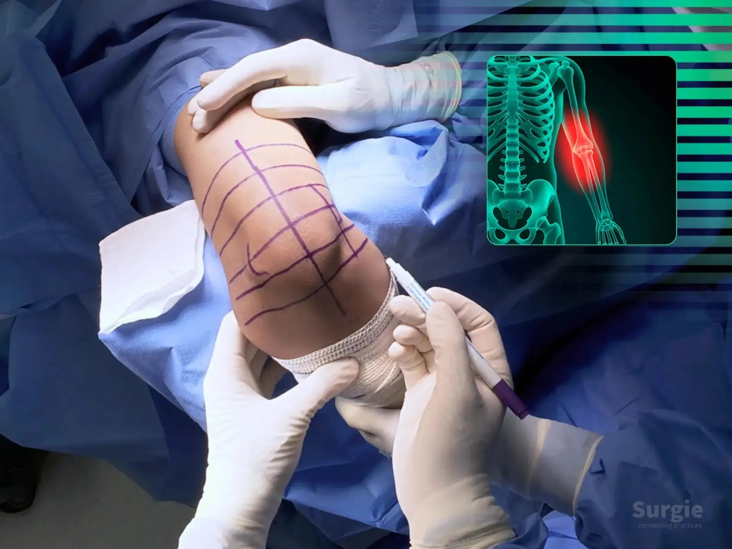
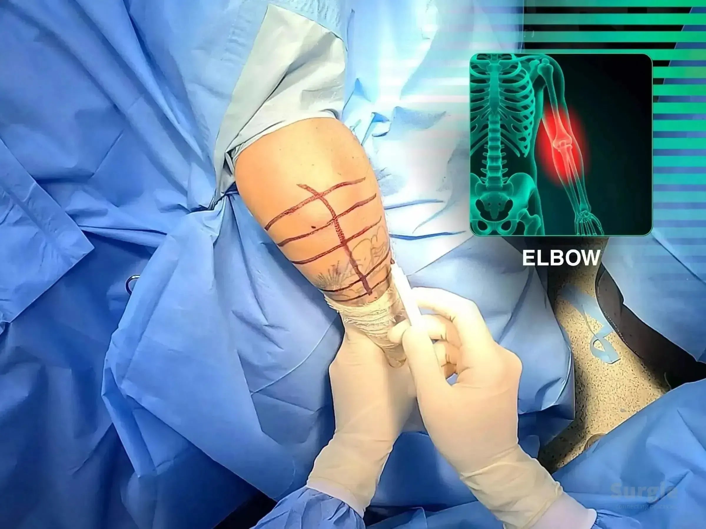
Surgeon marking a patient's elbow for distal humerus osteotomy with a digital skeletal overlay showing the humerus area.
50% preview 5.7K Elbow
28:54
Cubitus Varus: Distal Humerus Corrective Osteotomy

3D-guided corrective osteotomy with ulnar nerve transposition

Dr. Sávio Chami Dr. Sávio ChamiOrthopedic Surgeon
9 languages
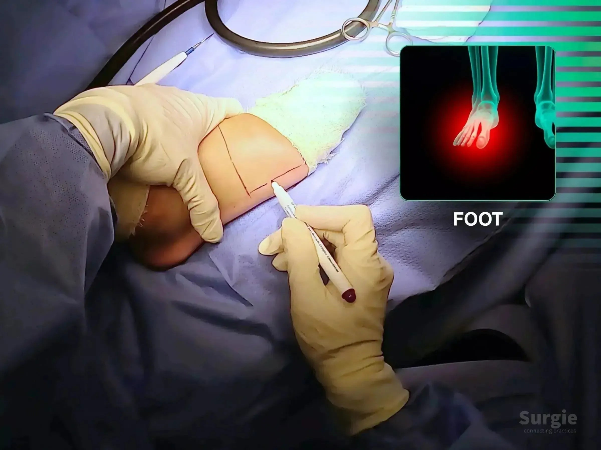
Surgeon marking a fifth metatarsal incision site on a patient's foot with an anatomical reference overlay.
50% preview 4K Foot
04:59
Displaced Fifth Metatarsal Fracture: Plate Fixation

Anatomic reduction and low-profile plate fixation of the fifth metatarsal

Dr. Sávio Chami Dr. Sávio ChamiOrthopedic Surgeon
9 languages
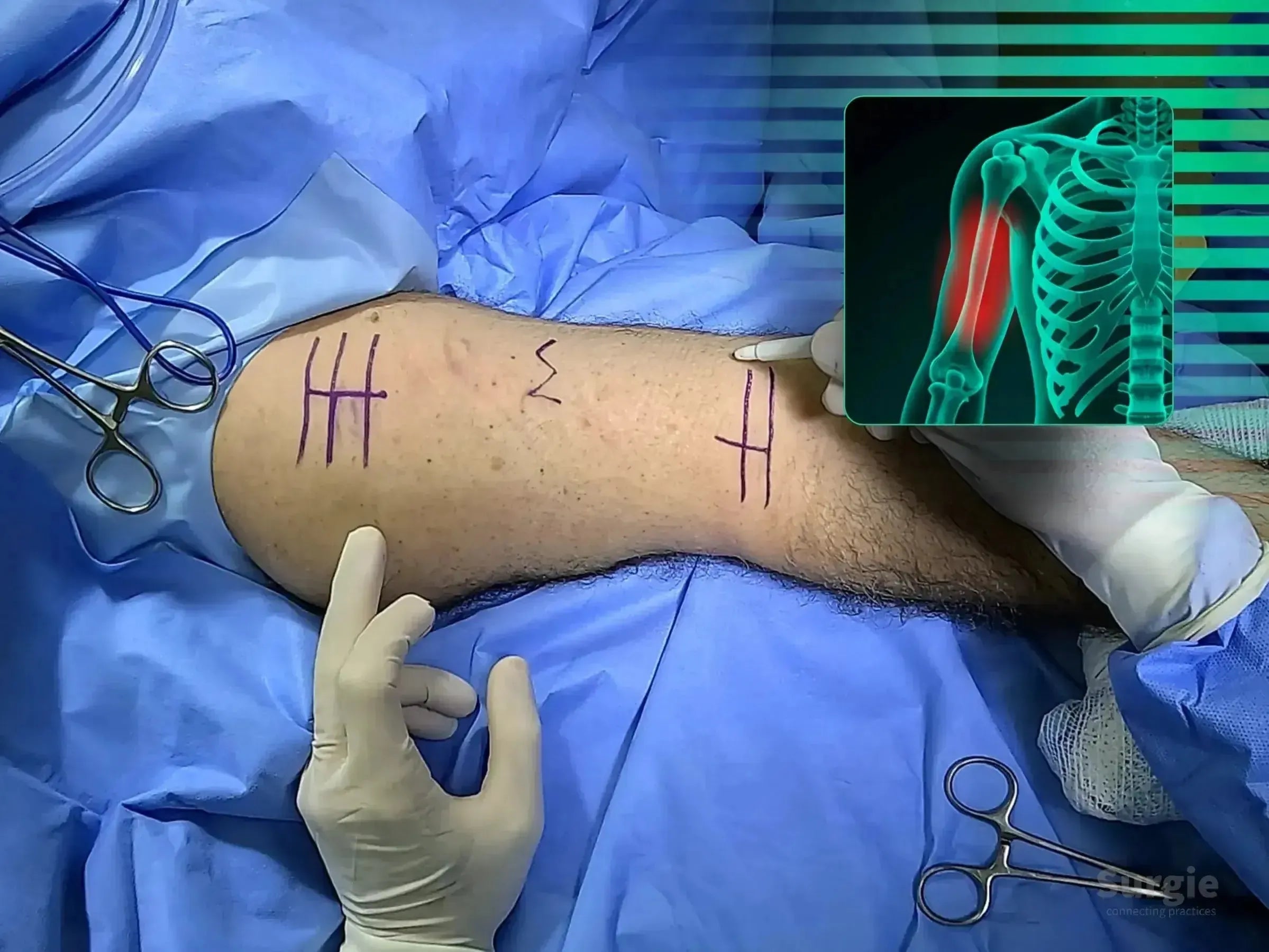
Surgeon marking a patient's arm for distal biceps tendon surgery with an anatomical elbow diagram overlay.
50% preview 4K Elbow
05:02
Distal Biceps Tendon Reconstruction: TightRope Fixation

TightRope reinsertion at the radial tuberosity with tissue preservation

Dr. Sávio Chami Dr. Sávio ChamiOrthopedic Surgeon
9 languages
Surgeon marking a patient's arm for distal biceps tendon reconstruction with an anatomical elbow reference overlay.
50% preview 4K Elbow
09:30
Distal Biceps Tendon Repair: TightRope with Nerve Protection

TightRope tendon repair with focused neurovascular protection

Dr. Sávio Chami Dr. Sávio ChamiOrthopedic Surgeon
9 languages
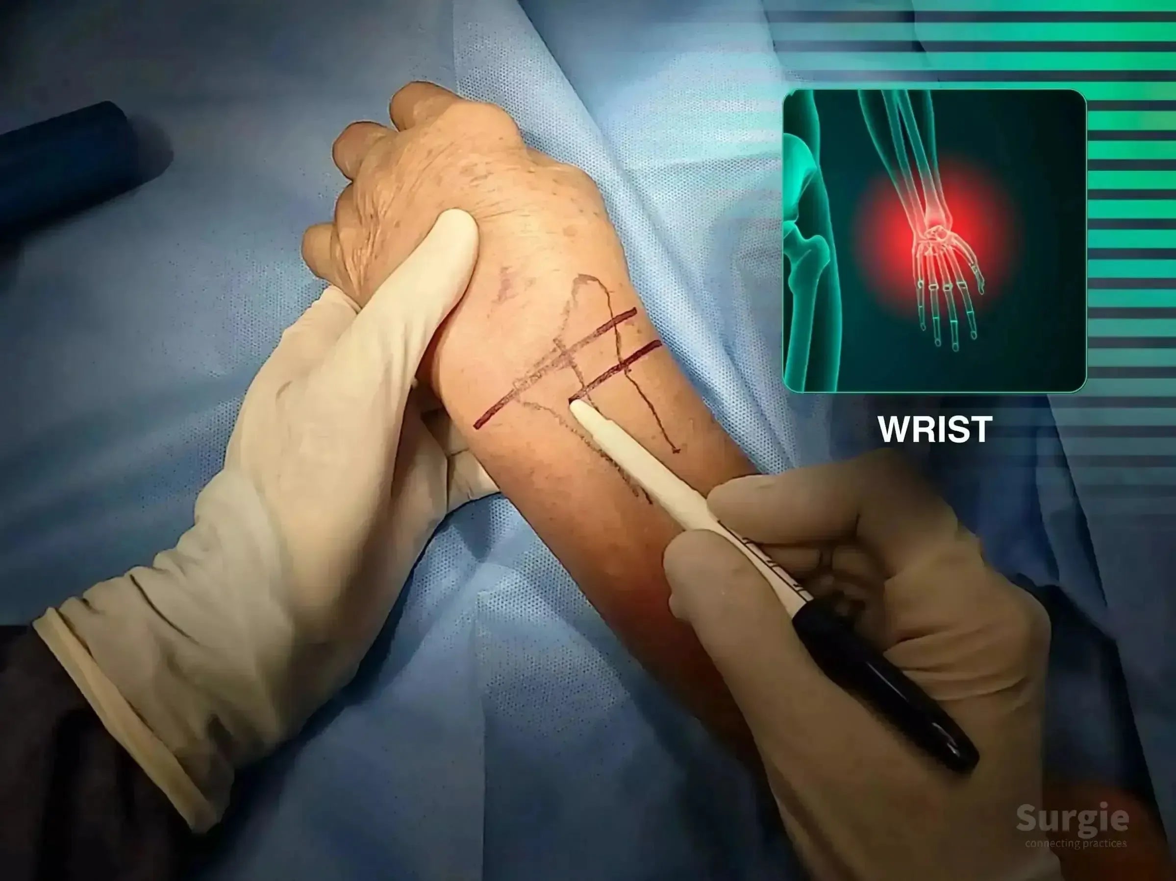
Surgeon marking a patient's wrist for a volar approach surgery with an anatomical X-ray overlay showing a fracture.
50% preview 4K Wrist
05:12
Distal Radius Fracture: Volar Plate Fixation

Volar exposure, provisional reduction, and plate stabilization

Dr. Sávio Chami Dr. Sávio ChamiOrthopedic Surgeon
9 languages
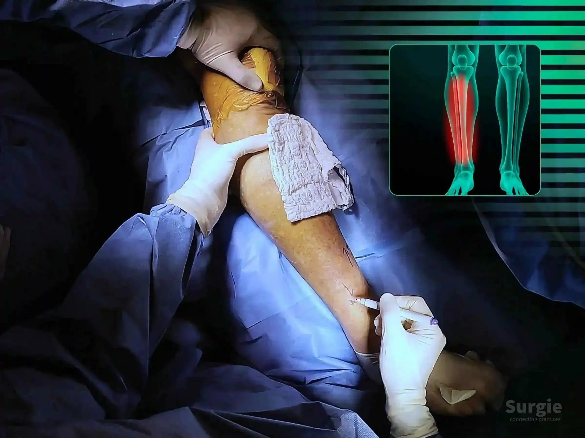
Surgeon marking a distal tibia fracture site for minimally invasive surgery with a digital X-ray overlay of the leg.
50% preview 4K Tibia
04:28
Distal Tibial Shaft Fracture: Locked Plate Fixation

Percutaneous reduction and minimally invasive locked plate fixation

Dr. Sávio Chami Dr. Sávio ChamiOrthopedic Surgeon
9 languages
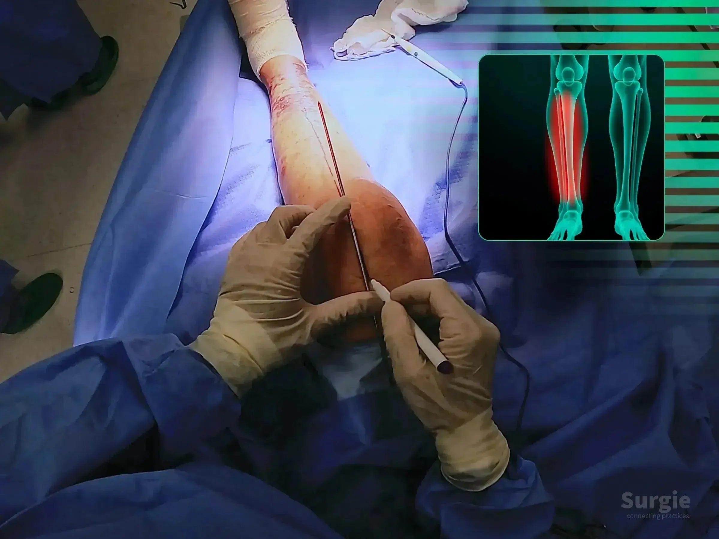
Surgeon marking a suprapatellar portal on a patient's leg for tibial intramedullary nail surgery with an X-ray overlay.
50% preview 4K Tibia
08:31
Distal Tibial Shaft Fracture: Suprapatellar Intramedullary Nail

Suprapatellar nailing with alignment control and soft-tissue protection

Dr. Sávio Chami Dr. Sávio ChamiOrthopedic Surgeon
9 languages
Surgeon marking a dorsal Barton's fracture site on a patient's wrist with an anatomical overlay showing the injury.
50% preview 4K Wrist
04:42
Dorsal Barton Fracture: Dorsal Radius Fixation

Dorsal fragment reduction with targeted radius fixation

Dr. Sávio Chami Dr. Sávio ChamiOrthopedic Surgeon
9 languages
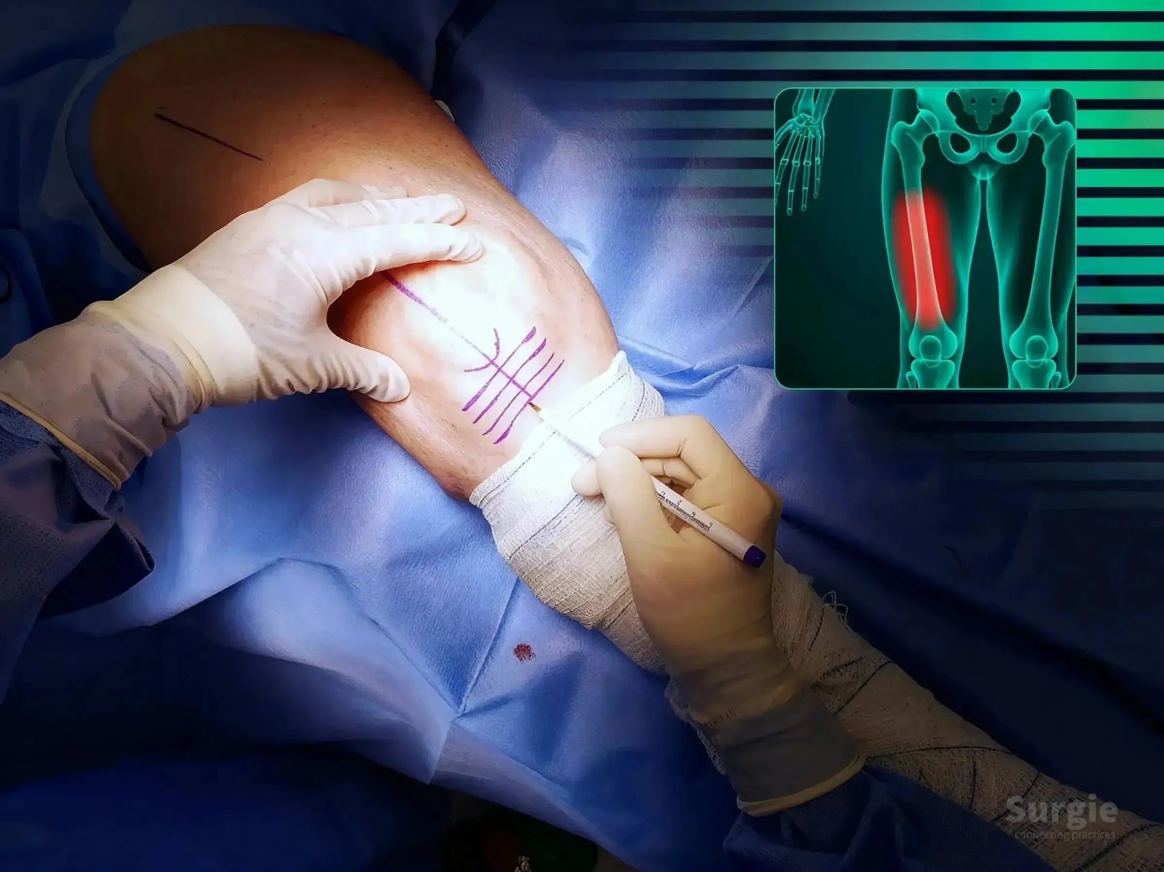
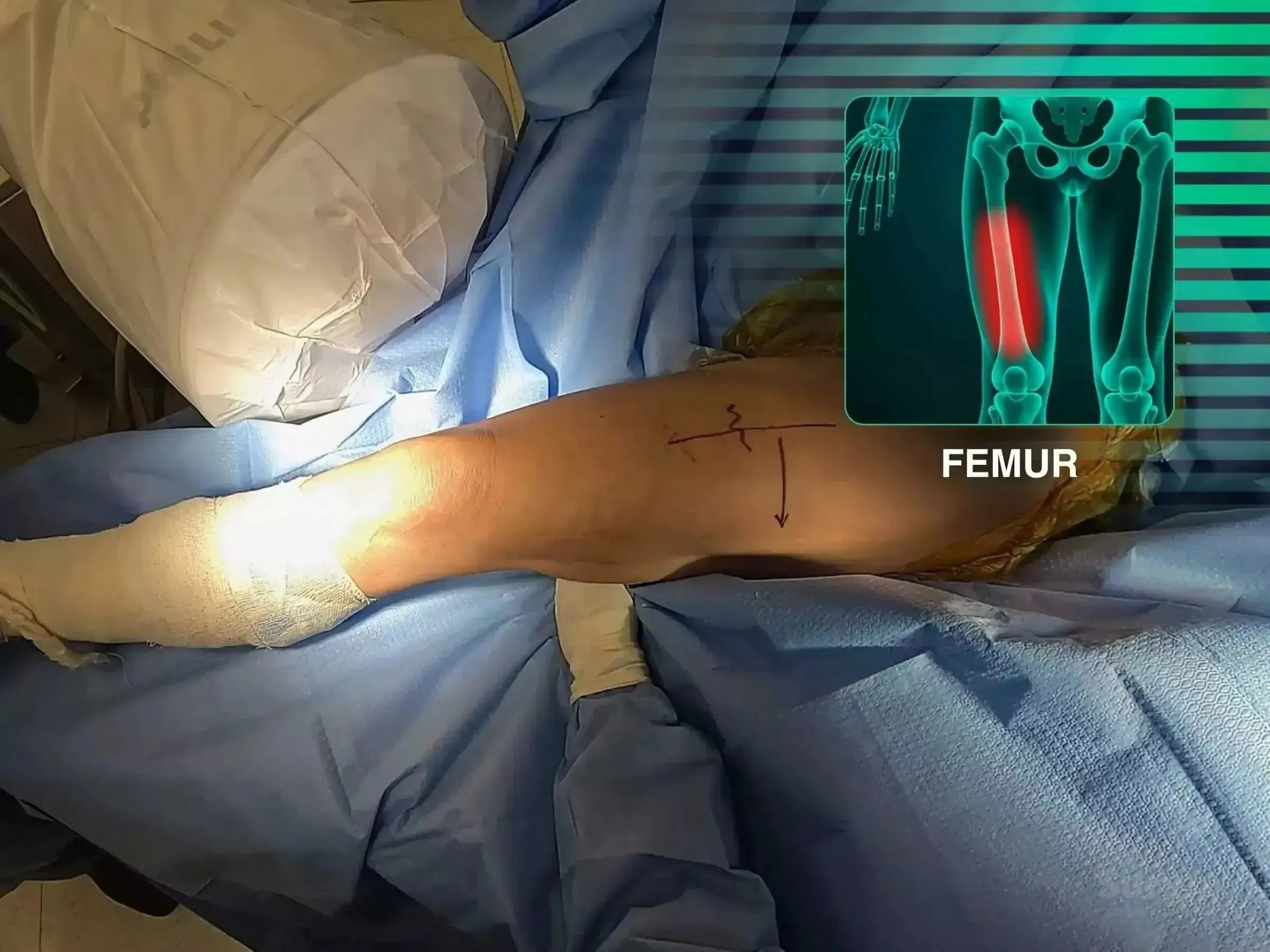
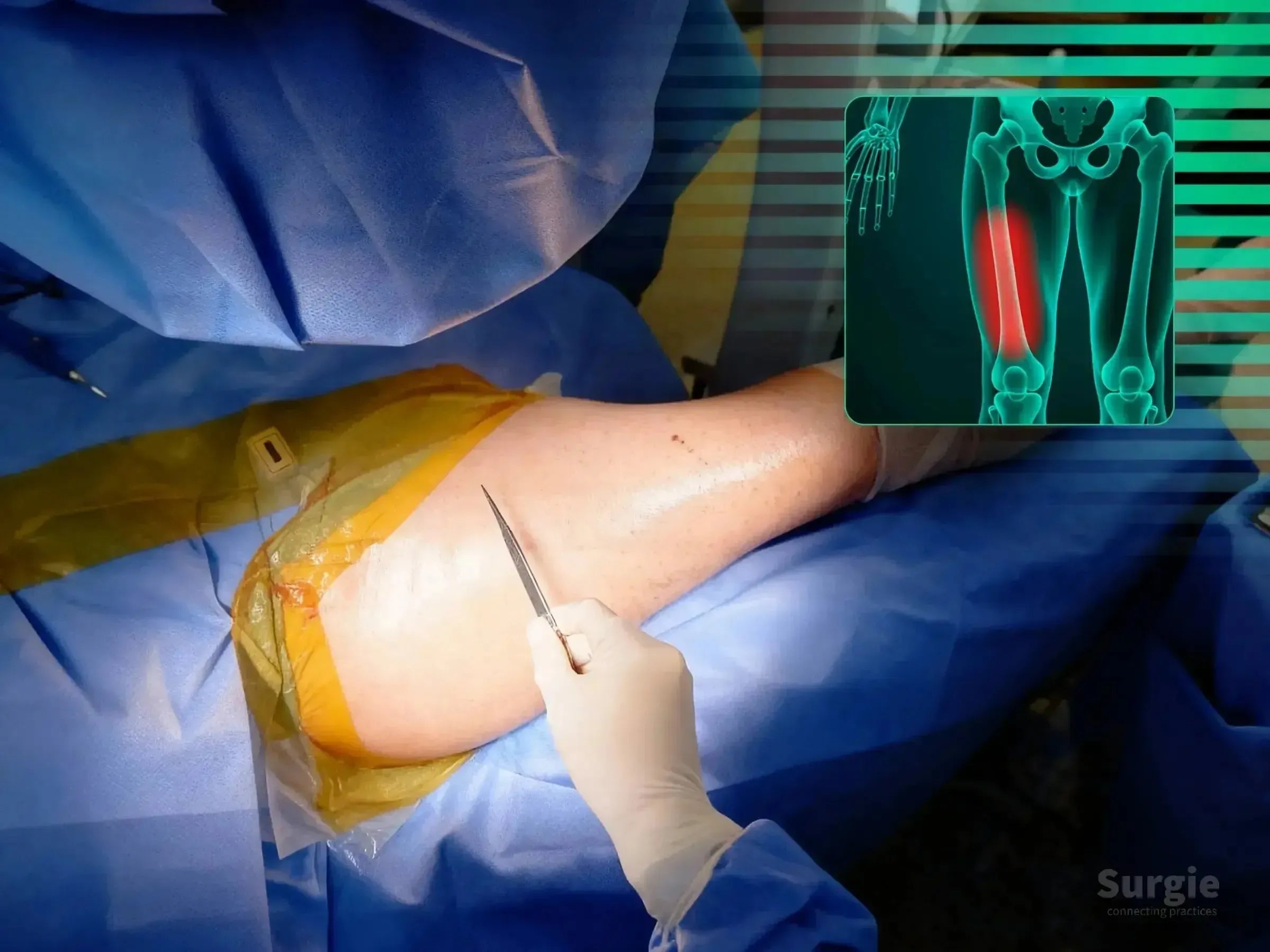
Surgeon marking a patient's leg for retrograde intramedullary nail surgery with a femoral fracture overlay.
50% preview 4K Femur
12:07
Femoral Shaft Fracture, Case 2: Retrograde Nail

Retrograde femoral nailing with rotational and fluoroscopic control

Dr. Sávio Chami Dr. Sávio ChamiOrthopedic Surgeon
9 languages
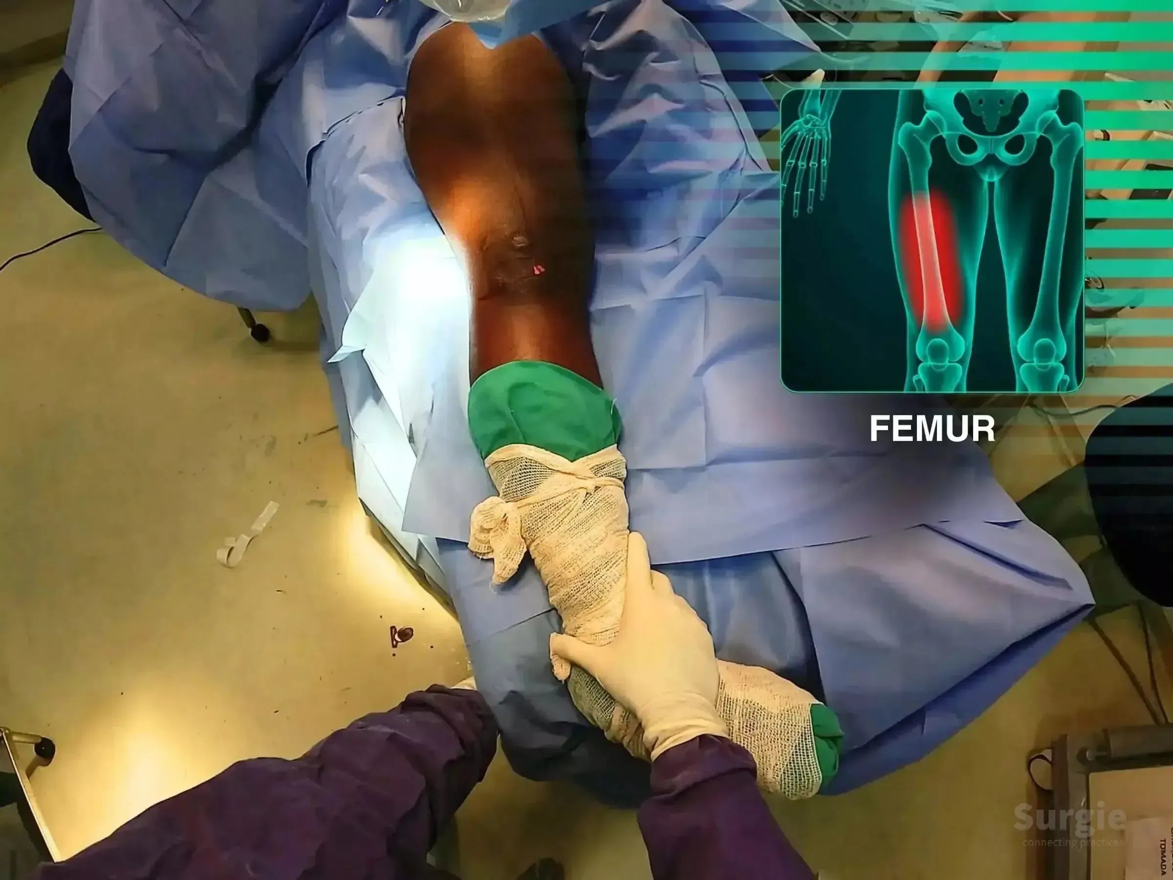
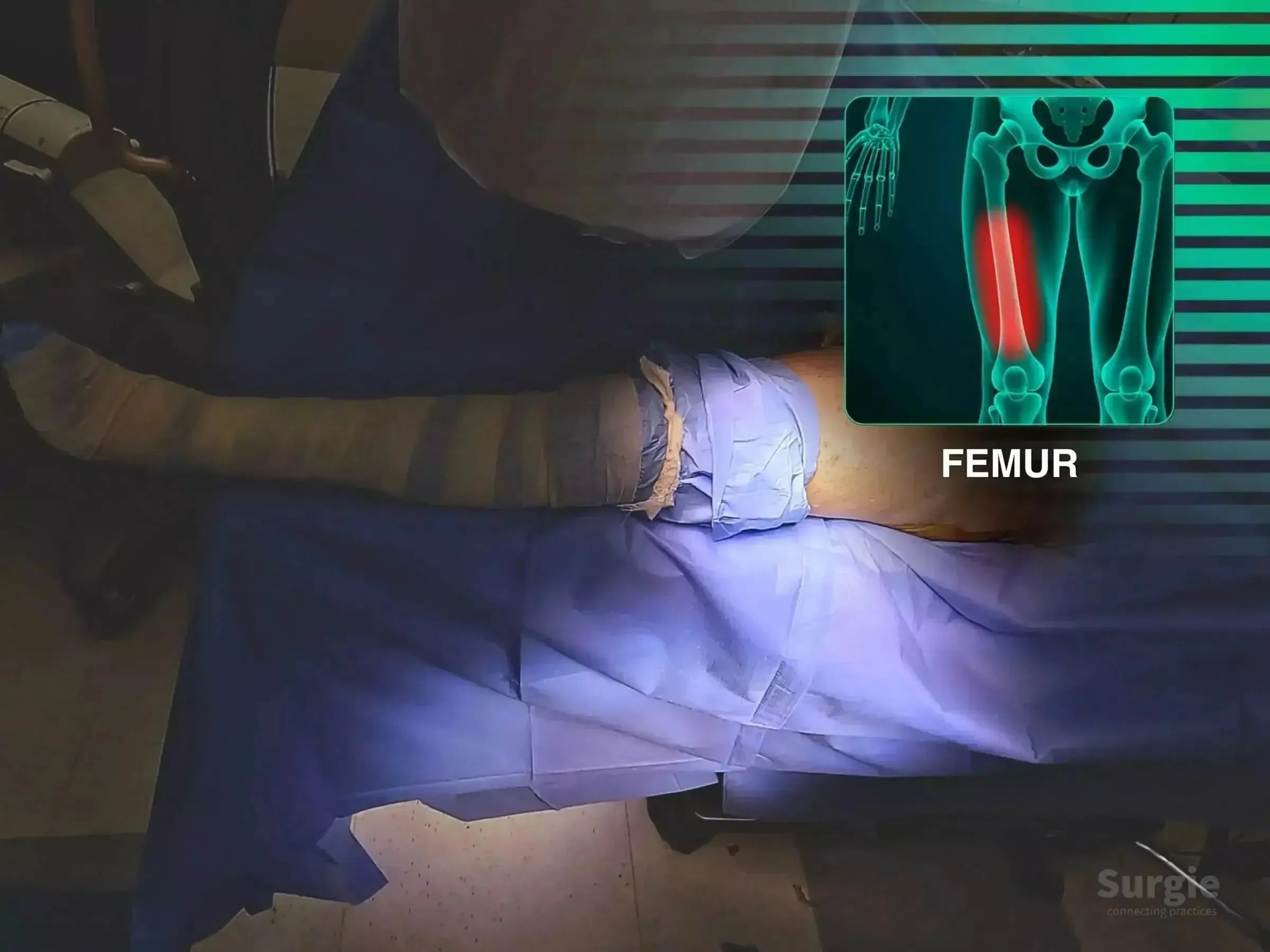
Surgeon performing femoral shaft fracture reduction with an anatomical overlay highlighting the femur on a blue surgical drape.
50% preview 4K Femur
04:23
Femoral Shaft Fracture: Retrograde Intramedullary Nail

Retrograde nailing with free-limb reduction and fluoroscopic control

Dr. Sávio Chami Dr. Sávio ChamiOrthopedic Surgeon
9 languages
Surgical preparation of a patient's thigh for cephalomedullary nail fixation with a femoral anatomy overlay.
50% preview 4K Femur
06:27
Femoral Shaft Refracture: Antegrade Cephalomedullary Nail

Antegrade nailing without a traction table for femoral shaft refracture

Dr. Sávio Chami Dr. Sávio ChamiOrthopedic Surgeon
9 languages
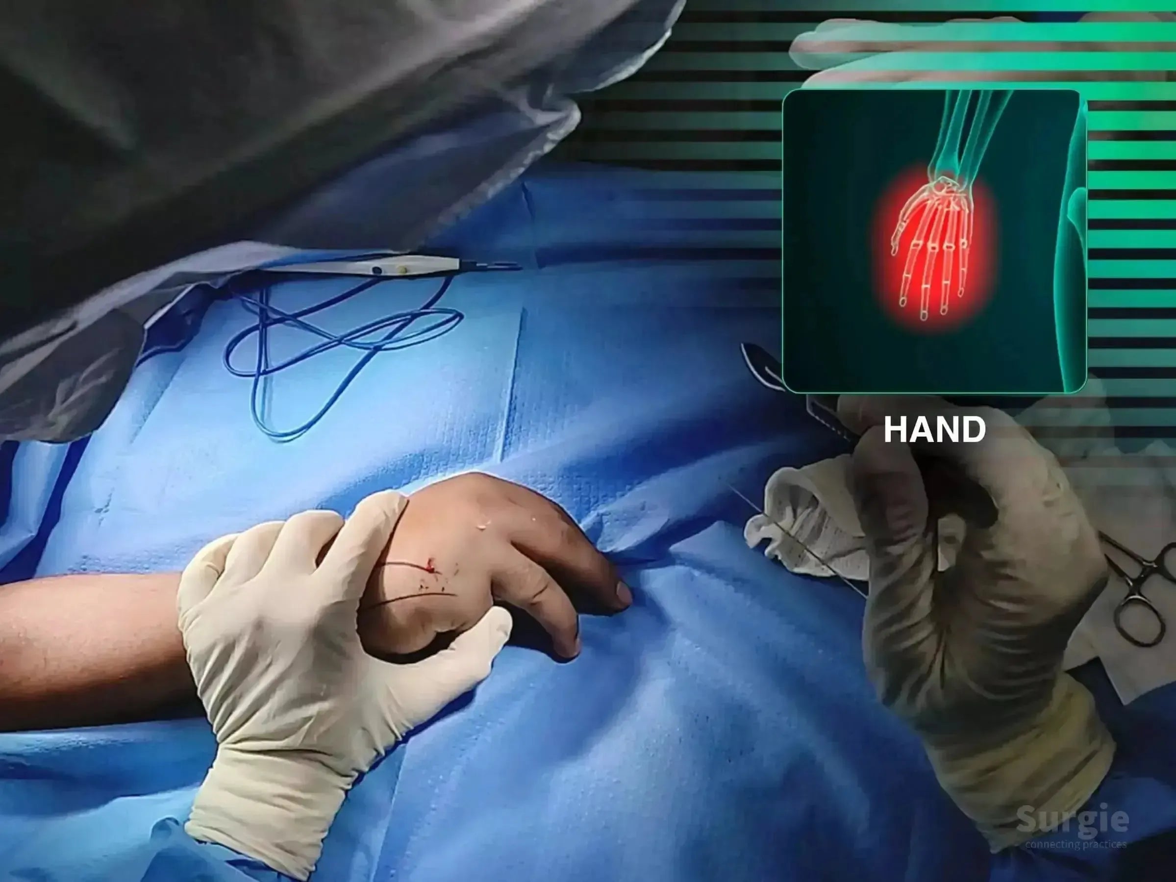
Surgeon performing minimally invasive surgery on a fifth metacarpal fracture with an anatomical hand diagram overlay.
50% preview 4K Hand
02:43
Fifth Metacarpal Neck Fracture: Double-Compression Screw

Minimally invasive double-compression fixation of the fifth metacarpal

Dr. Sávio Chami Dr. Sávio ChamiOrthopedic Surgeon
9 languages
Surgeon marking a shoulder for deltopectoral incision with an anatomical X-ray overlay showing a proximal humerus fracture.
50% preview 4K Shoulder
22:23
Four-Part Proximal Humerus Fracture, Case 5

Valgus-impacted head reduction with plate, calcar screws, and FiberTape

Dr. Sávio Chami Dr. Sávio ChamiOrthopedic Surgeon
9 languages
Surgeon marking a patient's shoulder for a deltopectoral approach with a skeletal diagram overlay showing a humerus fracture.
50% preview 4K Shoulder
05:37
Four-Part Proximal Humerus Fracture: Deltopectoral Fixation

Tuberosity reconstruction with plate-and-screw fixation

Dr. Sávio Chami Dr. Sávio ChamiOrthopedic Surgeon
9 languages
Surgeon marking a shoulder incision site with a grid, featuring an anatomical overlay of the glenoid joint.
50% preview 4K Shoulder
04:28
Glenoid Fracture Fixation: Combined Procedure Segment

Anterior glenoid exposure with compression screw fixation

Dr. Sávio Chami Dr. Sávio ChamiOrthopedic Surgeon
9 languages
Surgeon marking a shoulder for a deltopectoral approach with a digital anatomical overlay of a humeral fracture.
50% preview 4K Shoulder
07:22
Greater Tuberosity Fracture: Deltopectoral Approach

Direct tuberosity reduction with locking plate and suture reinforcement

Dr. Sávio Chami Dr. Sávio ChamiOrthopedic Surgeon
9 languages
Surgeon marking a humeral shaft fracture site with a guide wire and marker, featuring an anatomical bone overlay.
50% preview 4K Humerus
06:13
Humeral Shaft Fracture: Intramedullary Nail Fixation

Joystick-assisted reduction with proximal and distal nail locking

Dr. Sávio Chami Dr. Sávio ChamiOrthopedic Surgeon
9 languages
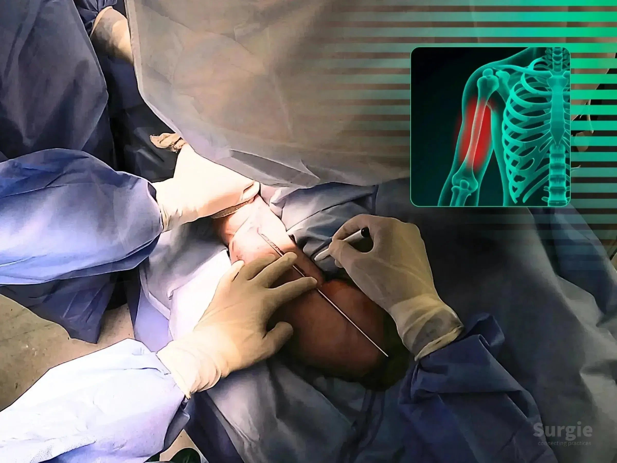
Surgical site on an arm with pre-operative markings and a digital X-ray overlay showing a humeral shaft fracture.
50% preview 4K Humerus
06:26
Humeral Shaft Fracture: Minimally Invasive Bridge Plating

Submuscular bridge plating with radial nerve protection

Dr. Sávio Chami Dr. Sávio ChamiOrthopedic Surgeon
9 languages
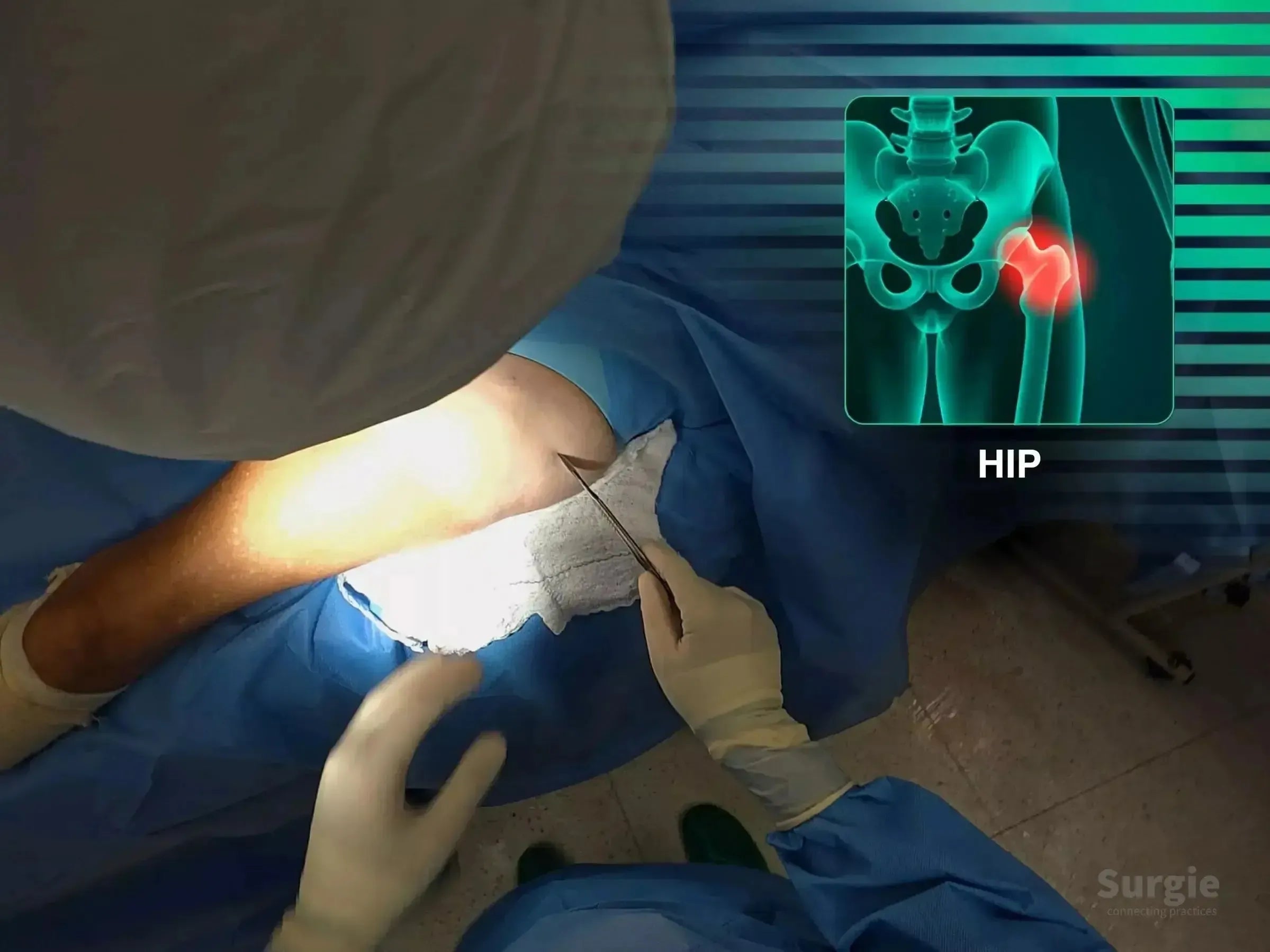
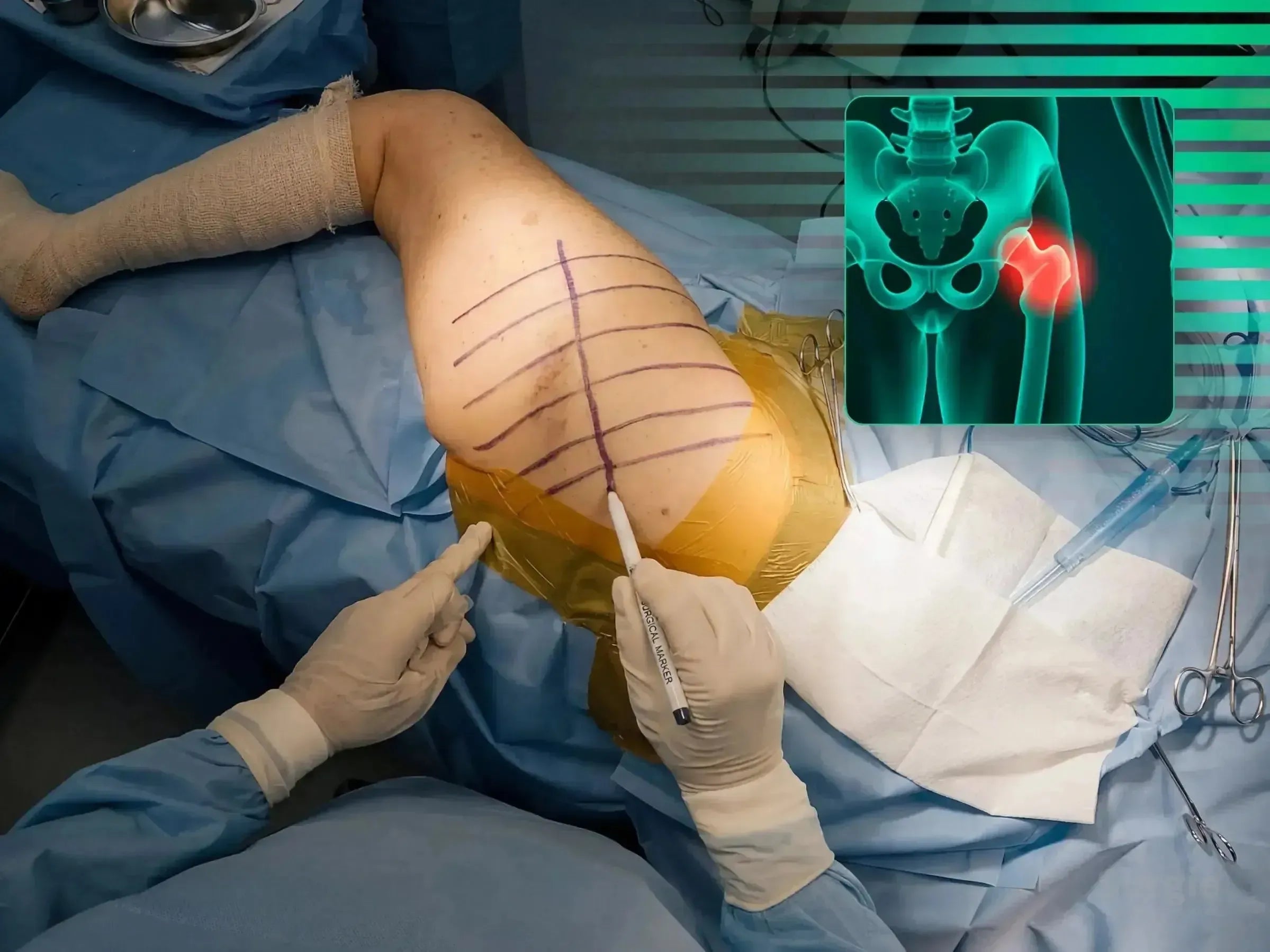
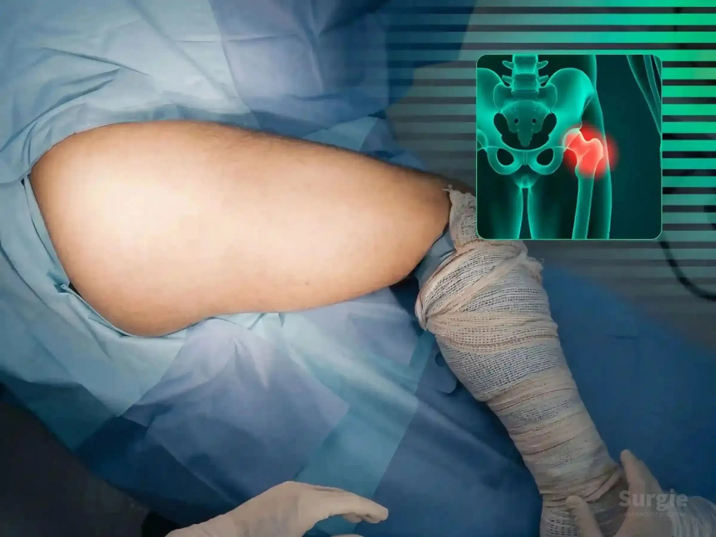
Surgeon performing a minimally invasive hip fracture procedure with a digital anatomical overlay of the femur.
50% preview 4K Hip
07:16
Intertrochanteric Fracture, Case 1: Intramedullary Nail

Guidewire precision and intramedullary fixation without traction

Dr. Sávio Chami Dr. Sávio ChamiOrthopedic Surgeon
9 languages
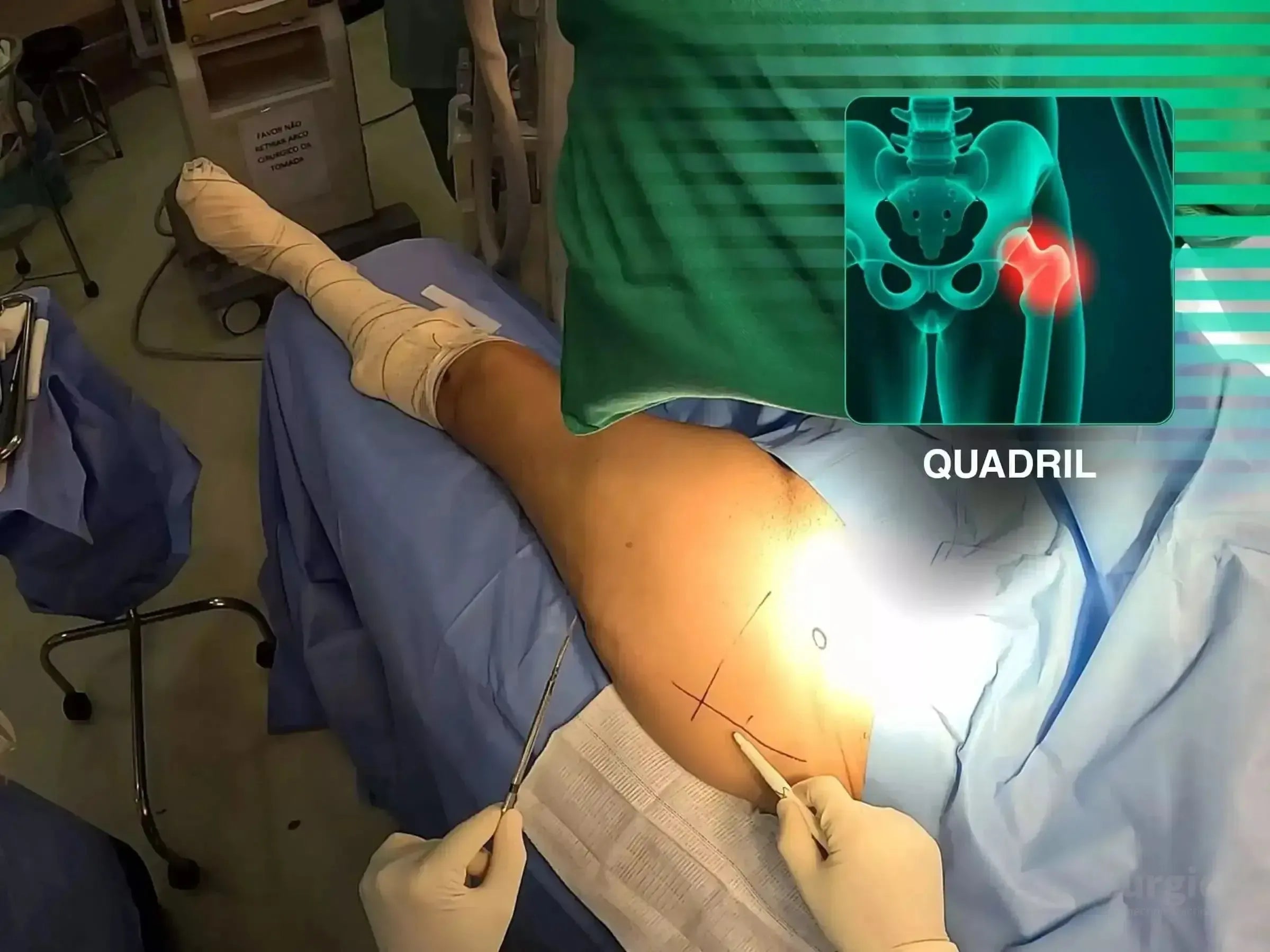
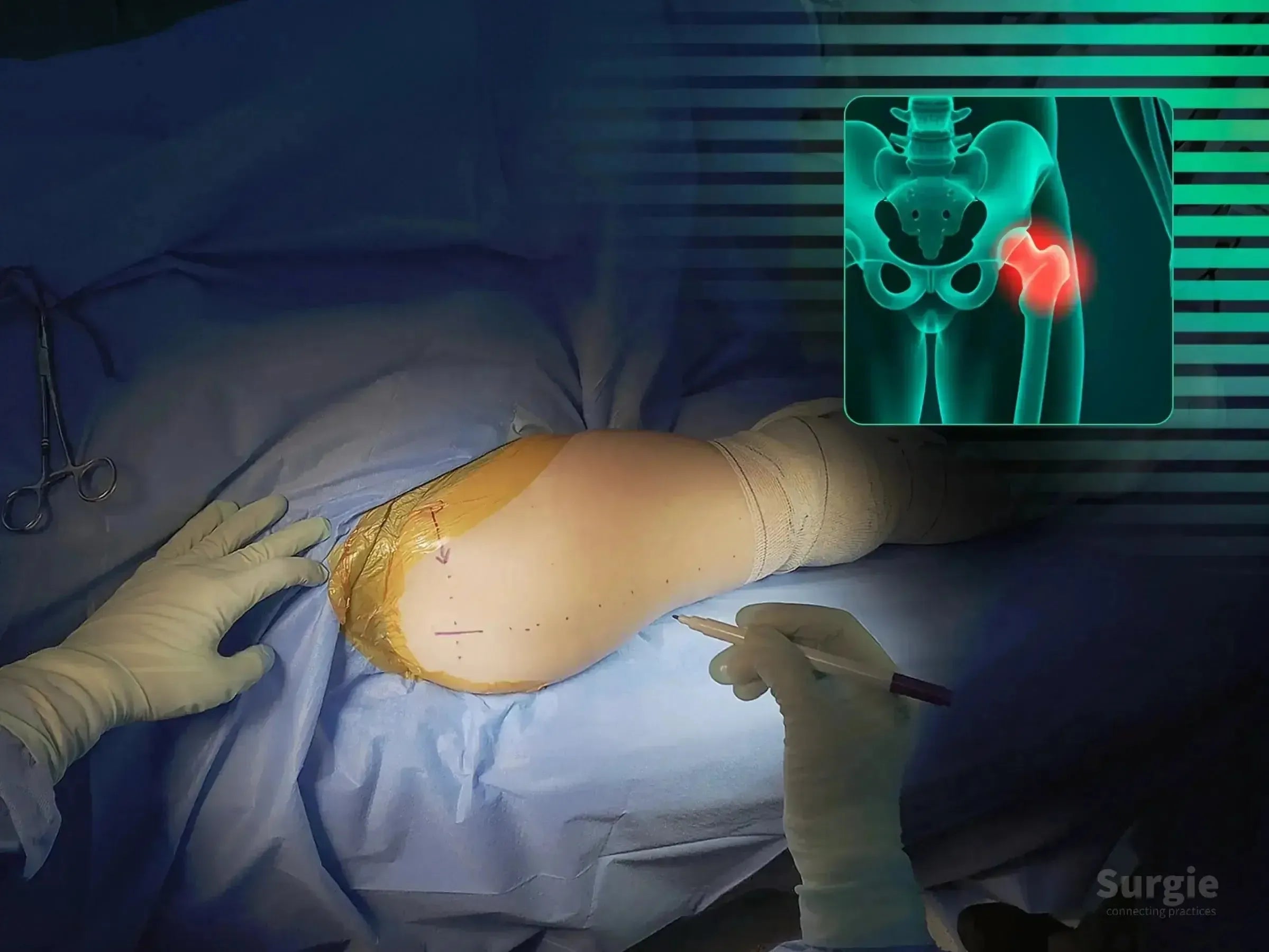
Surgeon marking a hip incision site for intramedullary nailing with a digital anatomical overlay of a transtrochanteric fracture.
50% preview 4K Hip
05:44
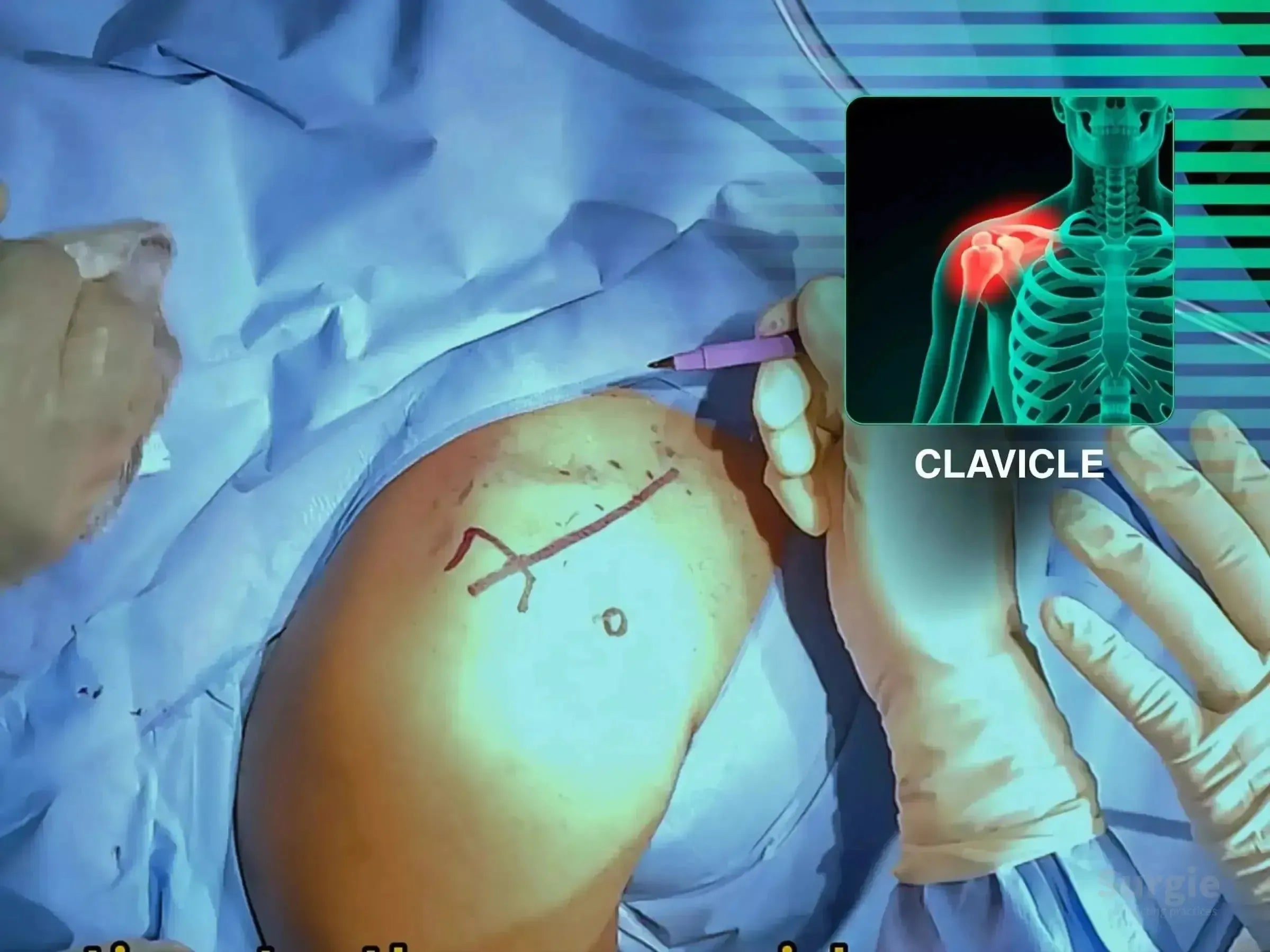
Surgeon marking a patient's shoulder for clavicle surgery with an anatomical overlay of a fractured collarbone.
50% preview 4K Shoulder
05:03
Lateral Clavicle Fracture: Coracoclavicular Fixation

Lateral clavicle reconstruction with coracoclavicular TightRope fixation

Dr. Sávio Chami Dr. Sávio ChamiOrthopedic Surgeon
9 languages
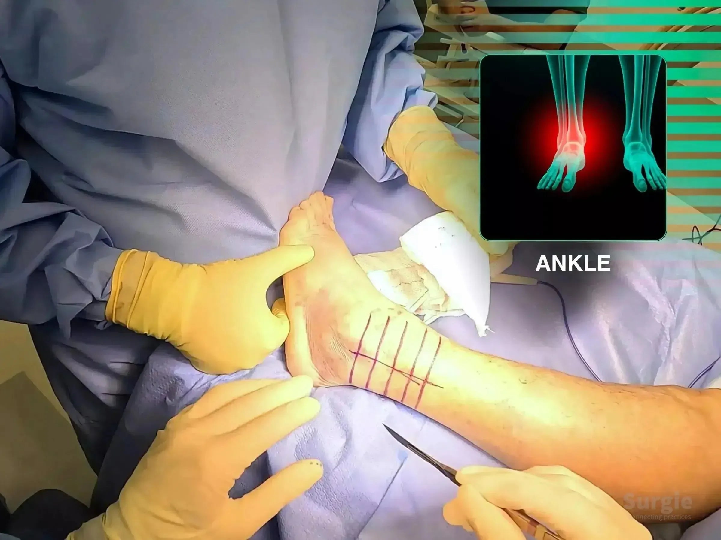
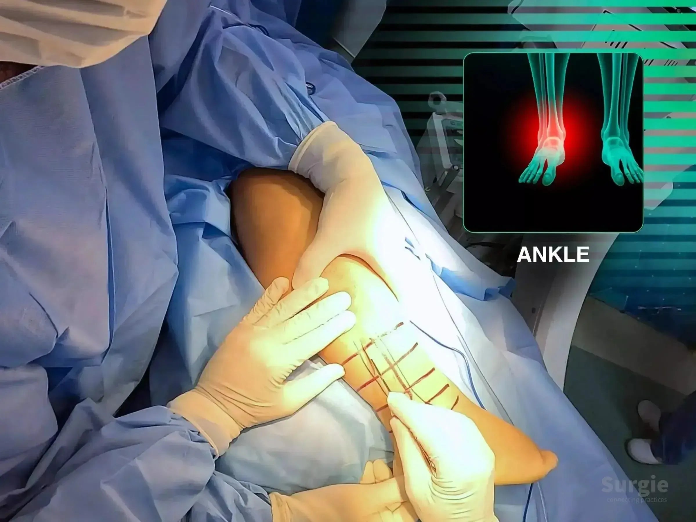
Surgeon marking a patient's ankle for lateral malleolus fracture surgery with an anatomical X-ray overlay.
50% preview 4K Ankle
02:29
Lateral Malleolus Fracture with Wagstaffe Fragment

Wagstaffe fragment compression with locked lateral malleolus fixation

Dr. Sávio Chami Dr. Sávio ChamiOrthopedic Surgeon
9 languages
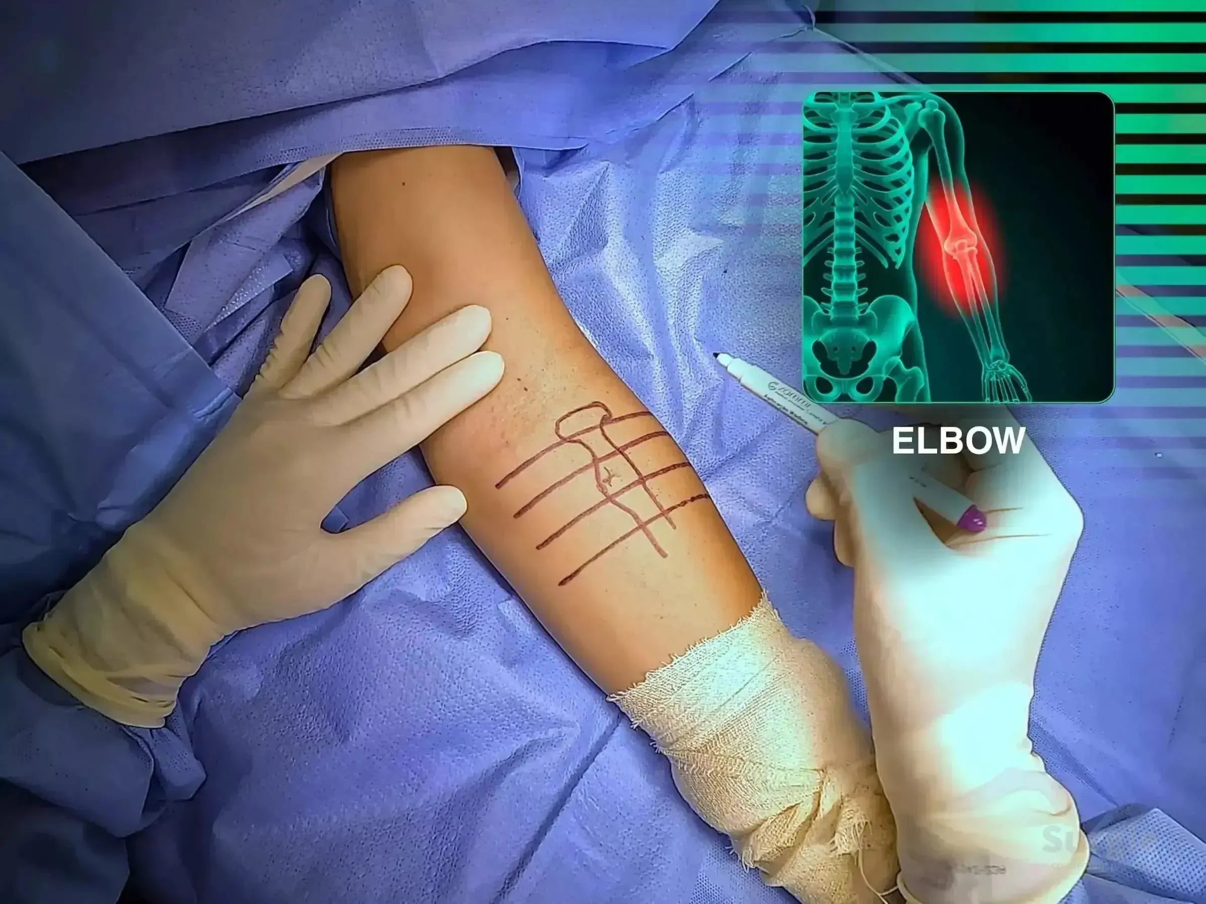
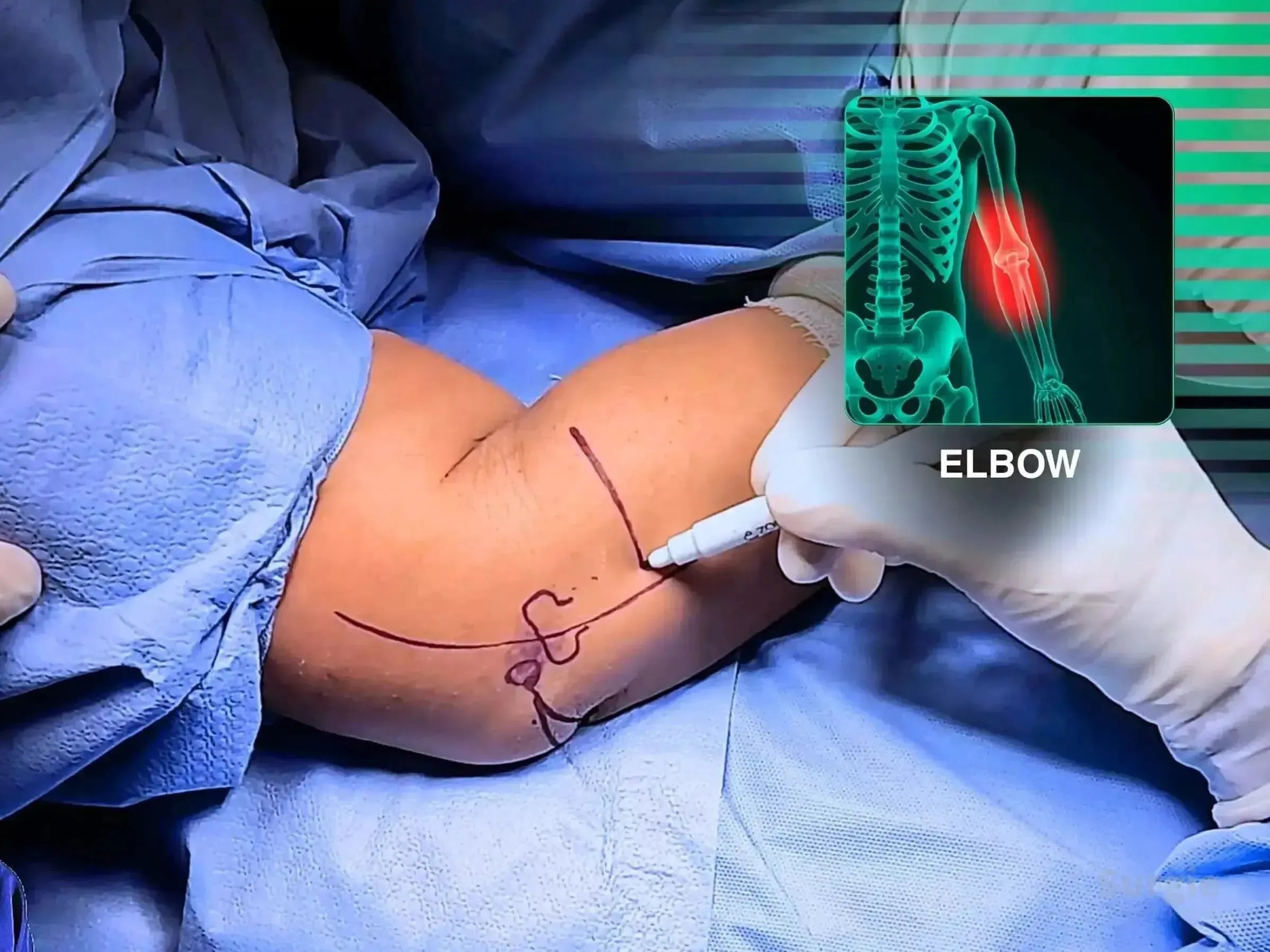
Surgeon marking a patient's elbow for surgery with an anatomical overlay showing a radial head fracture.
50% preview 4K Elbow
08:10
Mason Type III Radial Head Fracture with Elbow Instability

Mini-fragment fixation with coronoid and ligament reconstruction

Dr. Sávio Chami Dr. Sávio ChamiOrthopedic Surgeon
9 languages
Surgeon marking a posterior elbow incision site for Monteggia variant fracture repair with anatomical overlay.
50% preview 4K Elbow
14:59
Monteggia Variant: Proximal Ulna and Radial Head Fixation

Combined ulna and radial head fixation for Monteggia instability

Dr. Sávio Chami Dr. Sávio ChamiOrthopedic Surgeon
9 languages
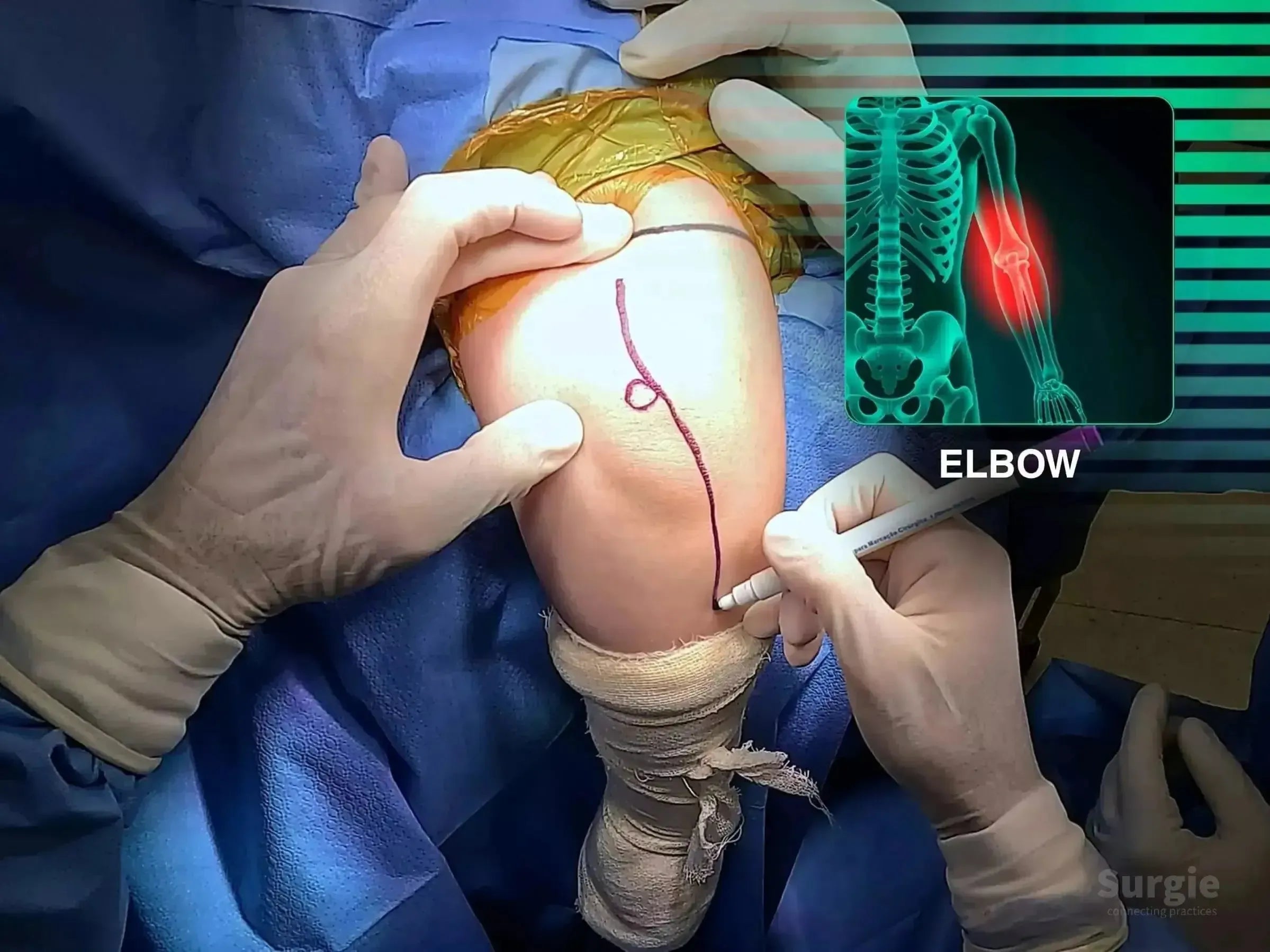
Surgeon marking an elbow for minimally invasive osteosynthesis with an anatomical skeletal overlay.
50% preview 4K Elbow
06:54
Olecranon Fracture: Minimally Invasive Plate Fixation

Minimally invasive olecranon plating with anatomic joint restoration

Dr. Sávio Chami Dr. Sávio ChamiOrthopedic Surgeon
9 languages
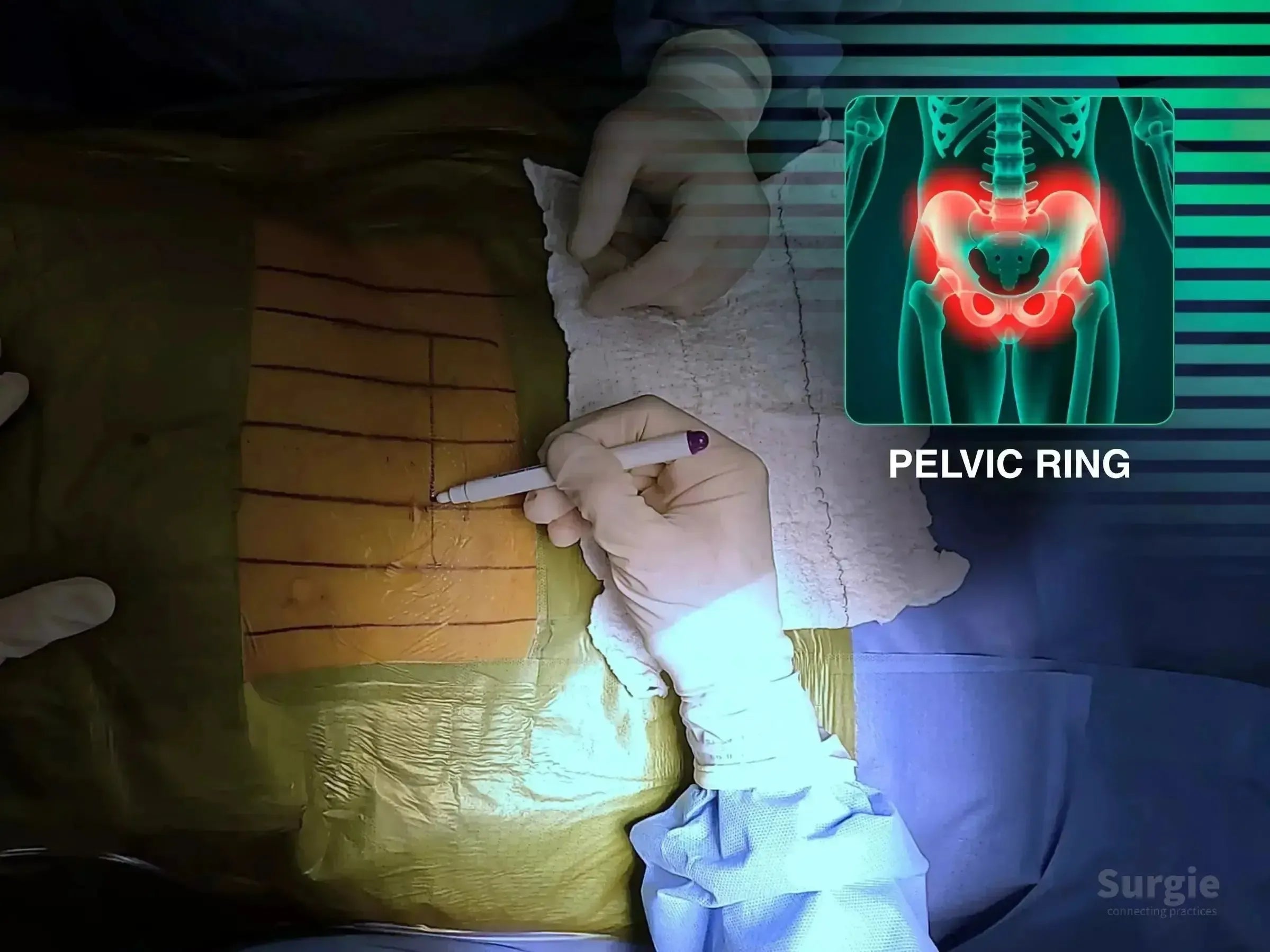
Surgeon marking a patient's abdomen for pelvic ring surgery with a digital overlay of a pubic symphysis dislocation.
50% preview 4K Pelvic Ring
06:13
Open-Book Pelvic Ring Injury: Pubic Symphysis Fixation

Controlled symphyseal reduction with anterior pelvic ring fixation

Dr. Sávio Chami Dr. Sávio ChamiOrthopedic Surgeon
9 languages
Surgeon marking a knee for patellar fracture surgery with an X-ray overlay showing a highlighted patella injury.
50% preview 4K Knee
24:55
Patellar Fracture, Case 2: Cannulated Screws and Cerclage

Cannulated screws and cerclage with extensor mechanism preservation

Dr. Sávio Chami Dr. Sávio ChamiOrthopedic Surgeon
9 languages
Surgeon marking a patient's hip for posterolateral surgery with a digital overlay of a periprosthetic hip fracture.
50% preview 4K Hip
19:45
Periprosthetic Hip Fracture: Trochanteric Plate and Cerclage

Trochanteric plating and cerclage without femoral component revision

Dr. Sávio Chami Dr. Sávio ChamiOrthopedic Surgeon
9 languages
Surgeon marking a patient's ankle for a posterolateral approach with an anatomical X-ray overlay of the fracture site.
50% preview 4K Ankle
04:14
Posterior and Lateral Malleolus Fractures: Posterolateral Approach

Single posterolateral exposure for posterior and lateral malleolus fixation

Dr. Sávio Chami Dr. Sávio ChamiOrthopedic Surgeon
9 languages
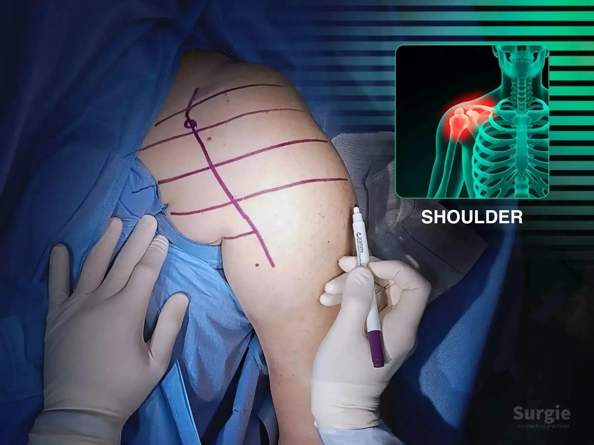
Surgeon marking a patient's shoulder for a deltopectoral approach with an anatomical diagram overlay.
50% preview 4K Shoulder
05:04
Posterior Shoulder Dislocation with Humeral Head Impaction

Reduction of a locked posterior dislocation with carbon-fiber plating

Dr. Sávio Chami Dr. Sávio ChamiOrthopedic Surgeon
9 languages
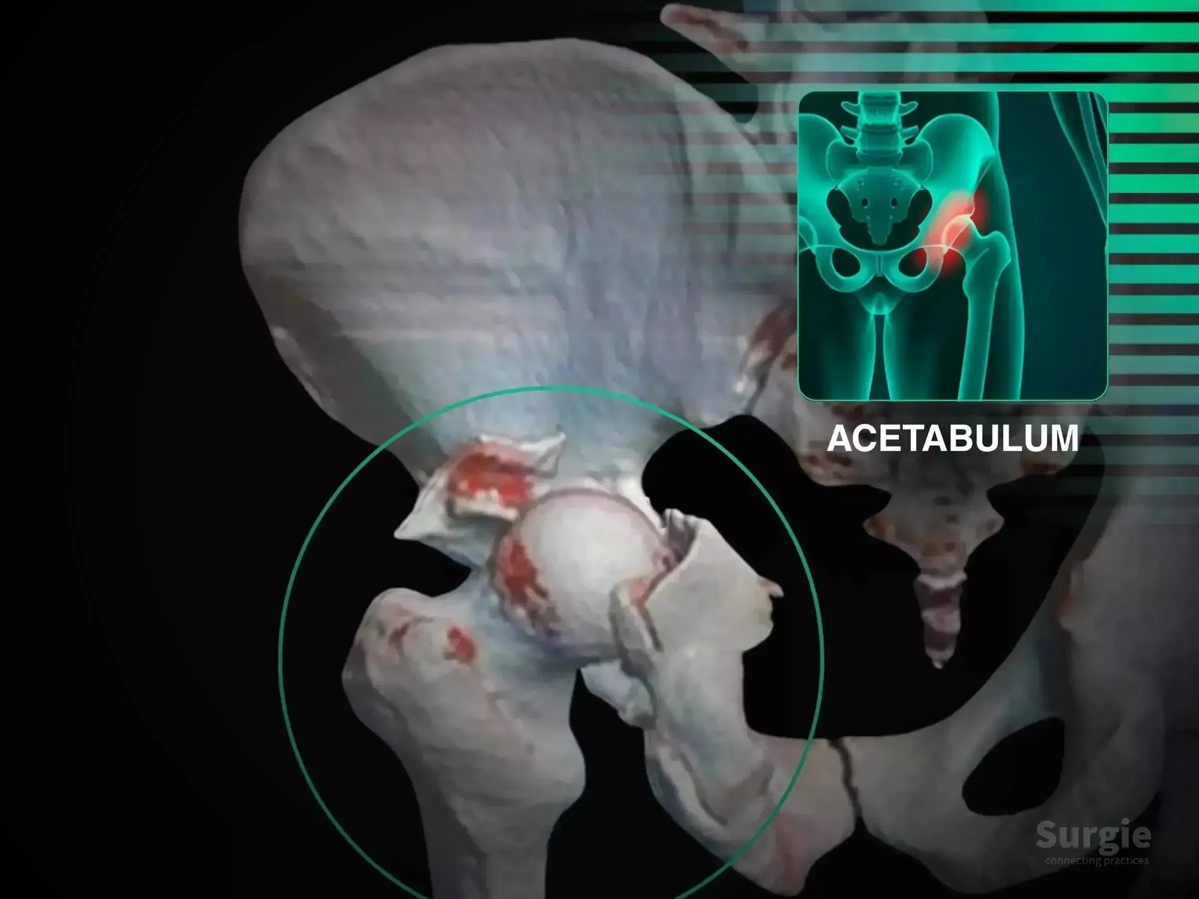
Surgical preparation for acetabular fracture with an anatomical 3D hip overlay showing posterior rim and articular impaction.
50% preview 4K Acetabulum
22:49
Posterior Wall Acetabular Fracture with Articular Impaction

Articular disimpaction and posterior wall reconstruction

Dr. Sávio Chami Dr. Sávio ChamiOrthopedic Surgeon
9 languages
3D medical illustration showing a posterior acetabular wall fracture with hip dislocation and highlighted bone fragments.
50% preview 4K Acetabulum
05:35
Posterior Wall Acetabular Fracture with Hip Dislocation

Kocher-Langenbeck exposure with posterior wall fixation and nerve protection

Dr. Sávio Chami Dr. Sávio ChamiOrthopedic Surgeon
9 languages
Surgeon marking a patient's hip for proximal femur intramedullary nail surgery with a digital anatomical overlay.
50% preview 4K Hip
10:24
Proximal Femur Fracture, Case 3: Rocker Maneuver

Rocker-assisted calcar reduction without a traction table

Dr. Sávio Chami Dr. Sávio ChamiOrthopedic Surgeon
9 languages
Surgeon performing percutaneous reduction on a patient's thigh with a digital femoral fracture overlay.
50% preview 4K Hip
13:34
Proximal Femur Fracture, Case 4: Double See-Saw Technique

Double see-saw reduction for shaft and calcar realignment

Dr. Sávio Chami Dr. Sávio ChamiOrthopedic Surgeon
9 languages
Surgical setup for proximal femur fracture with blue drapes and an anatomical overlay highlighting the femur.
50% preview 4K Hip
07:16
Proximal Femur Fracture: Intramedullary Nail and See-Saw Maneuver

See-saw reduction of an unstable proximal femur fracture

Dr. Sávio Chami Dr. Sávio ChamiOrthopedic Surgeon
9 languages