Passer au contenu

Panier

Votre panier est vide

Reconstruction osseuse

Trier par

64 produits

Filtres

Filter

Refine

view all

Sous-spécialité

Par le Docteur

Filter

Refine

view all

Sous-spécialité

Par le Docteur

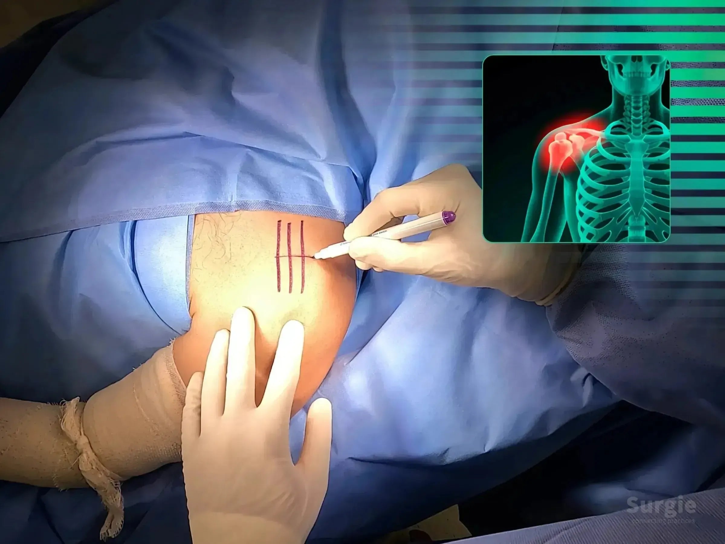
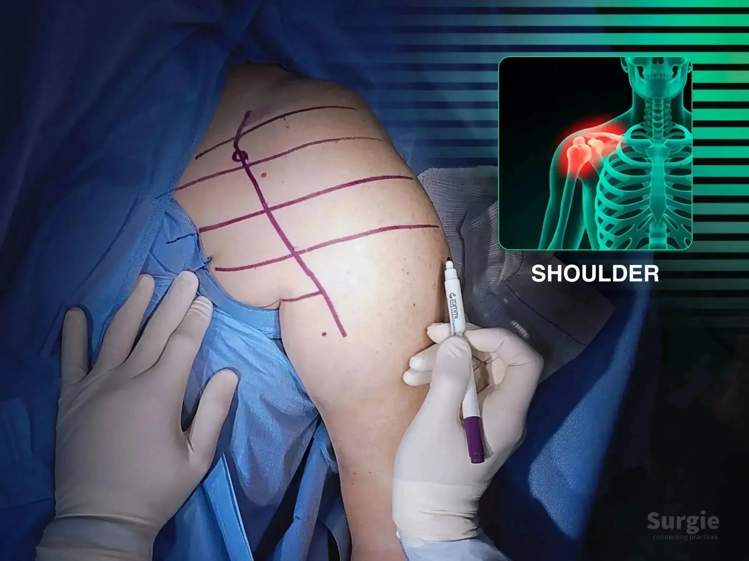
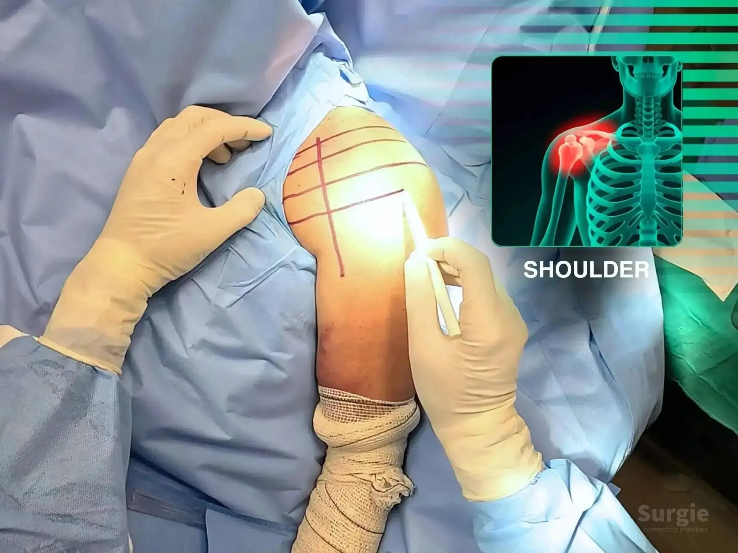
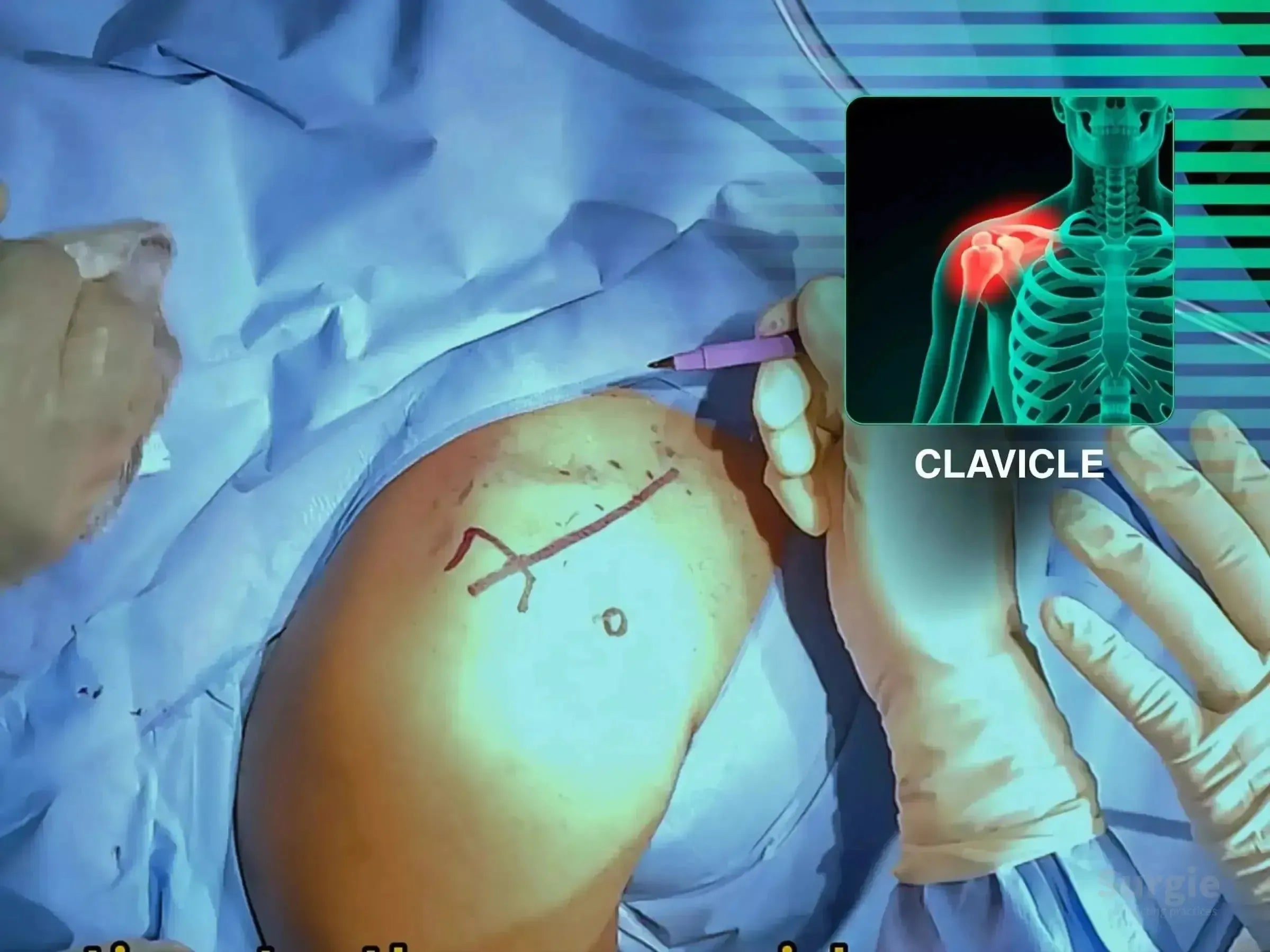
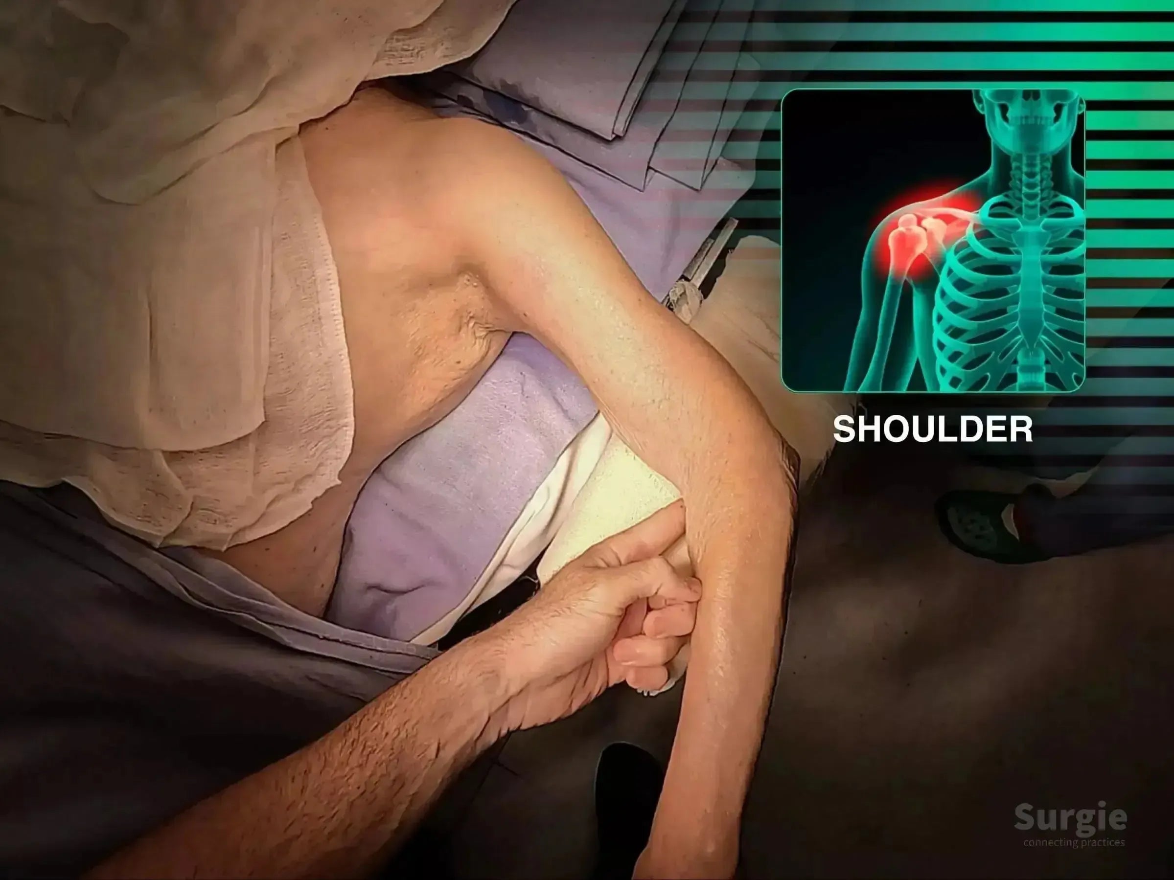
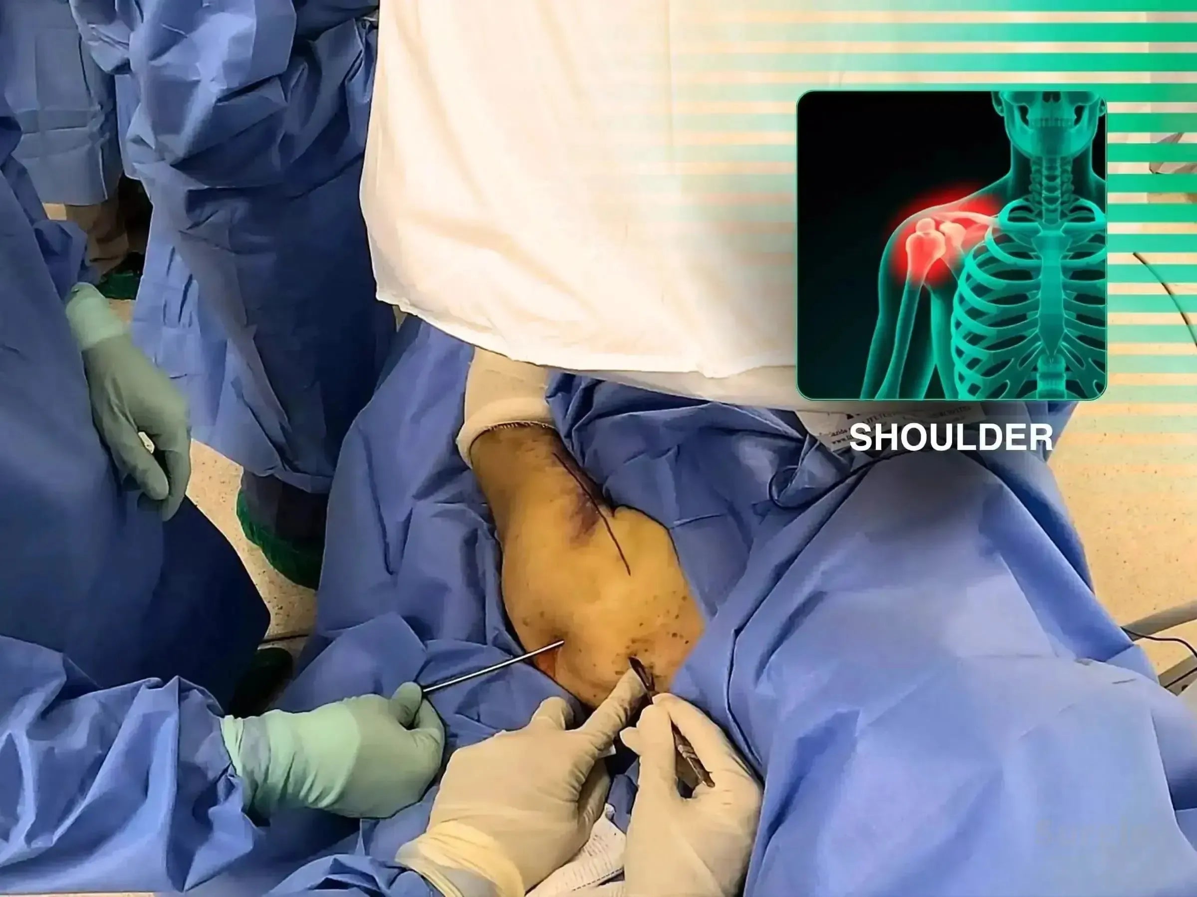
Surgeon marking shoulder skin for TightRope AC joint reconstruction with an anatomical overlay showing dislocation.
50% preview 4K Épaule
07:08
Luxation de l'articulation acromio-claviculaire, Cas 2 : Technique du tunnel TightRope

Tunneling précis de la clavicule et du coracoïde pour la fixation TightRope

Dr. Sávio Chami Dr. Sávio ChamiOrthopédiste
9 languages
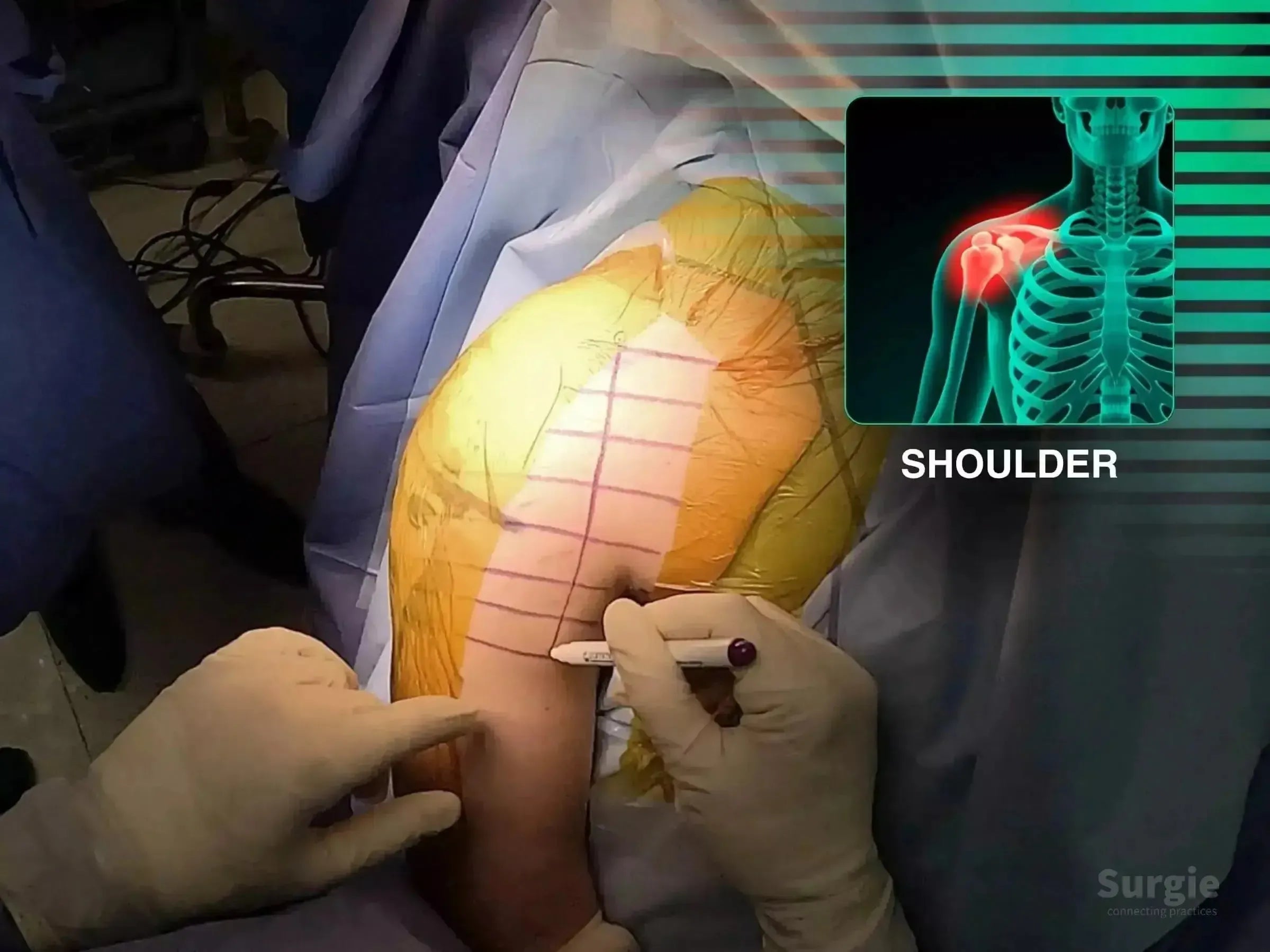
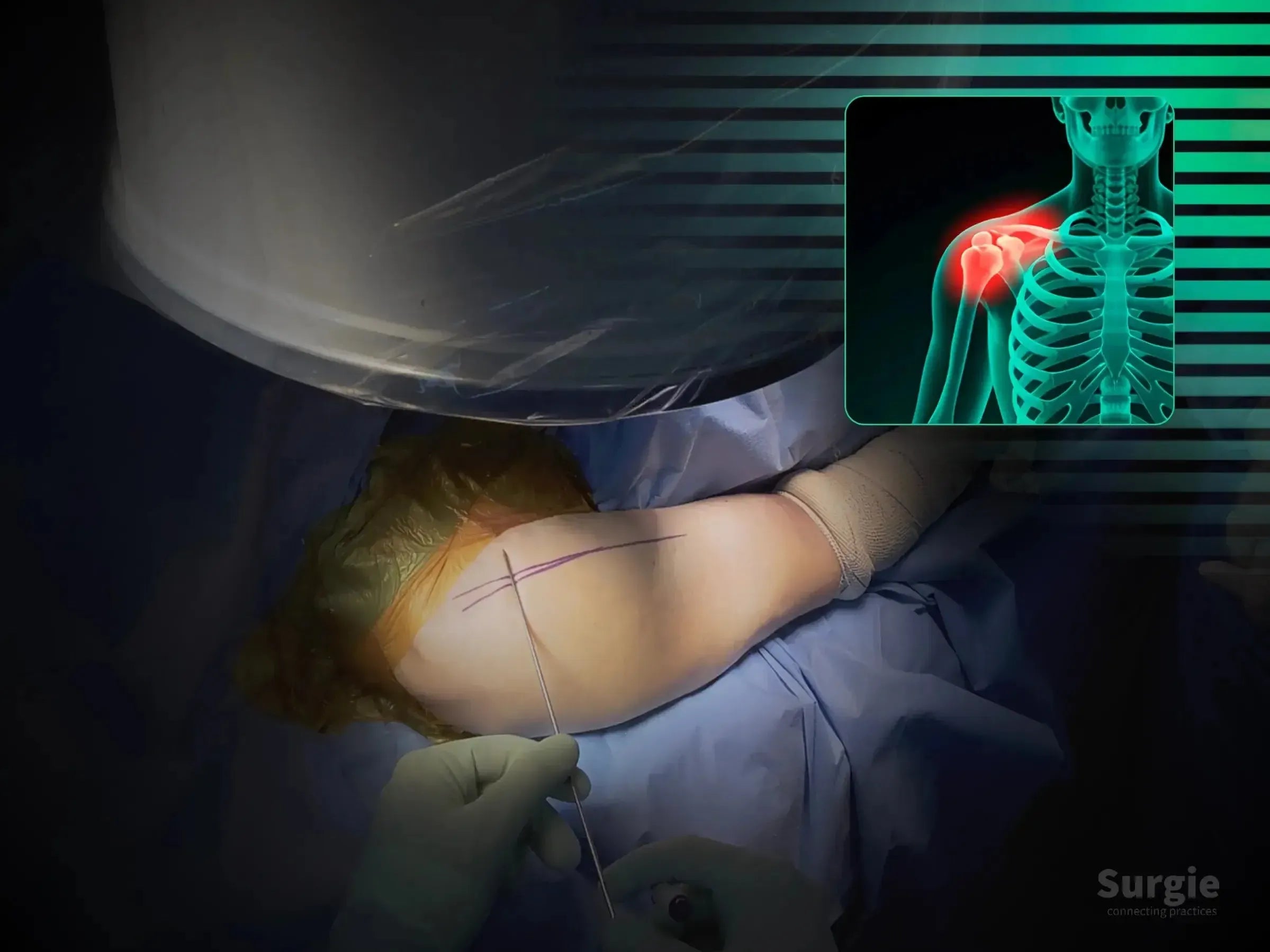
Surgeon marking a patient's shoulder for AC joint surgery with a TightRope device, featuring a skeletal X-ray overlay.
50% preview 4K Épaule
06:42
Luxation de l'articulation AC : Fixation ouverte par TightRope

Open coracoclavicular tunnel creation and TightRope stabilization

Dr. Sávio Chami Dr. Sávio ChamiOrthopédiste
9 languages
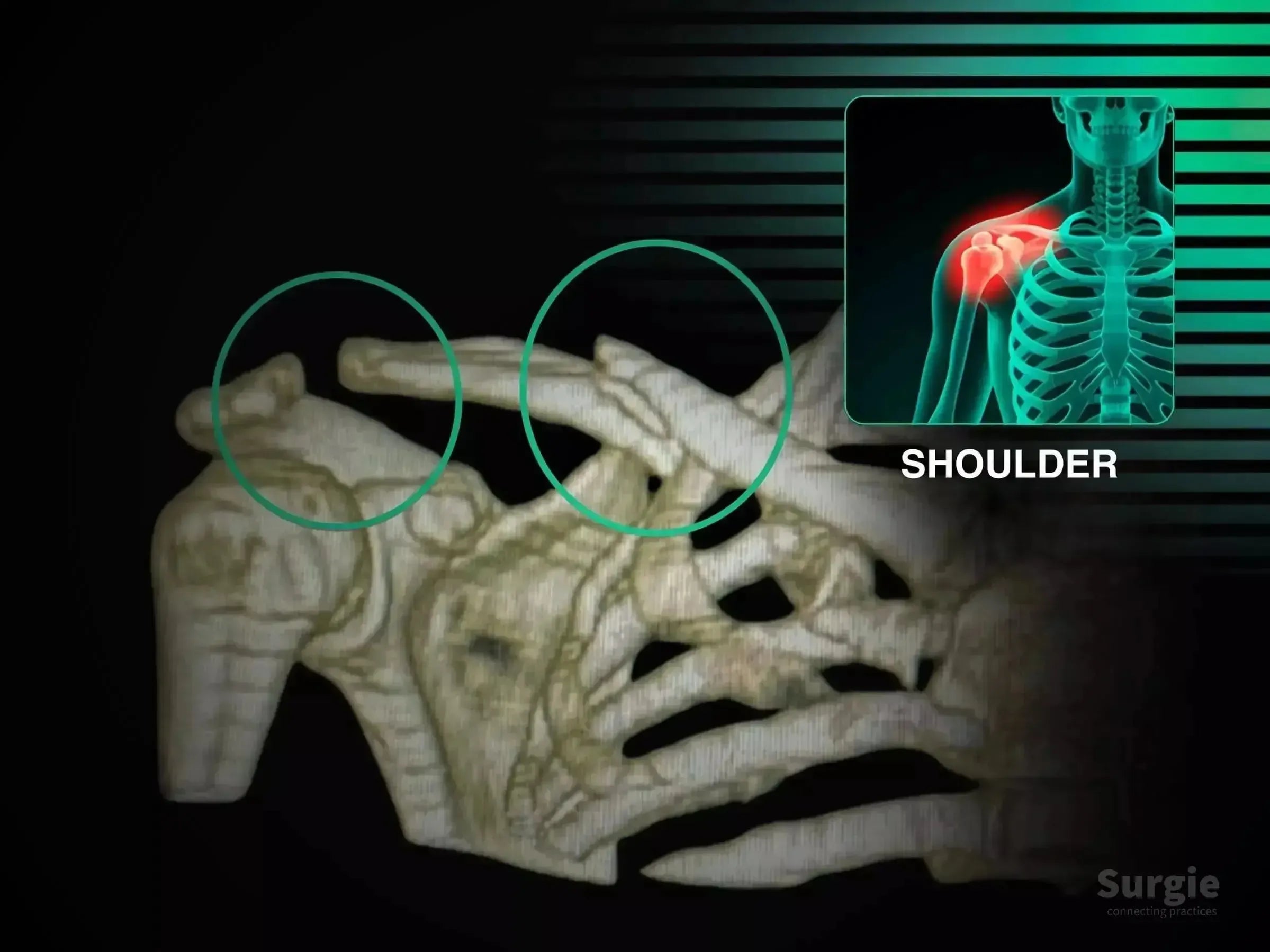
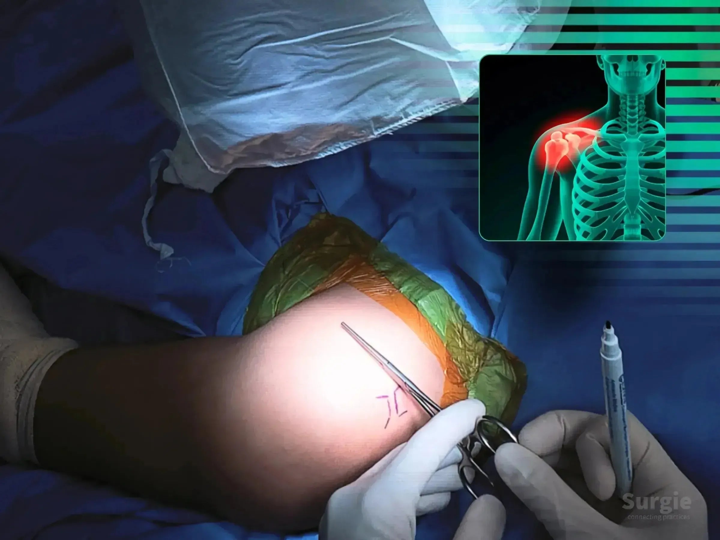
3D skeletal model showing clavicle fracture and acromioclavicular dislocation with highlighted surgical zones.
50% preview 4K Épaule
05:26
Fracture de la clavicule avec luxation postérieure de l'articulation acromio-claviculaire

Combined clavicle fixation and multidirectional AC joint stabilization

Dr. Sávio Chami Dr. Sávio ChamiOrthopédiste
9 languages
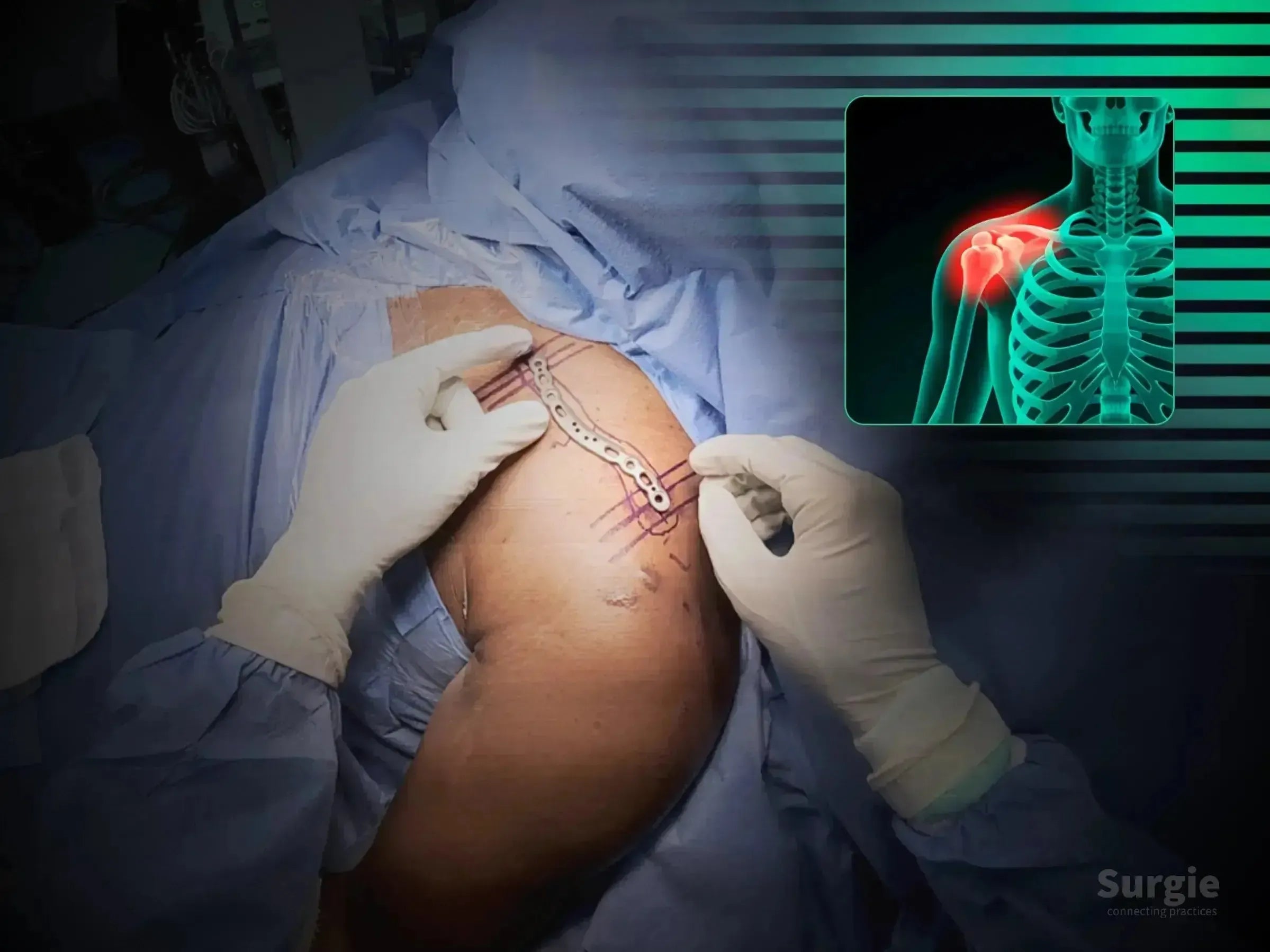
Surgeon placing a metallic anatomic clavicle plate on a patient with a skeletal diagram showing a shoulder fracture.
50% preview 4K Épaule
15:50
Fracture de la clavicule, Cas 2 : Fixation mini-invasive

Fixation de la clavicule à deux portails avec réduction fluoroscopique

Dr. Sávio Chami Dr. Sávio ChamiOrthopédiste
9 languages
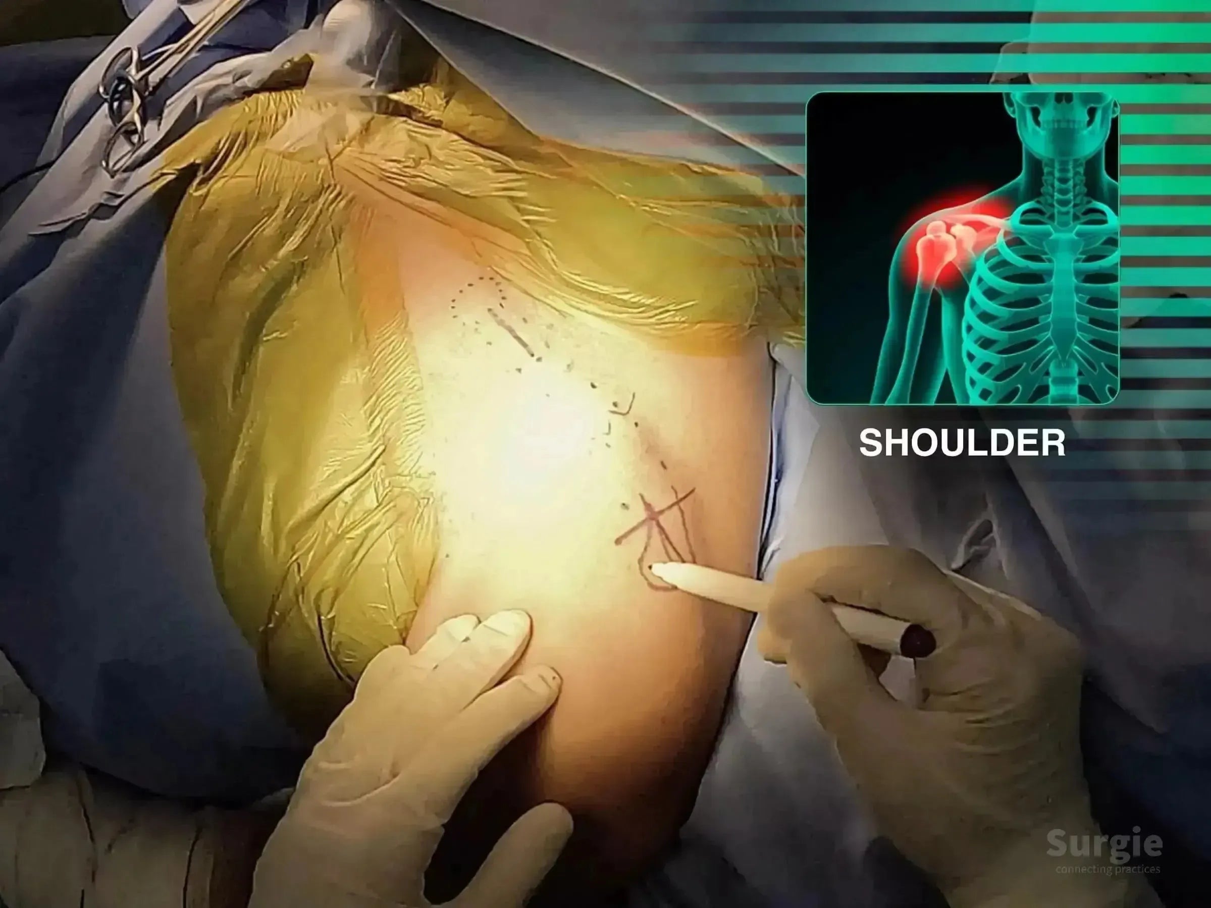
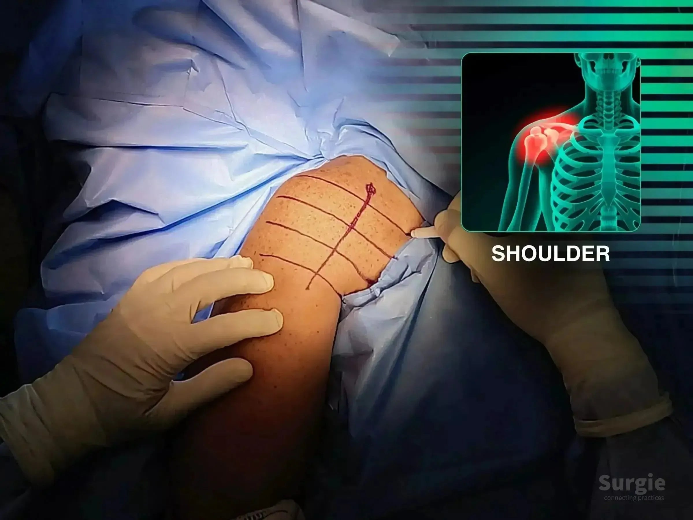
Surgeon marking skin for minimally invasive clavicle bridge plating with anatomical shoulder diagram overlay.
50% preview 4K Épaule
02:22
Fracture de la clavicule : Plaque de pontage mini-invasive

Two-portal bridge plating with soft-tissue preservation

Dr. Sávio Chami Dr. Sávio ChamiOrthopédiste
9 languages
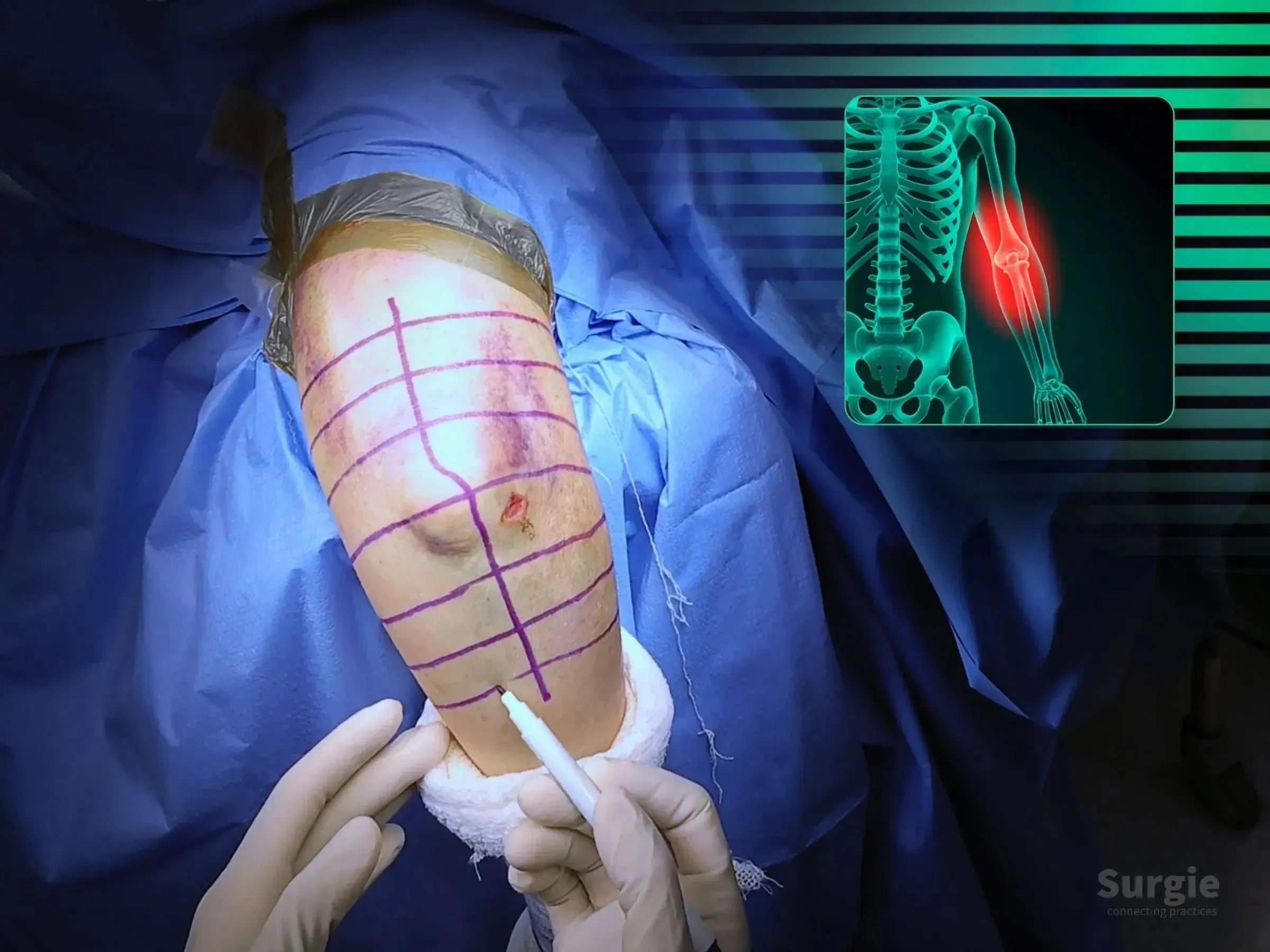
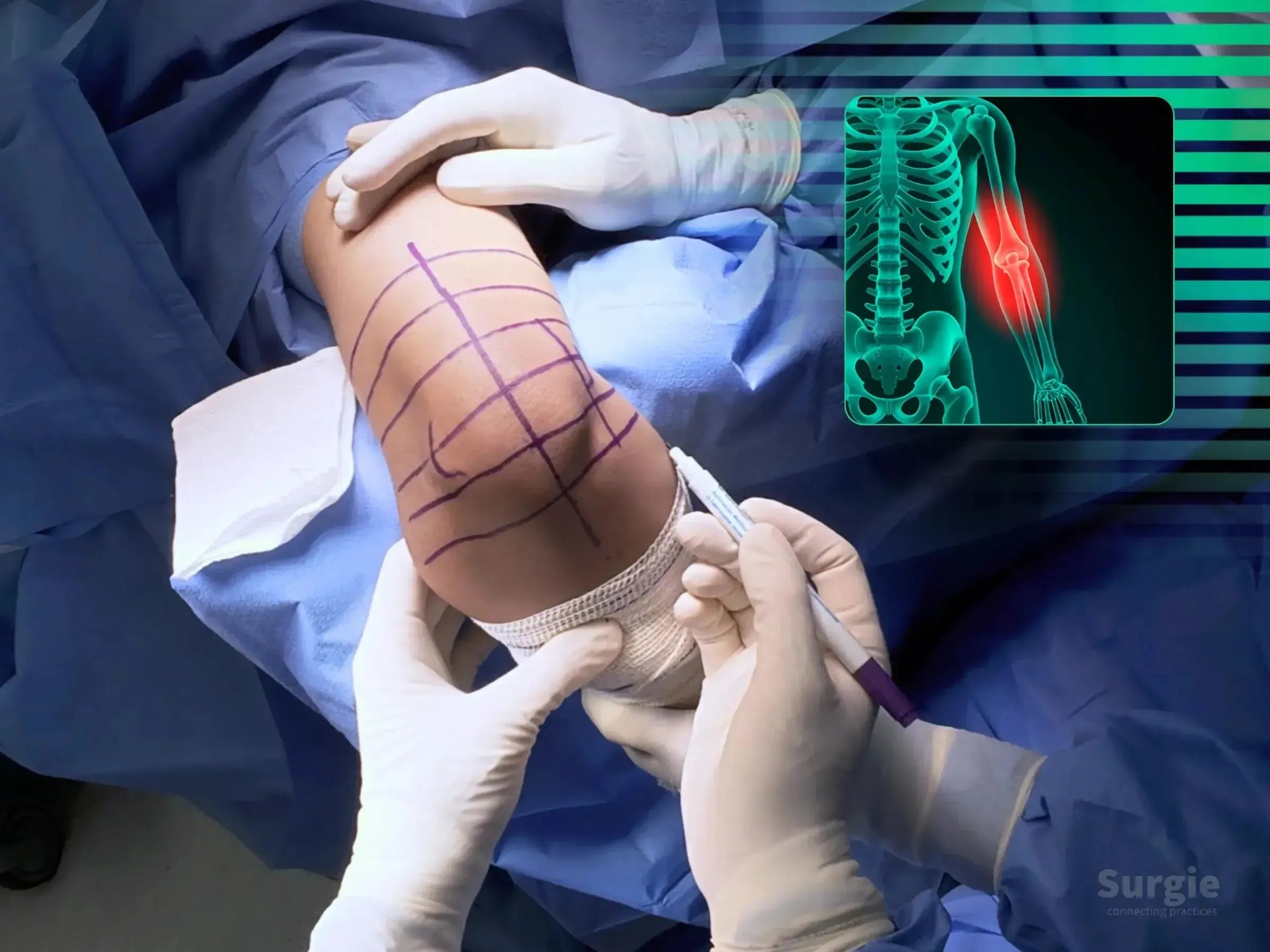
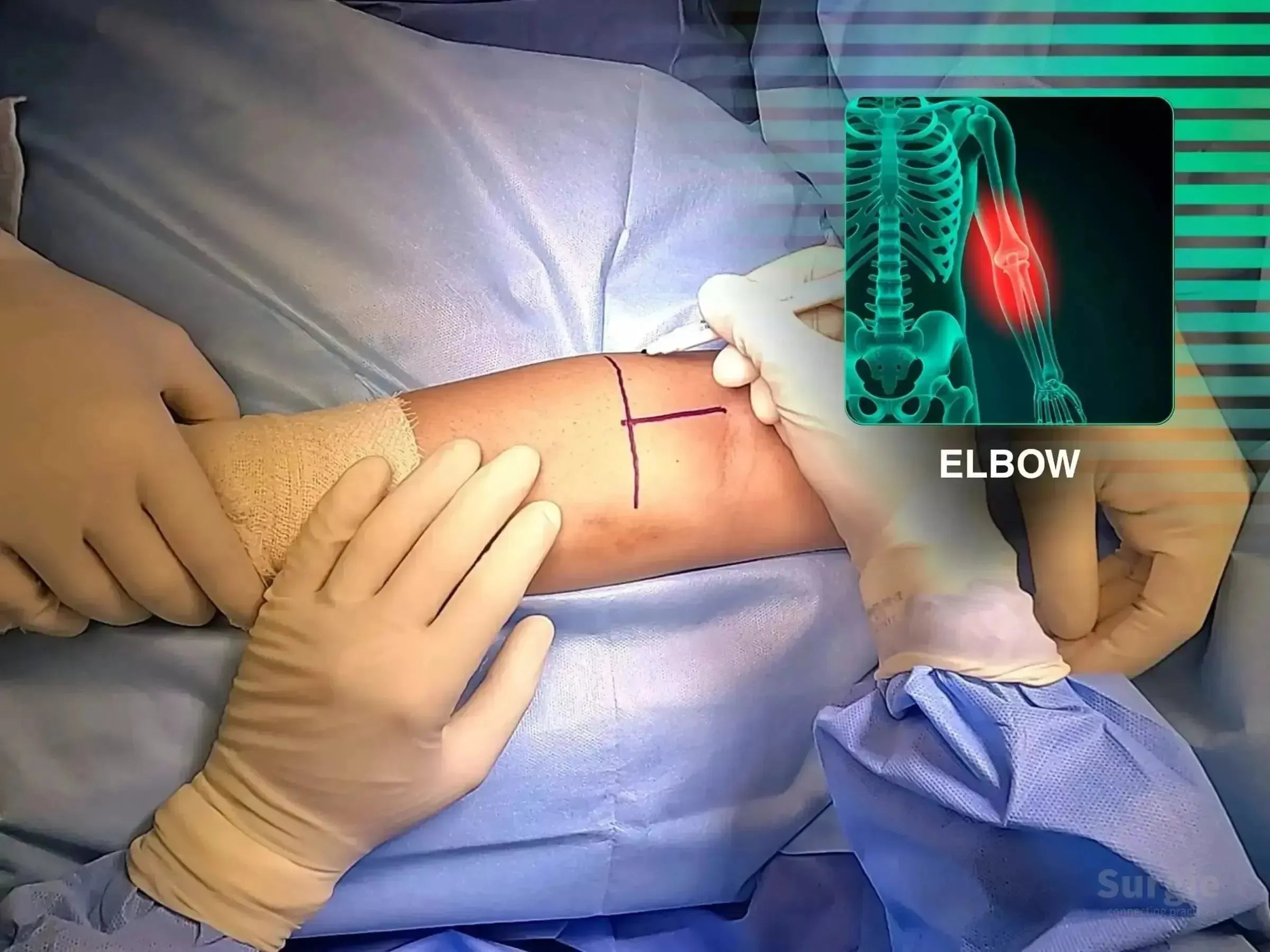
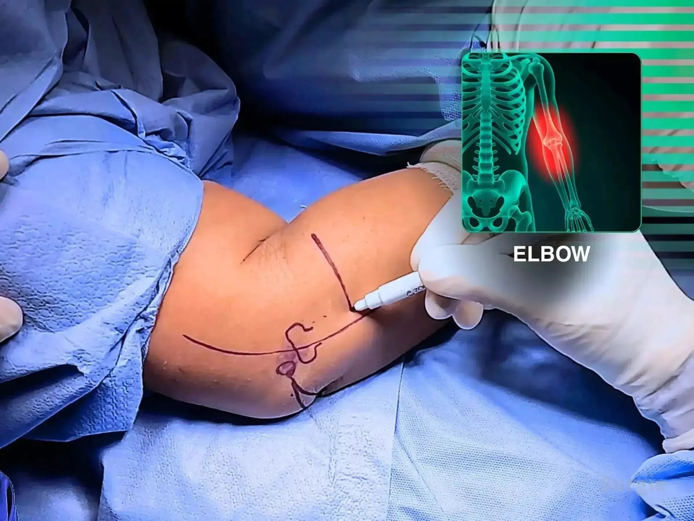
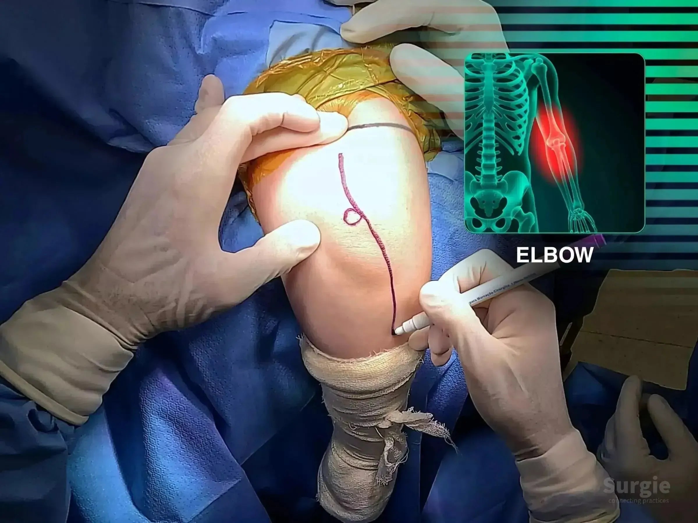
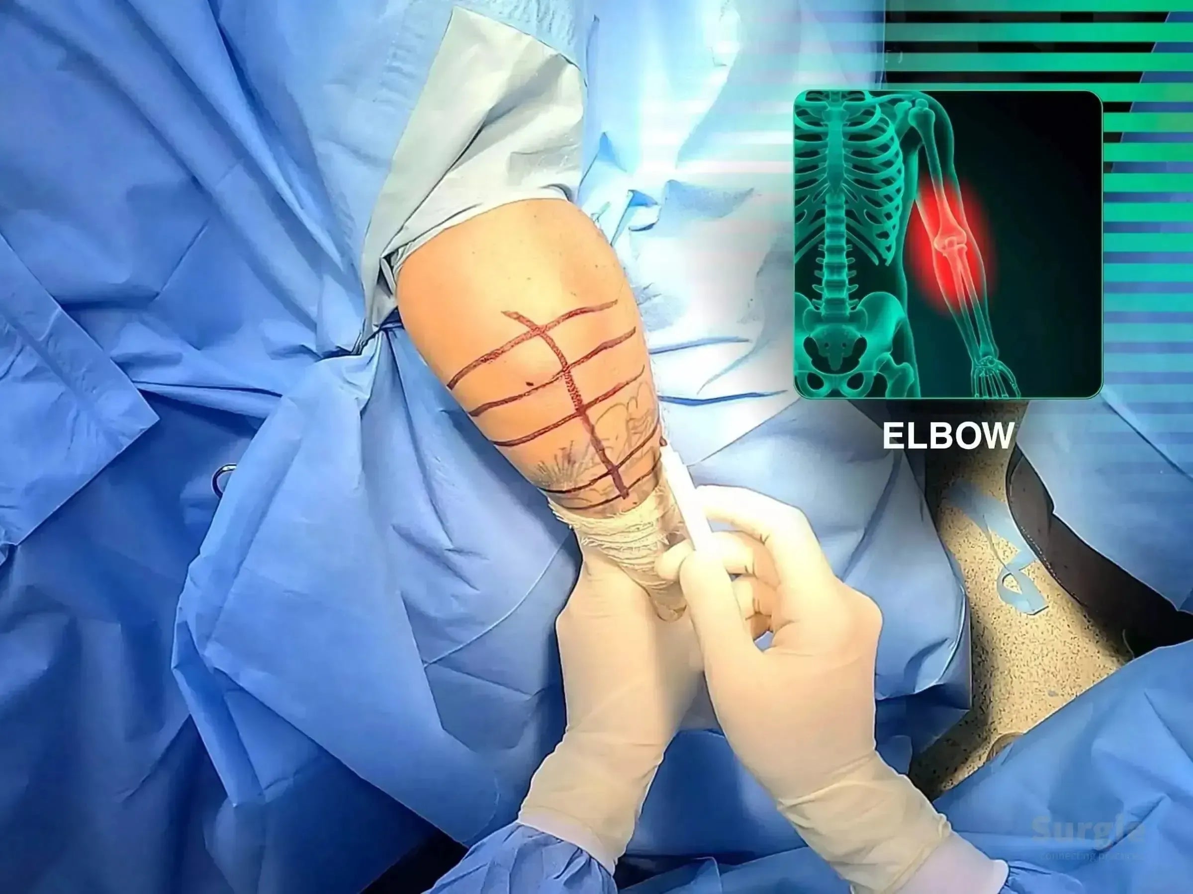
Surgical marking on a patient's elbow with a distal humerus fracture overlay showing the injury site in a clinical setting.
50% preview 4K Coude
36:05
Fracture articulaire comminutive de l'humérus distal : fixation

Olecranon osteotomy, articular reduction, and dual-column fixation

Dr. Sávio Chami Dr. Sávio ChamiOrthopédiste
9 languages
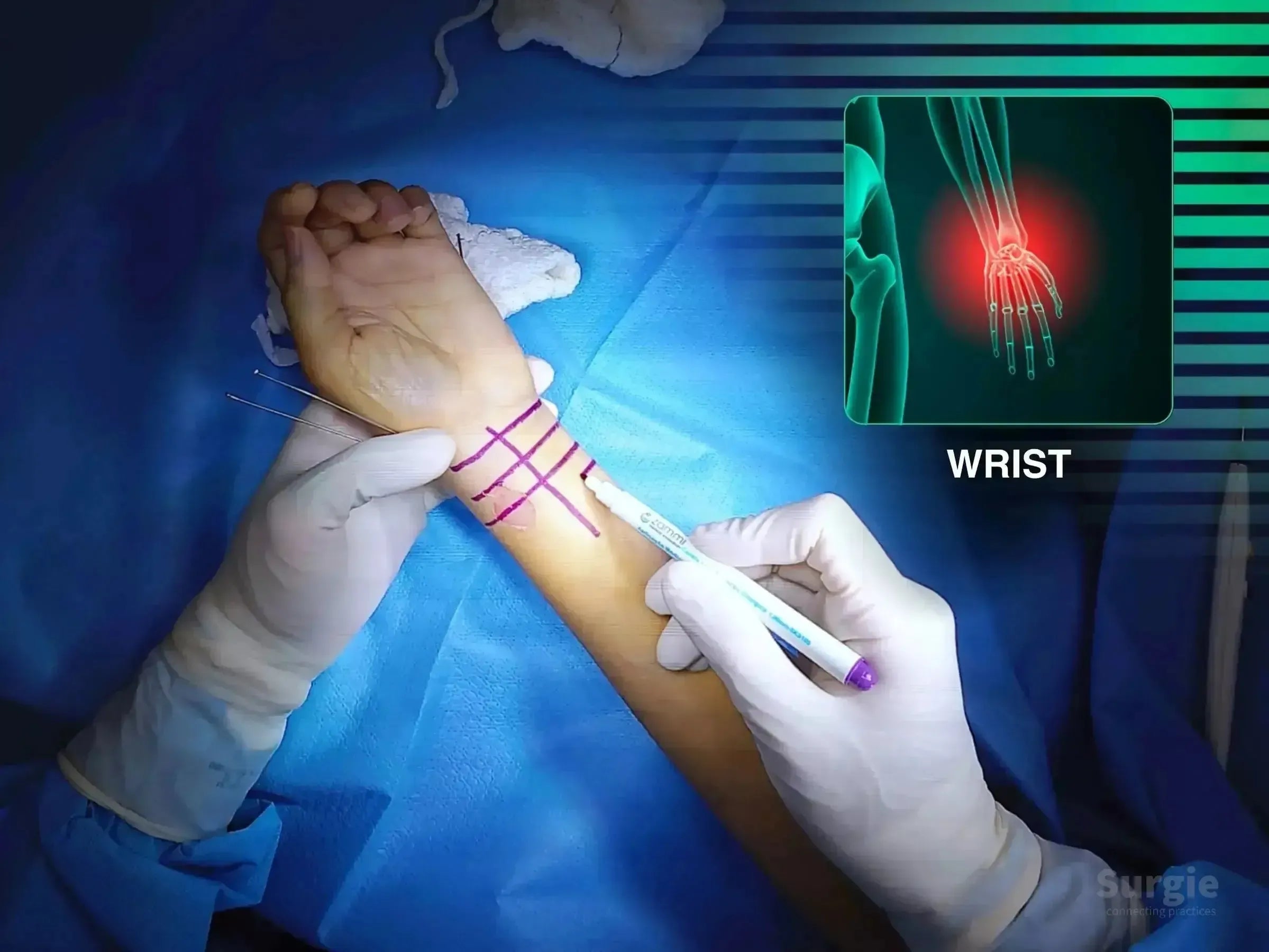
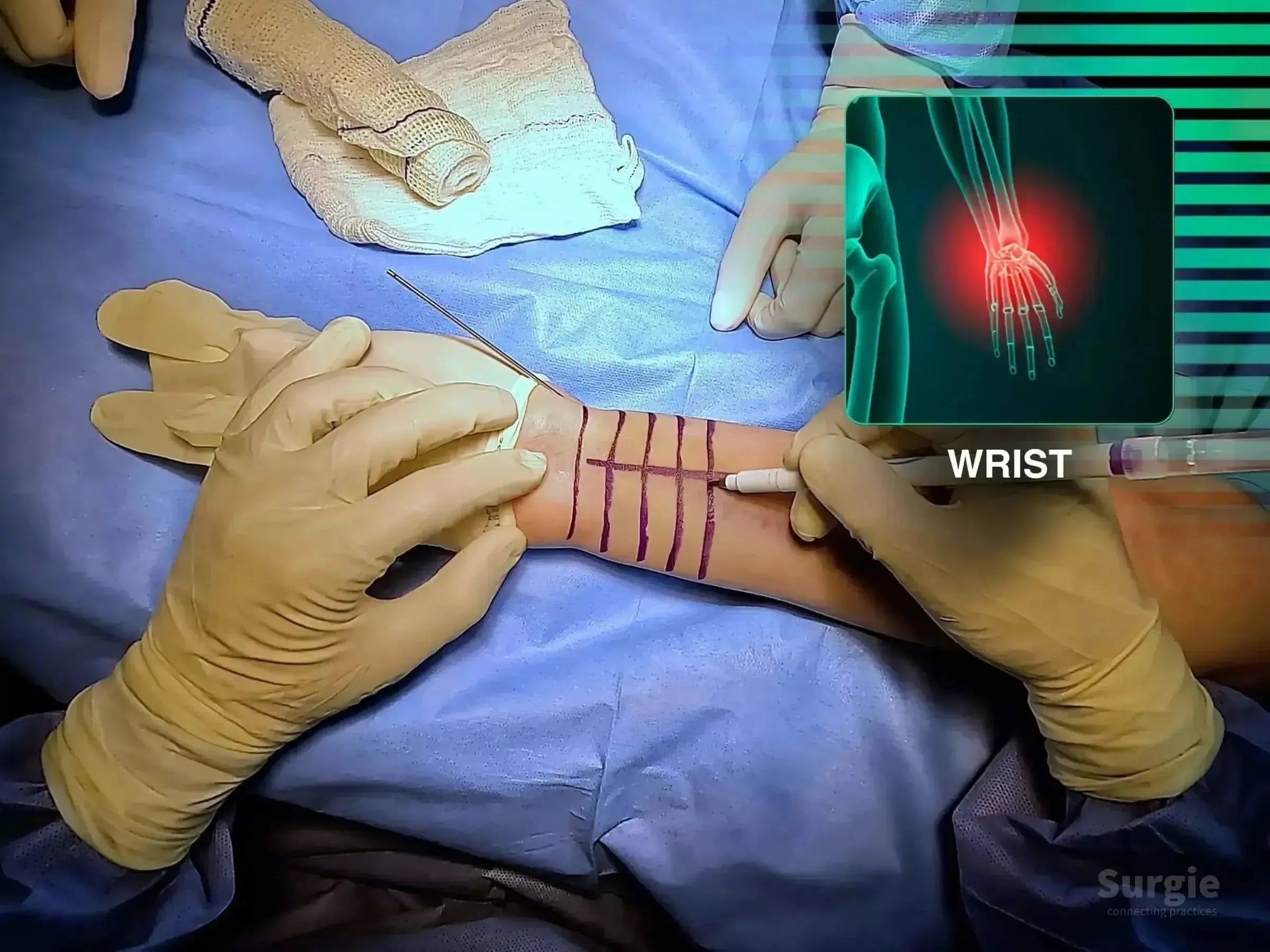
Surgeon marking a patient's wrist for volar approach surgery with a digital anatomical overlay of the distal radius.
50% preview 4K Poignet
11:08
Fracture comminutive du radius distal : fixation par plaque palmaire

Restore radial length and articular congruity through volar plating

Dr. Sávio Chami Dr. Sávio ChamiOrthopédiste
9 languages
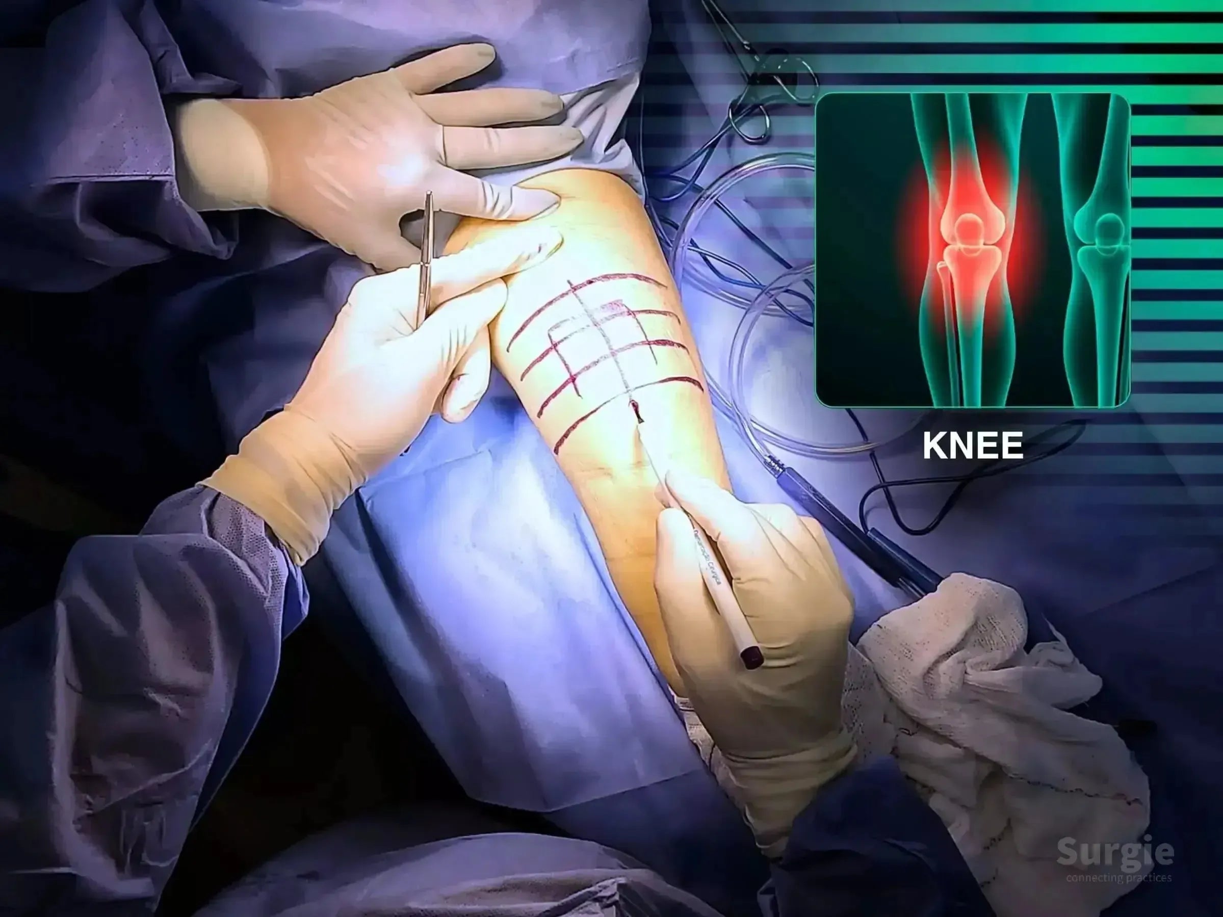
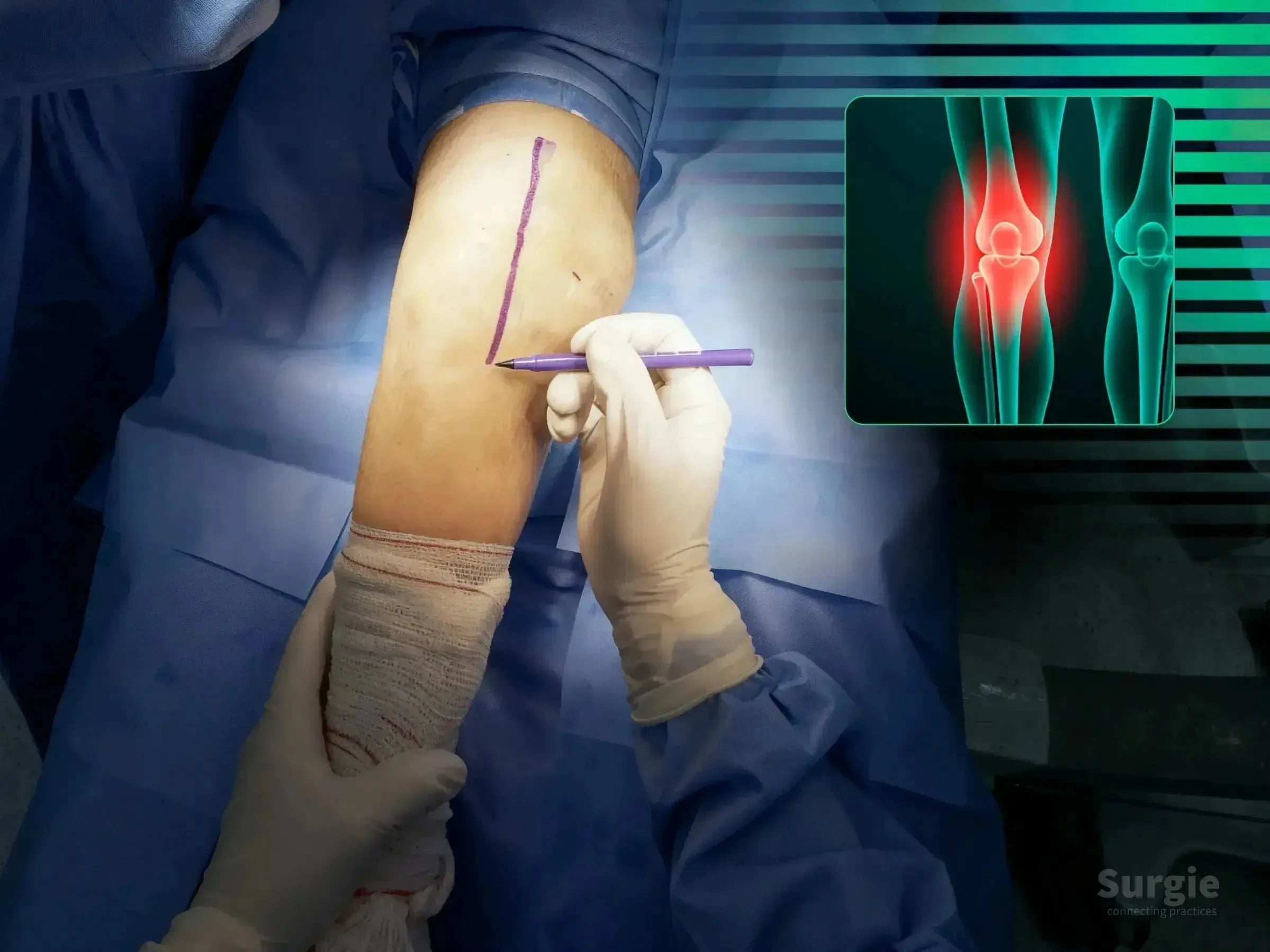
Surgeon marking a patient's knee for patella fracture surgery with an anatomical diagram overlay.
50% preview 4K Genou
13:28
Fracture comminutive de la rotule : fixation chirurgicale

Cannulated screws and cerclage for extensor mechanism reconstruction

Dr. Sávio Chami Dr. Sávio ChamiOrthopédiste
9 languages
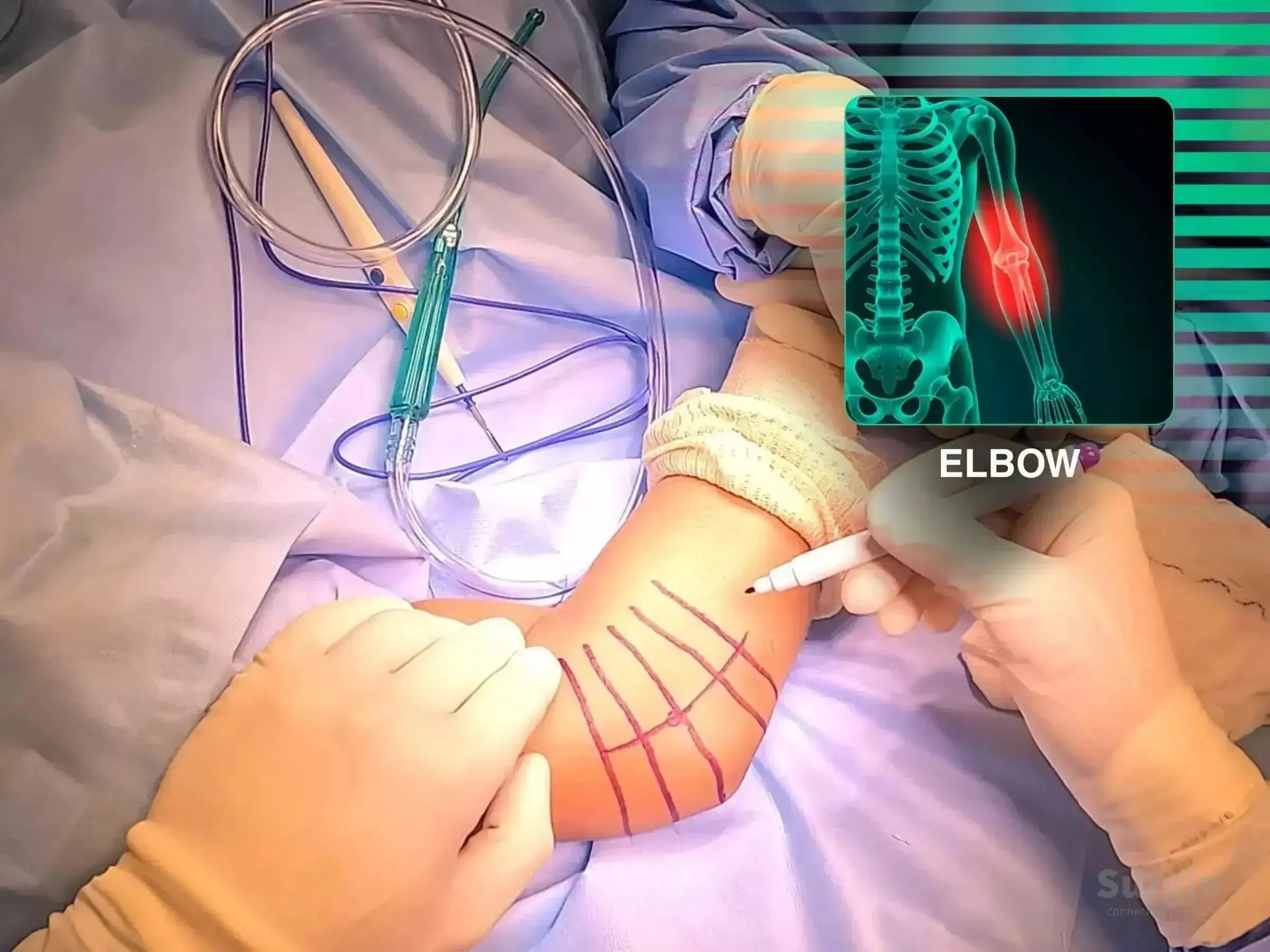
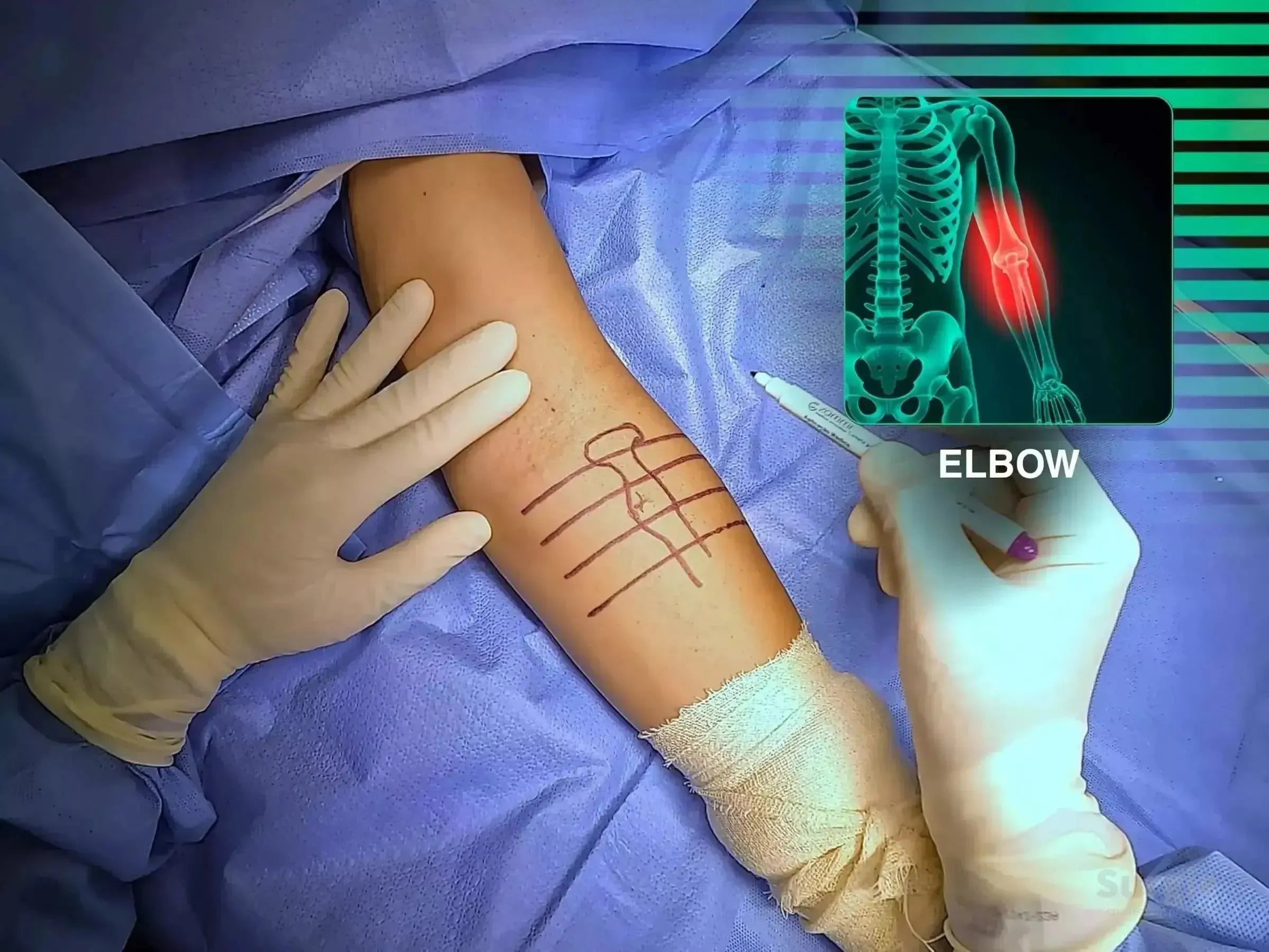
Surgeon marking a patient's elbow for radial head arthroplasty with an anatomical X-ray overlay highlighting the joint.
50% preview 4K Coude
05:35
Fracture comminutive de la tête radiale : Arthroplastie

Remplacement de la tête radiale avec réparation des ligaments et tests de stabilité

Dr. Sávio Chami Dr. Sávio ChamiOrthopédiste
9 languages
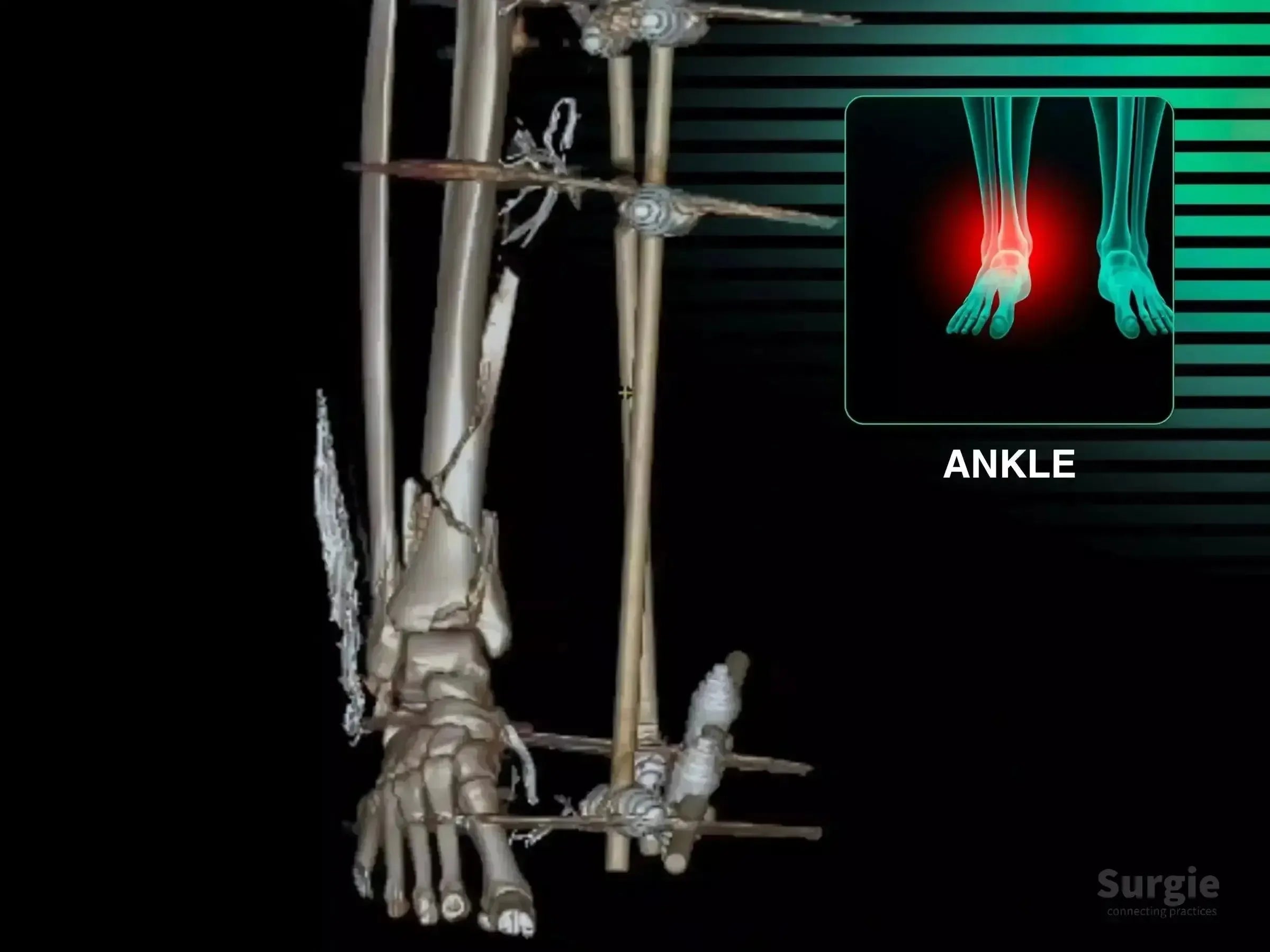
3D CT scan of a multi-fragmentary tibial pilon fracture with external fixation and an ankle pain highlight.
50% preview 4K Cheville
19:13
Fracture comminutive du pilon tibial : fixation en deux temps

Staged pilon reconstruction with articular reduction and multiplanar fixation

Dr. Sávio Chami Dr. Sávio ChamiOrthopédiste
9 languages
Lésion complexe de la cheville : reconstruction à 360 degrés
50% preview 4K Cheville
08:17
Lésion complexe de la cheville : reconstruction à 360 degrés

Achilles, posterior tibial tendon, deltoid, and syndesmosis reconstruction

Dr. Sávio Chami Dr. Sávio ChamiOrthopédiste
9 languages
Surgeon marking a patient's shoulder for a deltopectoral approach with an anatomical diagram overlay.
50% preview 4K Épaule
16:11
Reconstruction complexe d'une fracture proximale de l'humérus en quatre parties

Four-part reconstruction with valgus correction, plate, and suture fixation

Dr. Sávio Chami Dr. Sávio ChamiOrthopédiste
9 languages
Surgeon marking a patient's elbow for distal humerus osteotomy with a digital skeletal overlay showing the humerus area.
50% preview 5,7K Coude
28:54
Cubitus varus : ostéotomie corrective de l'humérus distal

3D-guided corrective osteotomy with ulnar nerve transposition

Dr. Sávio Chami Dr. Sávio ChamiOrthopédiste
9 languages
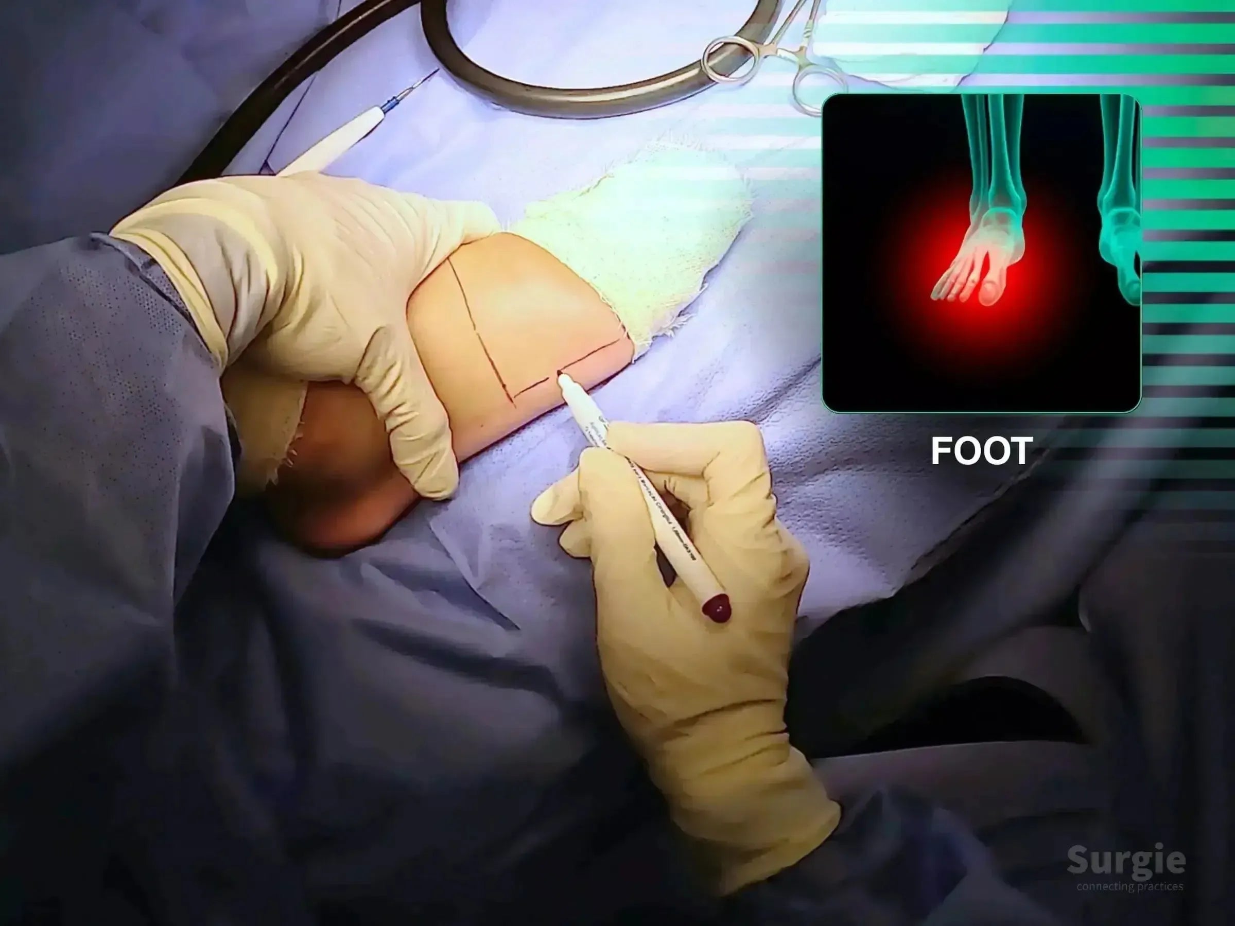
Surgeon marking a fifth metatarsal incision site on a patient's foot with an anatomical reference overlay.
50% preview 4K Pied
04:59
Fracture déplacée du cinquième métatarsien : fixation par plaque

Anatomic reduction and low-profile plate fixation of the fifth metatarsal

Dr. Sávio Chami Dr. Sávio ChamiOrthopédiste
9 languages
Surgeon marking a patient's arm for distal biceps tendon surgery with an anatomical elbow diagram overlay.
50% preview 4K Coude
05:02
Reconstruction du tendon distal du biceps : fixation TightRope

TightRope reinsertion at the radial tuberosity with tissue preservation

Dr. Sávio Chami Dr. Sávio ChamiOrthopédiste
9 languages
Surgeon marking a patient's arm for distal biceps tendon reconstruction with an anatomical elbow reference overlay.
50% preview 4K Coude
09:30
Réparation du tendon distal du biceps : TightRope avec protection nerveuse

TightRope tendon repair with focused neurovascular protection

Dr. Sávio Chami Dr. Sávio ChamiOrthopédiste
9 languages
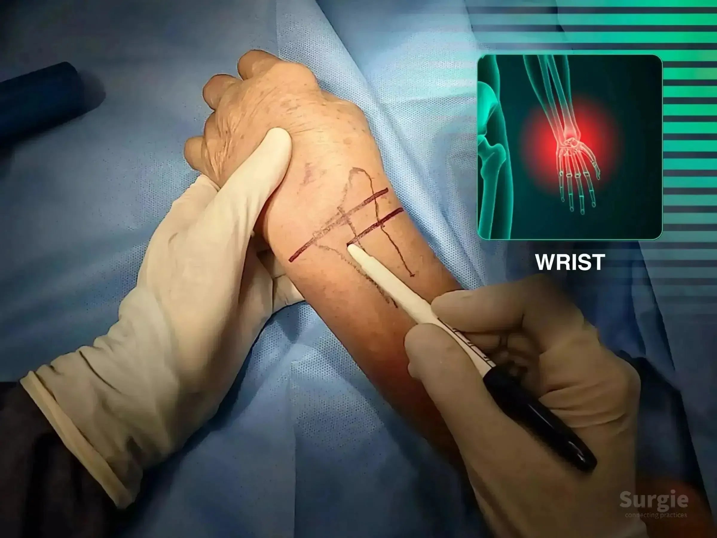
Surgeon marking a patient's wrist for a volar approach surgery with an anatomical X-ray overlay showing a fracture.
50% preview 4K Poignet
05:12
Fracture du radius distal : fixation par plaque palmaire

Volar exposure, provisional reduction, and plate stabilization

Dr. Sávio Chami Dr. Sávio ChamiOrthopédiste
9 languages
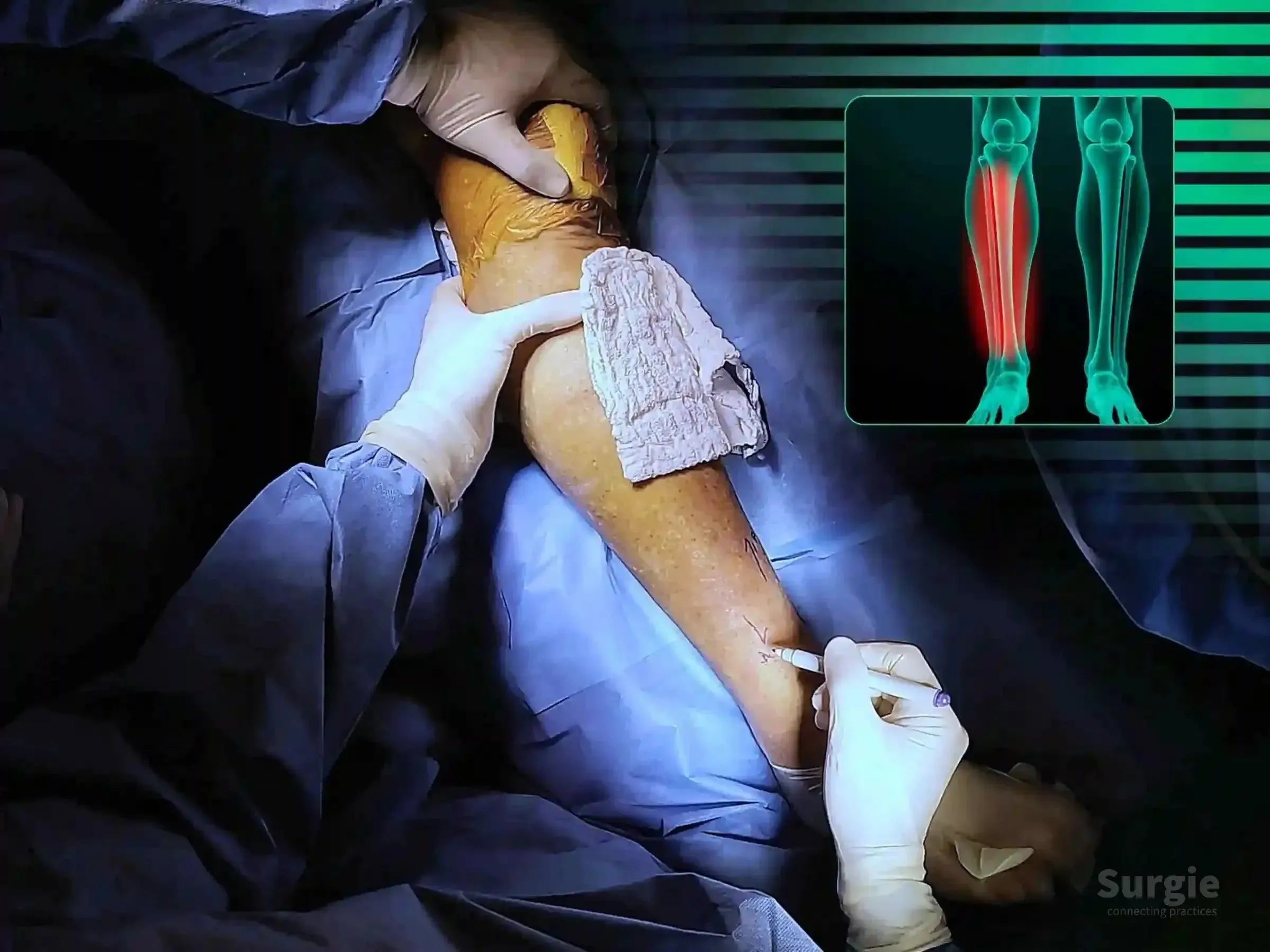
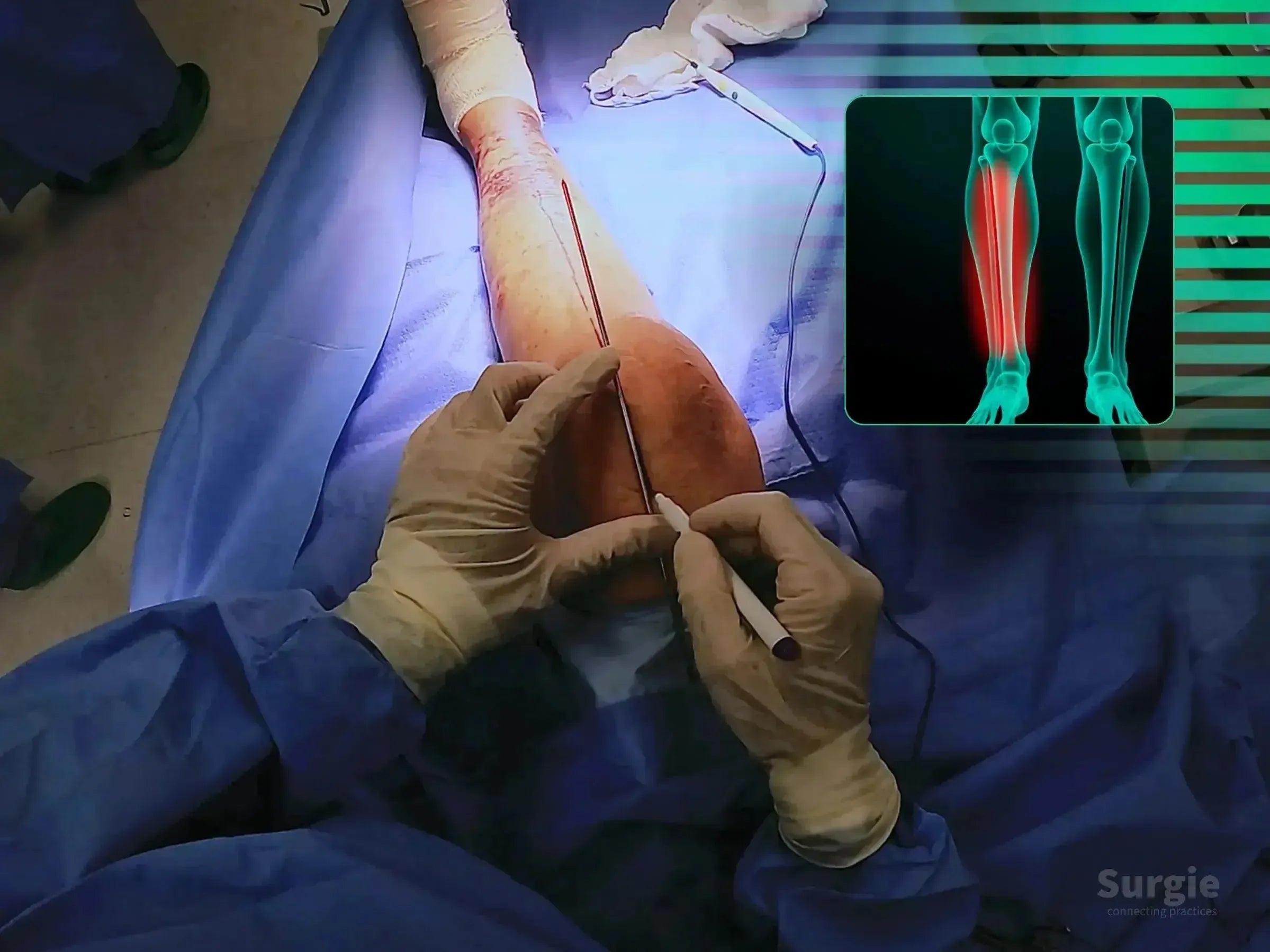
Surgeon marking a distal tibia fracture site for minimally invasive surgery with a digital X-ray overlay of the leg.
50% preview 4K Tibia
04:28
Fracture de la diaphyse tibiale distale : fixation par plaque verrouillée

Percutaneous reduction and minimally invasive locked plate fixation

Dr. Sávio Chami Dr. Sávio ChamiOrthopédiste
9 languages
Surgeon marking a suprapatellar portal on a patient's leg for tibial intramedullary nail surgery with an X-ray overlay.
50% preview 4K Tibia
08:31
Fracture de la diaphyse tibiale distale : clou intramédullaire suprapatellaire

Suprapatellar nailing with alignment control and soft-tissue protection

Dr. Sávio Chami Dr. Sávio ChamiOrthopédiste
9 languages
Surgeon marking a dorsal Barton's fracture site on a patient's wrist with an anatomical overlay showing the injury.
50% preview 4K Poignet
04:42
Fracture de Barton dorsale : Fixation du radius dorsal

Réduction du fragment dorsal avec fixation radiale ciblée

Dr. Sávio Chami Dr. Sávio ChamiOrthopédiste
9 languages
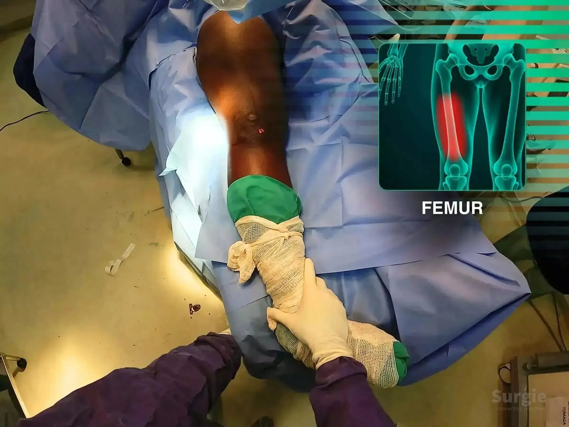
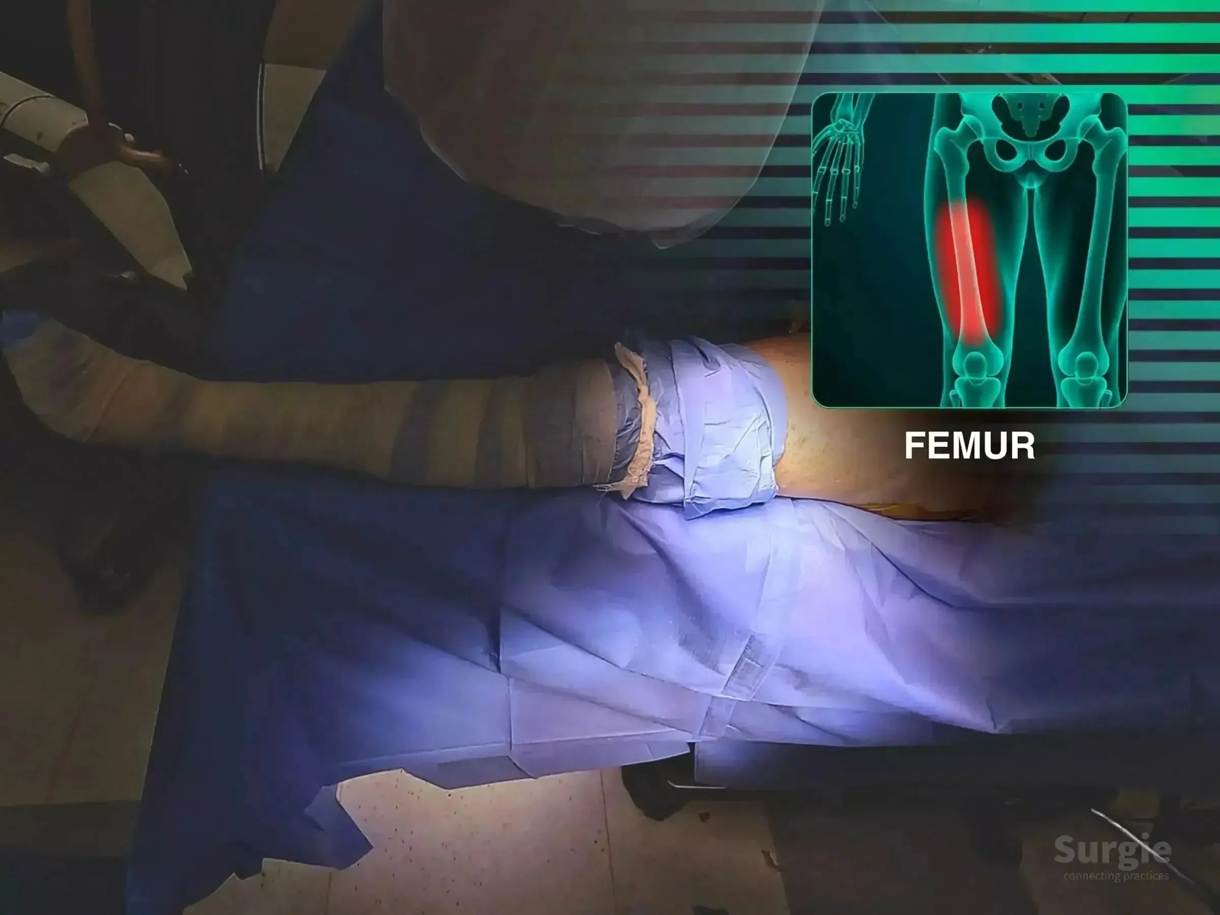
Surgeon performing femoral shaft fracture reduction with an anatomical overlay highlighting the femur on a blue surgical drape.
50% preview 4K Fémur
04:23
Fracture du fémur : clou intramédullaire rétrograde

Retrograde nailing with free-limb reduction and fluoroscopic control

Dr. Sávio Chami Dr. Sávio ChamiOrthopédiste
9 languages
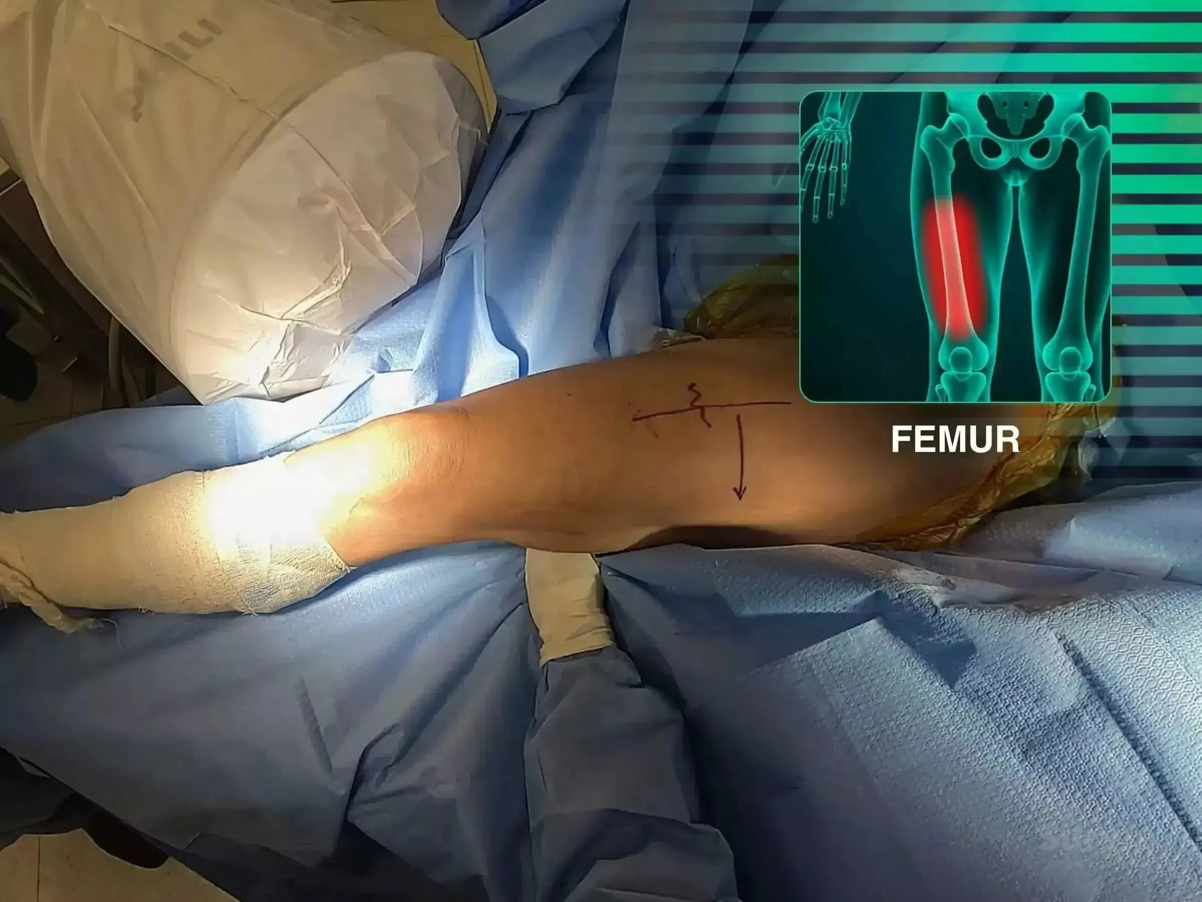
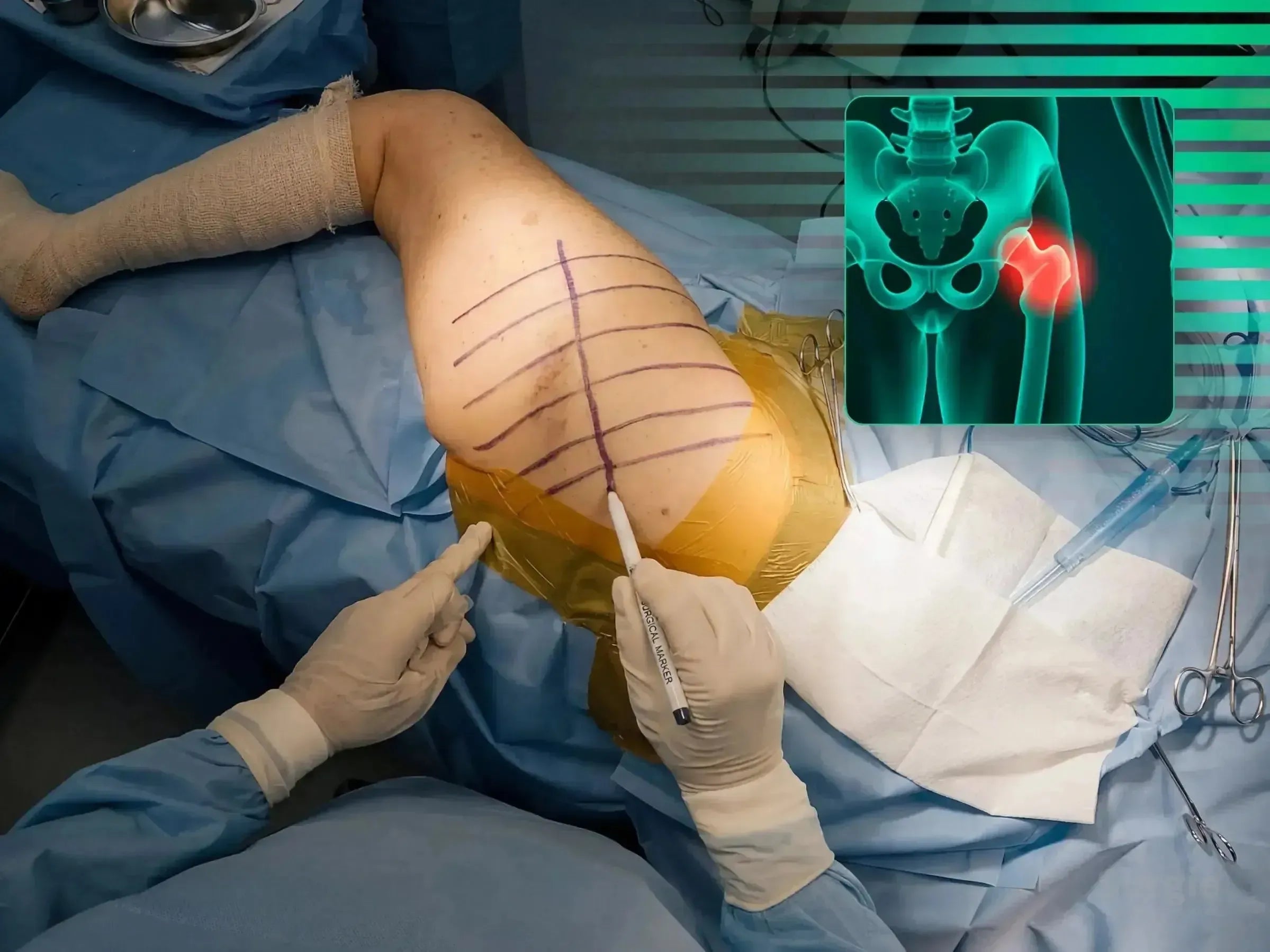
Surgical preparation of a patient's thigh for cephalomedullary nail fixation with a femoral anatomy overlay.
50% preview 4K Fémur
06:27
Refracture de la diaphyse fémorale : Clou céphalomédullaire antégrade

Antegrade nailing without a traction table for femoral shaft refracture

Dr. Sávio Chami Dr. Sávio ChamiOrthopédiste
9 languages
Surgeon performing minimally invasive surgery on a fifth metacarpal fracture with an anatomical hand diagram overlay.
50% preview 4K Main
02:43
Fracture du col du cinquième métacarpien : Vis à double compression

Minimally invasive double-compression fixation of the fifth metacarpal

Dr. Sávio Chami Dr. Sávio ChamiOrthopédiste
9 languages
Surgeon marking a patient's shoulder for a deltopectoral approach with a skeletal diagram overlay showing a humerus fracture.
50% preview 4K Épaule
05:37
Fracture en quatre parties de l'humérus proximal : fixation delto-pectorale

Reconstruction de la tubérosité avec fixation par plaque et vis

Dr. Sávio Chami Dr. Sávio ChamiOrthopédiste
9 languages
Surgeon marking a shoulder incision site with a grid, featuring an anatomical overlay of the glenoid joint.
50% preview 4K Épaule
04:28
Fixation des fractures glénoïdiennes : Segment de procédure combinée

Exposition antérieure de la glène avec fixation par vis de compression

Dr. Sávio Chami Dr. Sávio ChamiOrthopédiste
9 languages
Surgeon marking a shoulder for a deltopectoral approach with a digital anatomical overlay of a humeral fracture.
50% preview 4K Épaule
07:22
Fracture de la grande tubérosité : approche delto-pectorale

Direct tuberosity reduction with locking plate and suture reinforcement

Dr. Sávio Chami Dr. Sávio ChamiOrthopédiste
9 languages
Surgeon marking a humeral shaft fracture site with a guide wire and marker, featuring an anatomical bone overlay.
50% preview 4K Humerus
06:13
Fracture de la diaphyse humérale : fixation par clou intramédullaire

Joystick-assisted reduction with proximal and distal nail locking

Dr. Sávio Chami Dr. Sávio ChamiOrthopédiste
9 languages
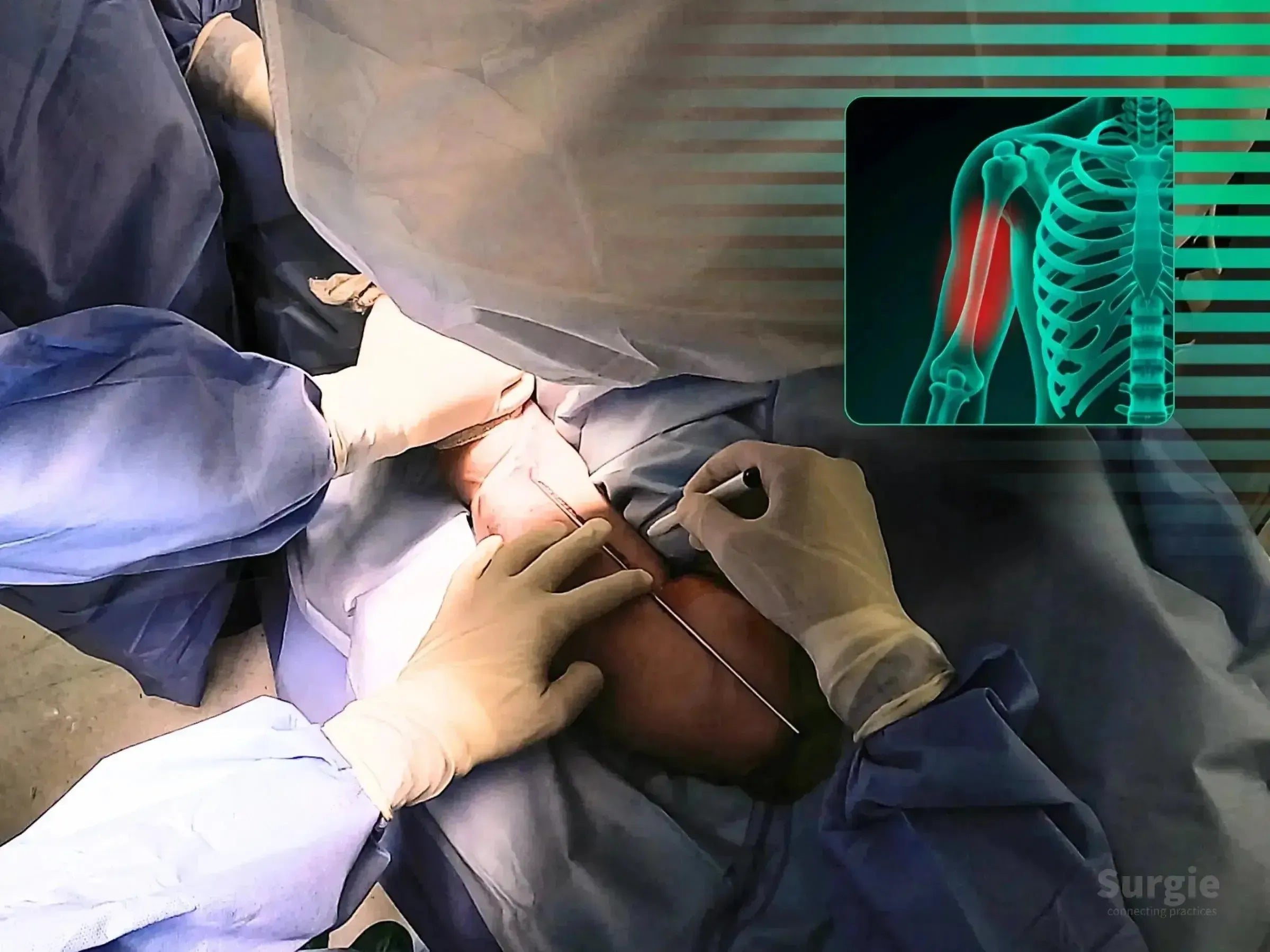
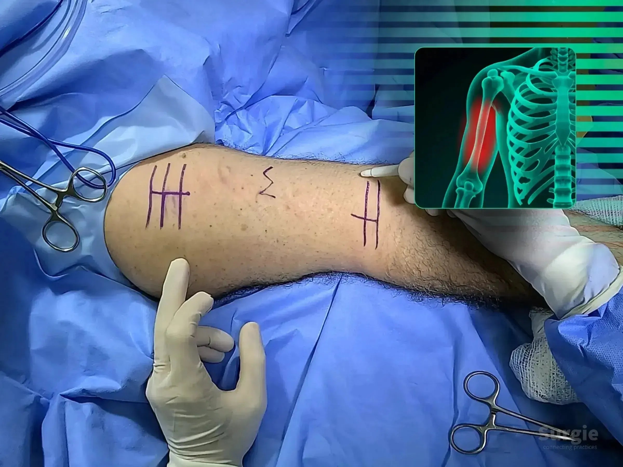
Surgical site on an arm with pre-operative markings and a digital X-ray overlay showing a humeral shaft fracture.
50% preview 4K Humerus
06:26
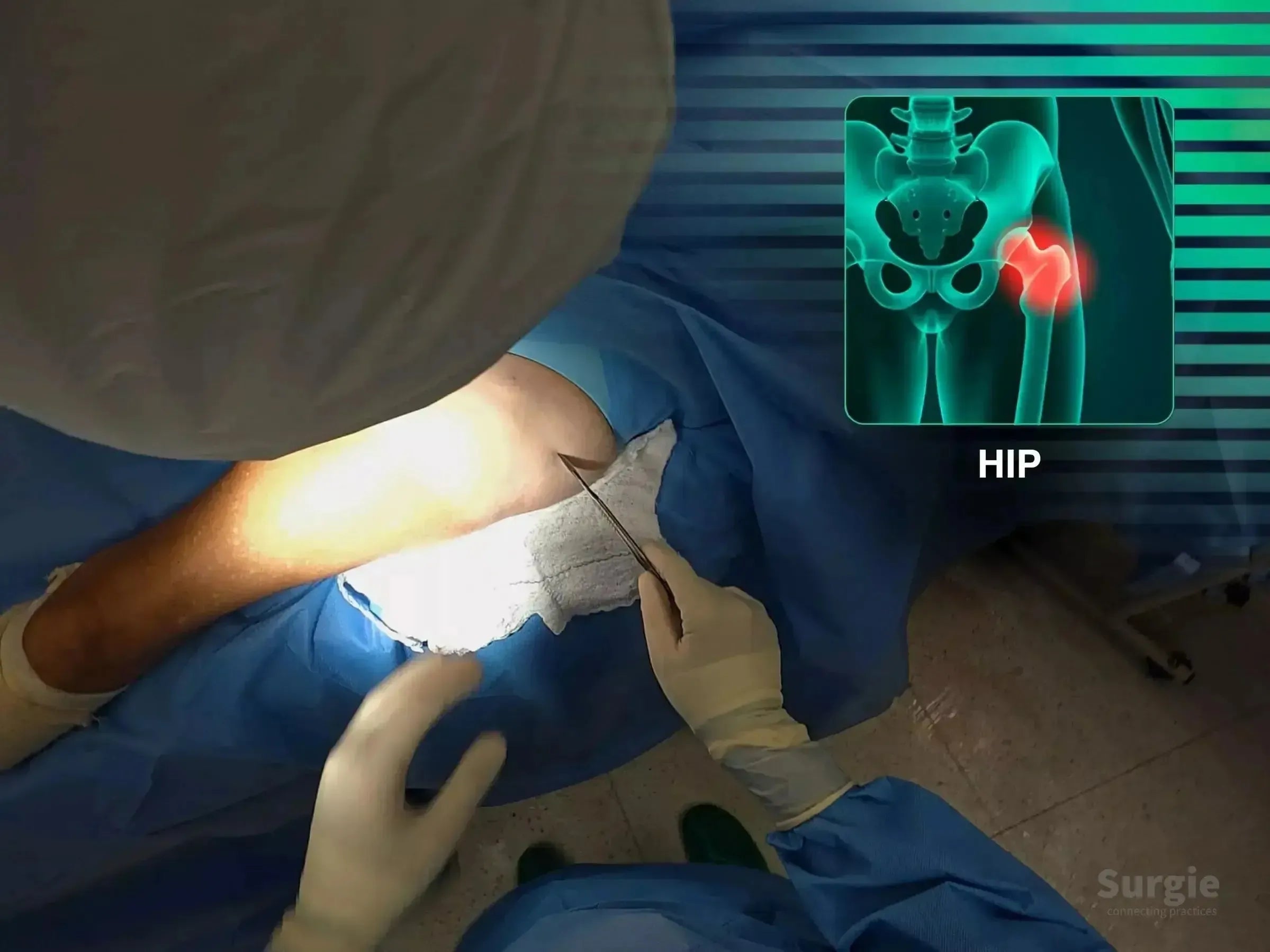
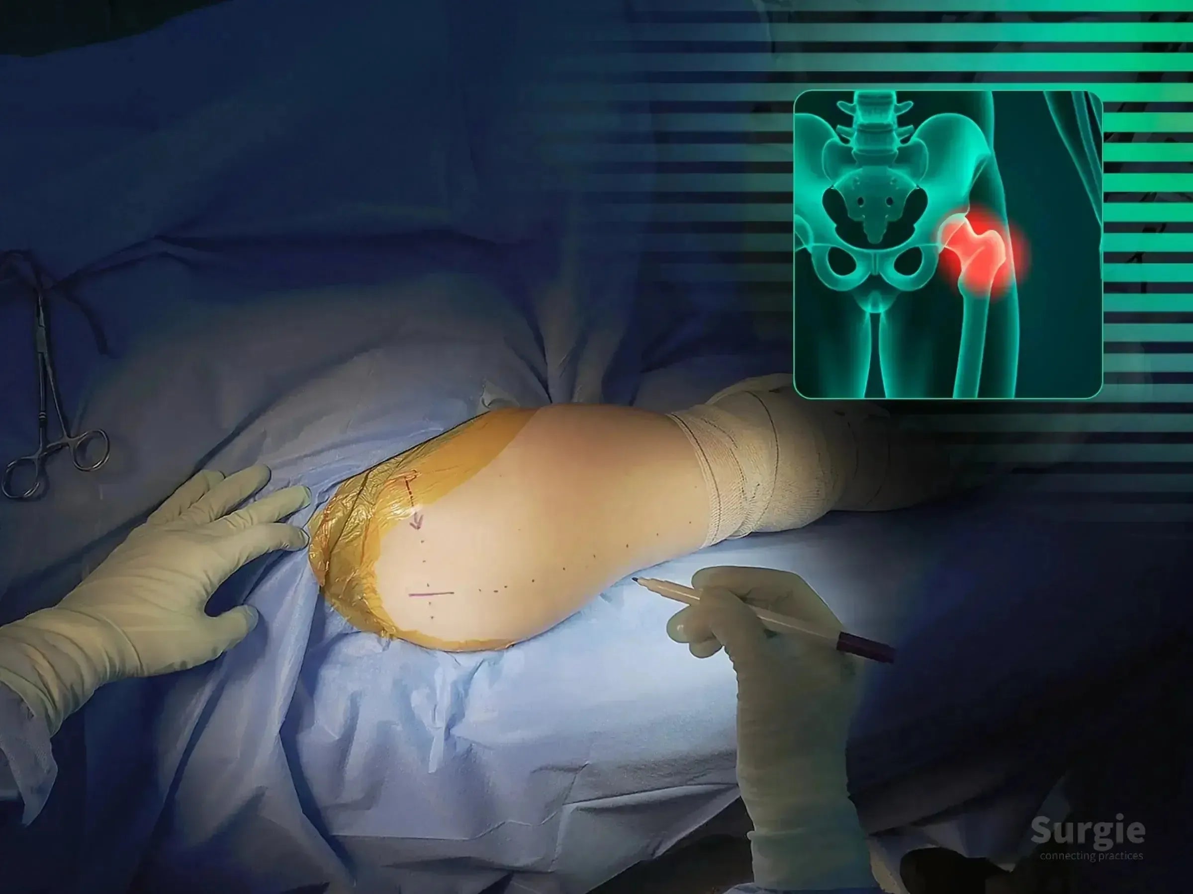
Surgeon performing a minimally invasive hip fracture procedure with a digital anatomical overlay of the femur.
50% preview 4K Hip
07:16
Fracture intertrochantérienne, Cas 1 : Clou intramédullaire

Guidewire precision and intramedullary fixation without traction

Dr. Sávio Chami Dr. Sávio ChamiOrthopédiste
9 languages
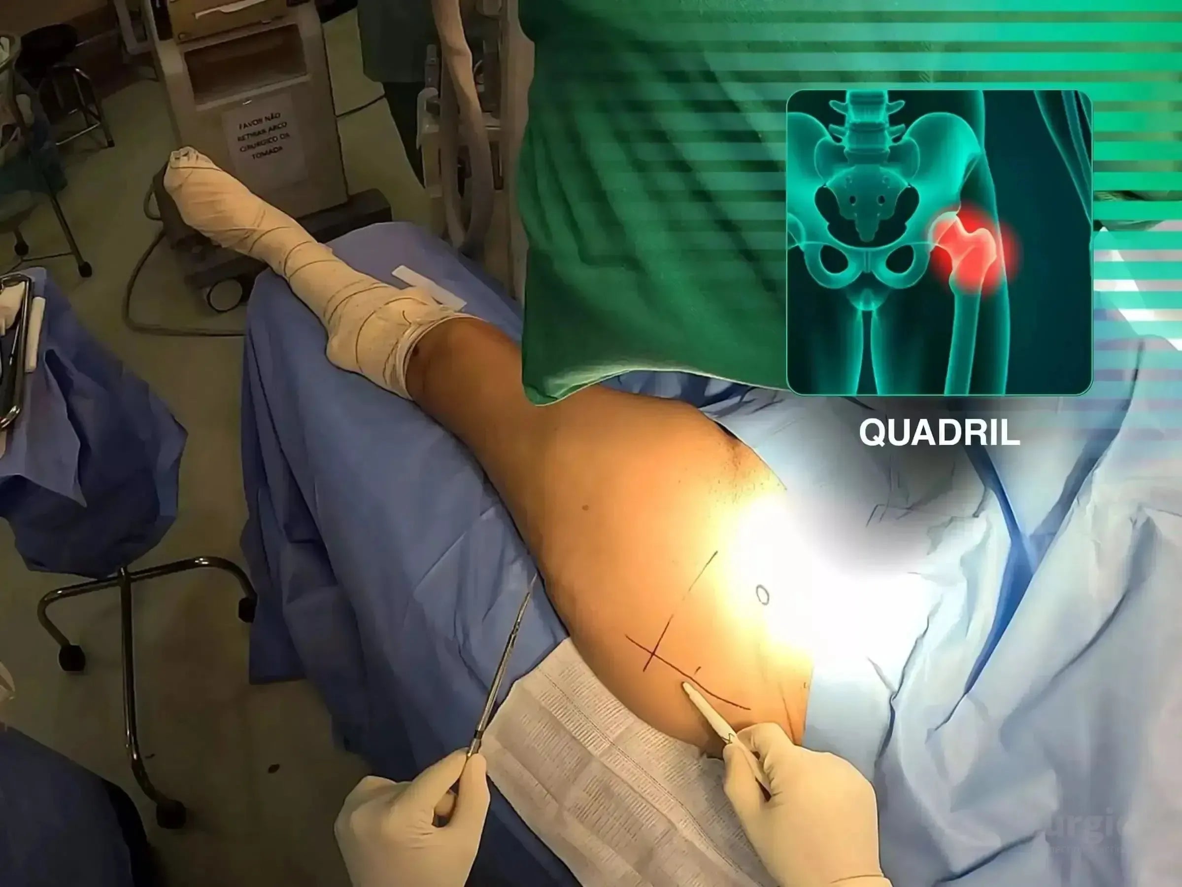
Surgeon marking a hip incision site for intramedullary nailing with a digital anatomical overlay of a transtrochanteric fracture.
50% preview 4K Hip
05:44
Surgeon marking a patient's shoulder for clavicle surgery with an anatomical overlay of a fractured collarbone.
50% preview 4K Épaule
05:03
Fracture latérale de la clavicule : Fixation coracoclaviculaire

Reconstruction de la clavicule latérale avec fixation TightRope coraco-claviculaire

Dr. Sávio Chami Dr. Sávio ChamiOrthopédiste
9 languages
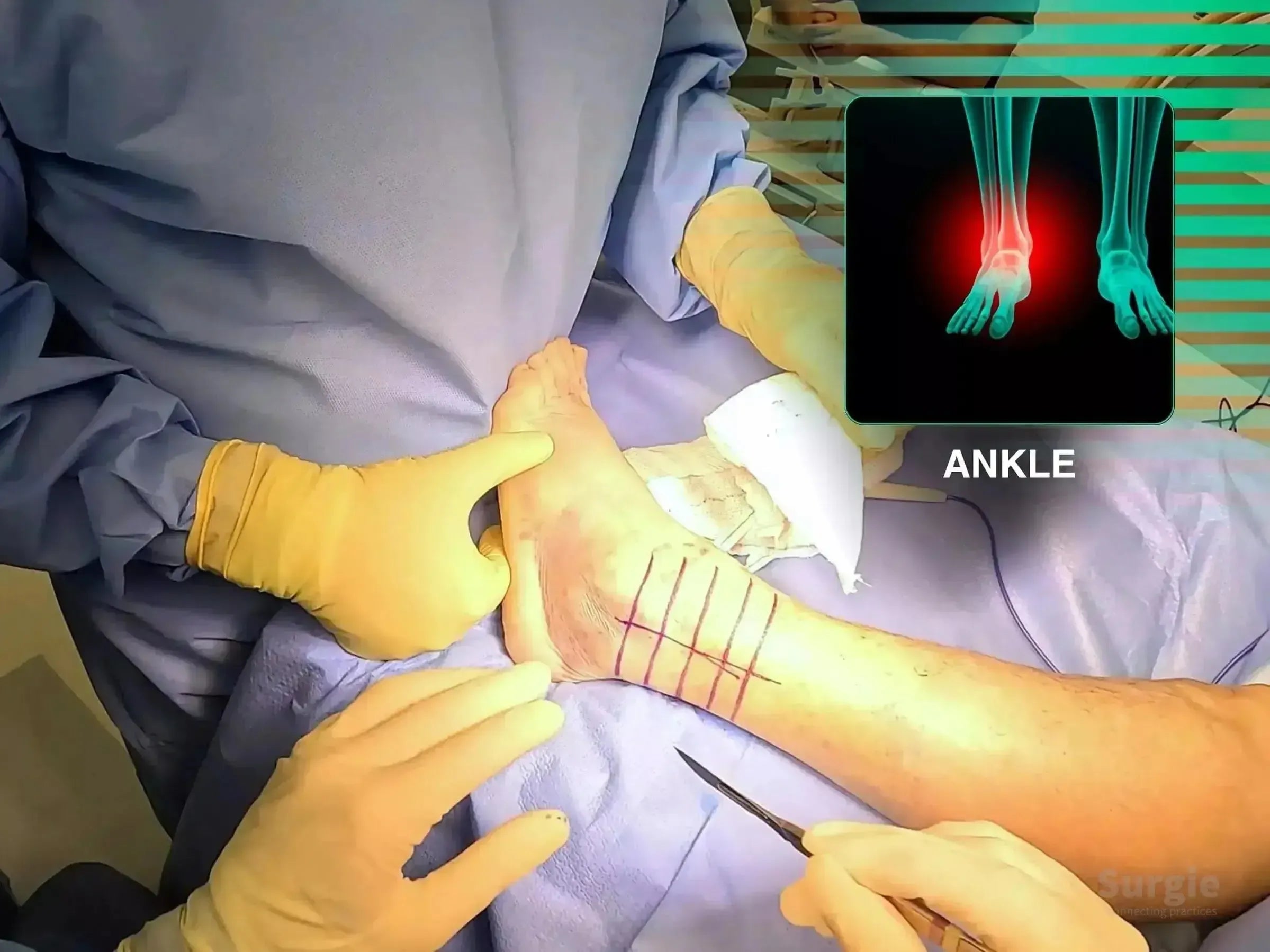
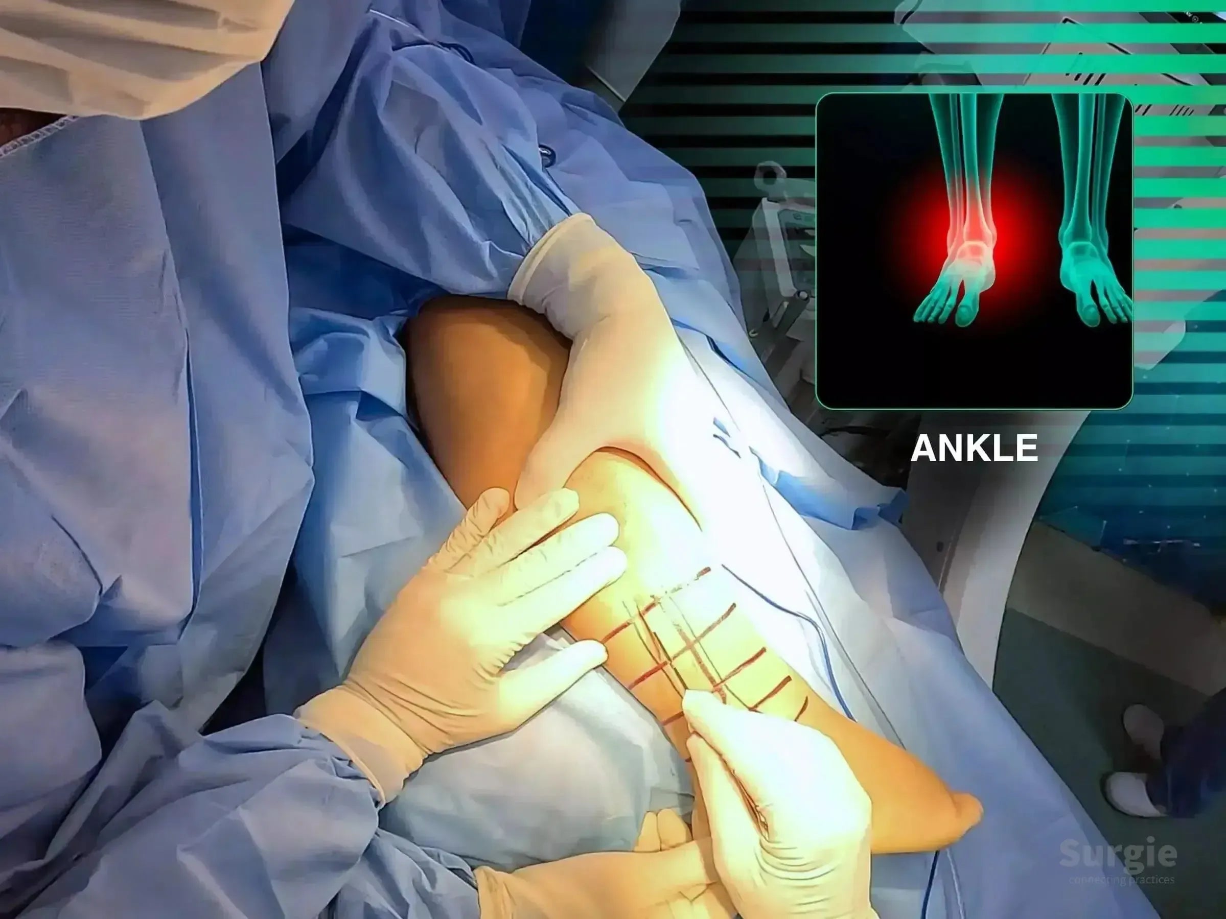
Surgeon marking a patient's ankle for lateral malleolus fracture surgery with an anatomical X-ray overlay.
50% preview 4K Cheville
02:29
Fracture de la malléole latérale avec fragment de Wagstaffe

Compression du fragment de Wagstaffe avec fixation du malléole latéral verrouillé

Dr. Sávio Chami Dr. Sávio ChamiOrthopédiste
9 languages
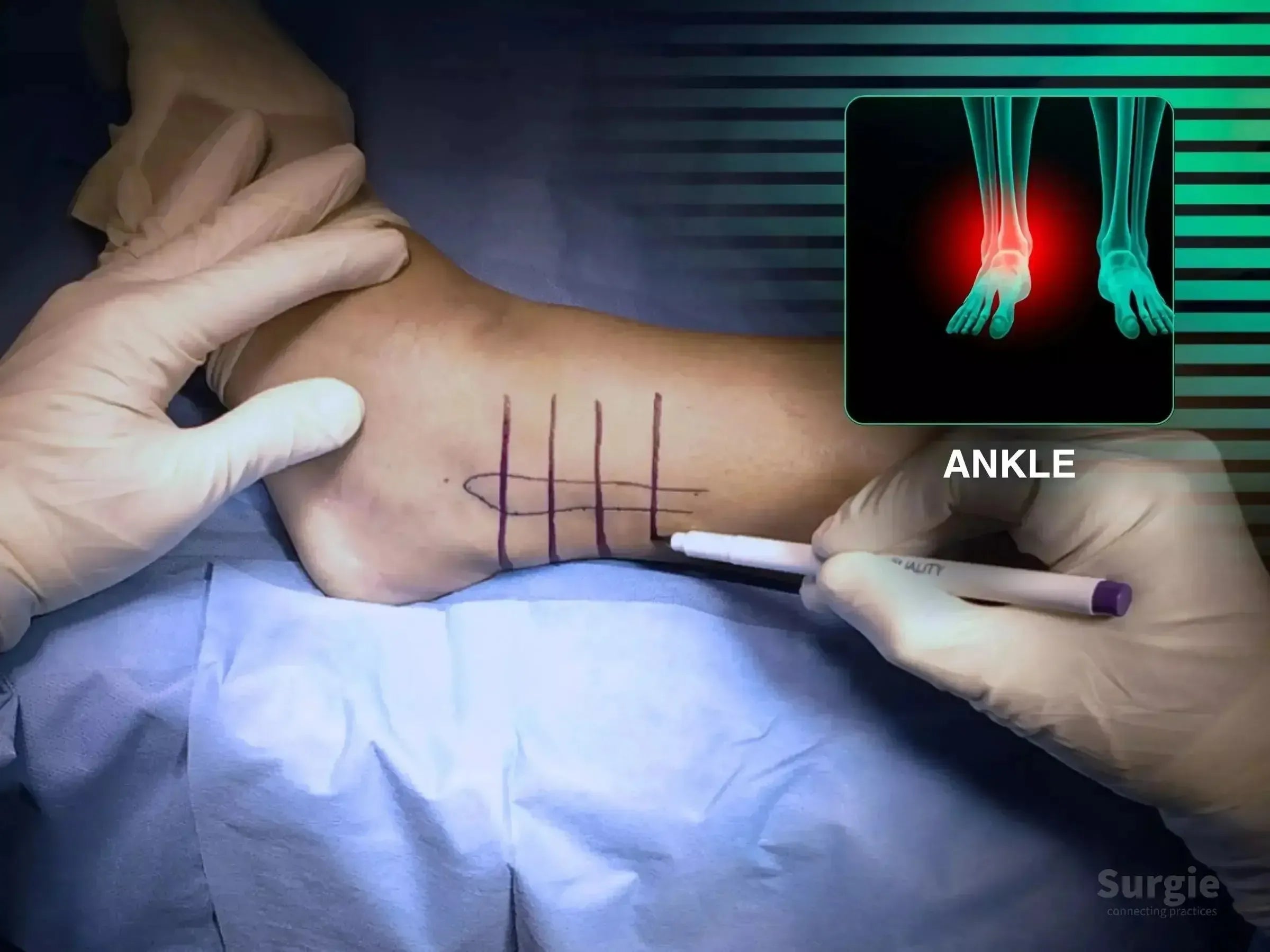
Surgeon marking a lateral malleolus fracture site on an ankle with a medical marker, featuring an anatomical overlay.
Free 4K Cheville
07:41
Fracture de la malléole latérale : Technique de plaque postérieure

Posterior antiglide plating for shear control and tissue preservation

Dr. Sávio Chami Dr. Sávio ChamiOrthopédiste
9 languages
Surgeon marking a patient's elbow for surgery with an anatomical overlay showing a radial head fracture.
50% preview 4K Coude
08:10
Fracture de la tête radiale de type III de Mason avec instabilité du coude

Mini-fragment fixation with coronoid and ligament reconstruction

Dr. Sávio Chami Dr. Sávio ChamiOrthopédiste
9 languages
Surgeon marking a posterior elbow incision site for Monteggia variant fracture repair with anatomical overlay.
50% preview 4K Coude
14:59
Variante de Monteggia : Fixation proximale de l’ulna et de la tête radiale

Fixation combinée de l'ulna et de la tête radiale pour l'instabilité de Monteggia

Dr. Sávio Chami Dr. Sávio ChamiOrthopédiste
9 languages
Surgeon marking an elbow for minimally invasive osteosynthesis with an anatomical skeletal overlay.
50% preview 4K Coude
06:54
Fracture de l’olécrâne : fixation par plaque mini-invasive

Plaquage minimalement invasif de l'olécrâne avec restauration anatomique de l'articulation

Dr. Sávio Chami Dr. Sávio ChamiOrthopédiste
9 languages
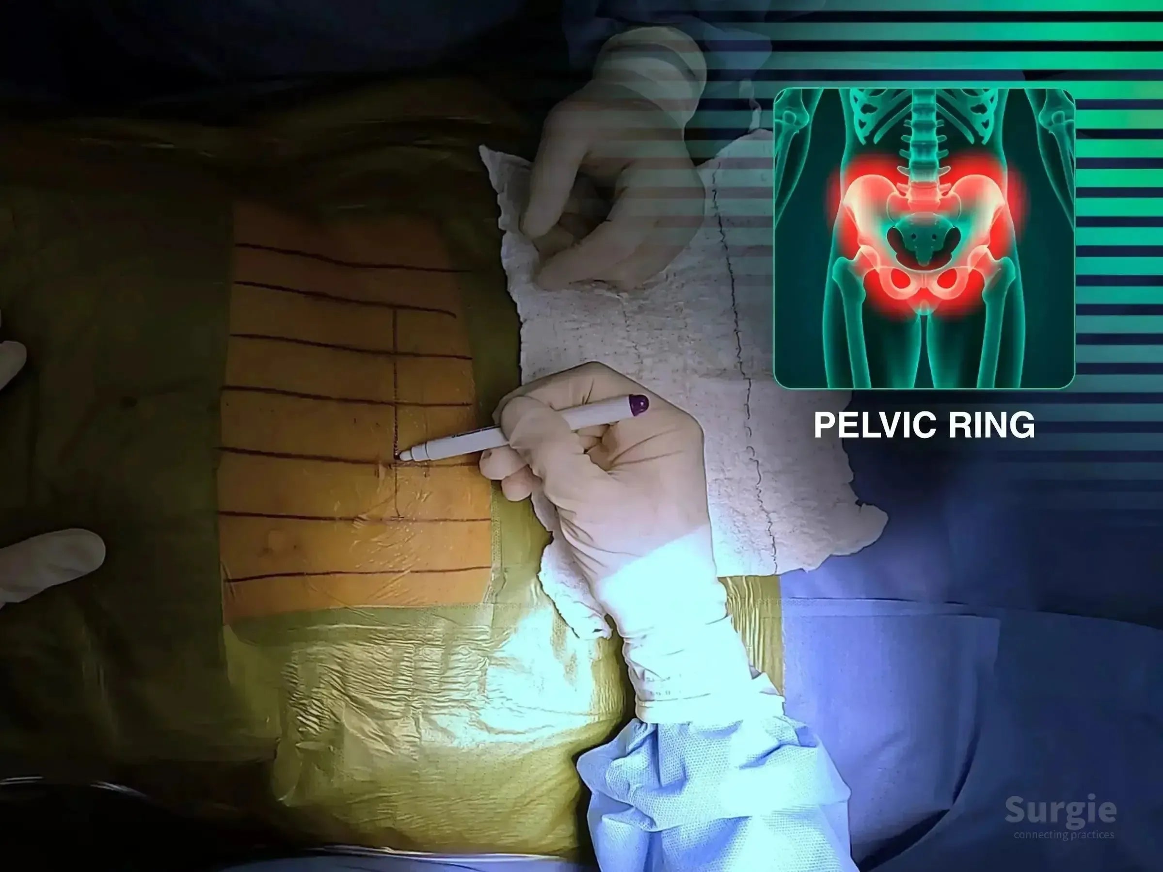
Surgeon marking a patient's abdomen for pelvic ring surgery with a digital overlay of a pubic symphysis dislocation.
50% preview 4K Anneau pelvien
06:13
Lésion ouverte de l’anneau pelvien : fixation de la symphyse pubienne

Controlled symphyseal reduction with anterior pelvic ring fixation

Dr. Sávio Chami Dr. Sávio ChamiOrthopédiste
9 languages
Surgeon marking a knee for patellar fracture surgery with an X-ray overlay showing a highlighted patella injury.
50% preview 4K Genou
24:55
Fracture de la rotule, Cas 2 : Vis canulées et cerclage

Cannulated screws and cerclage with extensor mechanism preservation

Dr. Sávio Chami Dr. Sávio ChamiOrthopédiste
9 languages
Surgeon marking a patient's hip for posterolateral surgery with a digital overlay of a periprosthetic hip fracture.
50% preview 4K Hip
19:45
Periprosthetic Hip Fracture: Trochanteric Plate and Cerclage

Trochanteric plating and cerclage without femoral component revision

Dr. Sávio Chami Dr. Sávio ChamiOrthopédiste
9 languages
Surgeon marking a patient's ankle for a posterolateral approach with an anatomical X-ray overlay of the fracture site.
50% preview 4K Cheville
04:14
Fractures du malléole postérieur et latéral : Approche postérolatérale

Exposition posterolatérale unique pour la fixation du malléole postérieur et latéral

Dr. Sávio Chami Dr. Sávio ChamiOrthopédiste
9 languages
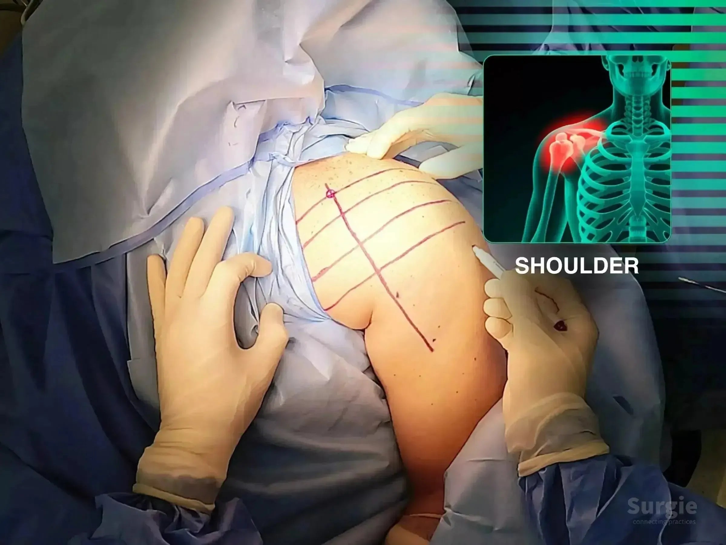
Surgeon marking a patient's shoulder for a deltopectoral approach with an anatomical diagram overlay.
50% preview 4K Épaule
05:04
Luxation postérieure de l'épaule avec impaction de la tête humérale

Réduction d'une luxation postérieure verrouillée avec un plaquage en fibre de carbone

Dr. Sávio Chami Dr. Sávio ChamiOrthopédiste
9 languages
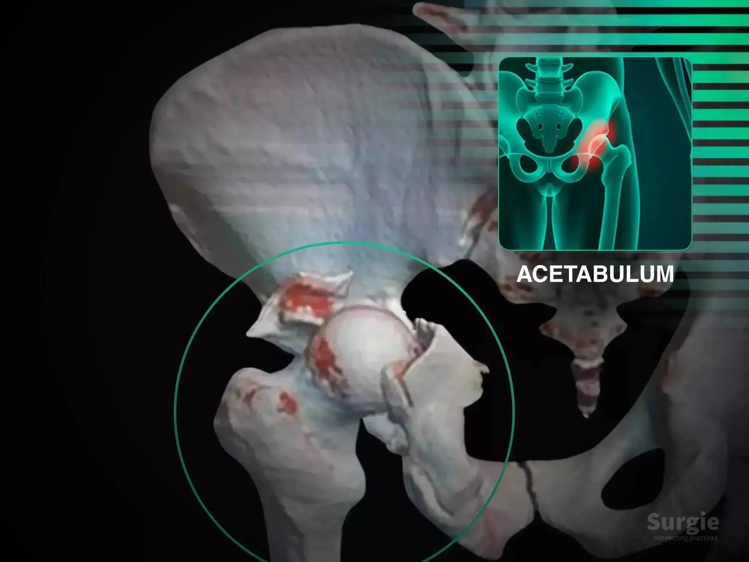
3D medical illustration showing a posterior acetabular wall fracture with hip dislocation and highlighted bone fragments.
50% preview 4K Acétabulum
05:35
Fracture de la paroi postérieure de l'acétabulum avec luxation de la hanche

Kocher-Langenbeck exposure with posterior wall fixation and nerve protection

Dr. Sávio Chami Dr. Sávio ChamiOrthopédiste
9 languages
Surgeon marking a patient's hip for proximal femur intramedullary nail surgery with a digital anatomical overlay.
50% preview 4K Hip
10:24
Fracture du fémur proximal, Cas 3 : Manœuvre du basculeur

Rocker-assisted calcar reduction without a traction table

Dr. Sávio Chami Dr. Sávio ChamiOrthopédiste
9 languages
Surgical setup for proximal femur fracture with blue drapes and an anatomical overlay highlighting the femur.
50% preview 4K Hip
07:16
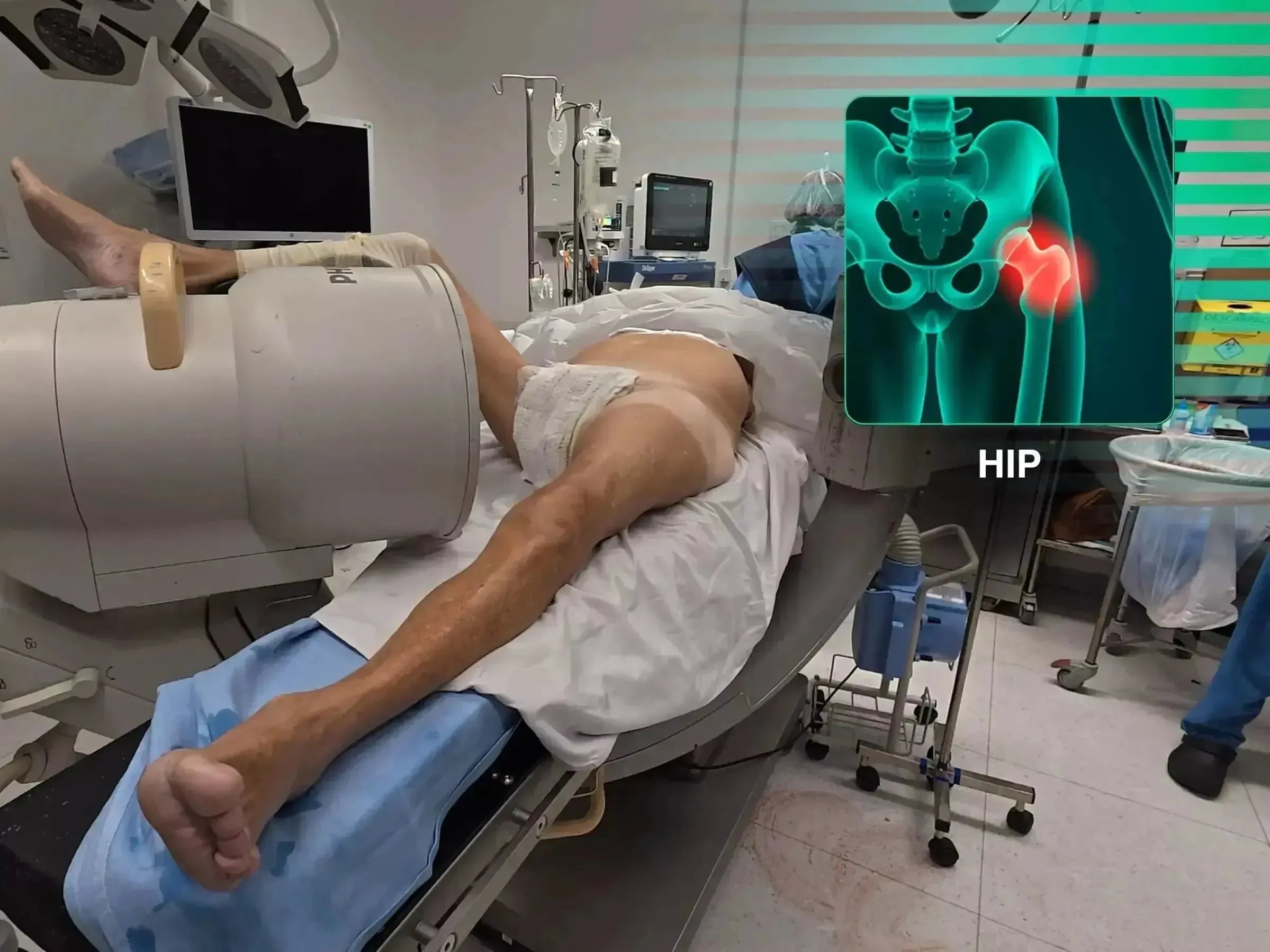
Patient positioned on a conventional surgical table for proximal femur fracture surgery with C-arm and hip anatomy overlay.
50% preview 4K Hip
04:12
Fractures du fémur proximal : chirurgie sans table de traction

Free-fragment manipulation using a conventional operating table

Dr. Sávio Chami Dr. Sávio ChamiOrthopédiste
9 languages
Surgeon performing a deltopectoral approach for a proximal humerus fracture with a shoulder anatomy overlay.
50% preview 4K Épaule
09:55
Fracture de l'humérus proximal : fixation par plaque deltopectorale

Deltopectoral exposure with cuff management and plate fixation

Dr. Sávio Chami Dr. Sávio ChamiOrthopédiste
9 languages
Surgeons performing minimally invasive intramedullary nailing on a humerus with a joystick technique and shoulder anatomy overlay.
50% preview 4K Épaule
04:10
Fracture proximale de l'humérus : enclouage assisté par joystick

Réduction guidée par joystick et fixation intramédullaire

Dr. Sávio Chami Dr. Sávio ChamiOrthopédiste
9 languages
Surgical guide wire insertion into a proximal humerus with an anatomical overlay showing a shoulder fracture.
50% preview 4K Épaule
15:40
Fracture proximale de l'humérus : clou intramédullaire long

Fixation par clou long avec préservation de la manchette et contrôle fluoroscopique

Dr. Sávio Chami Dr. Sávio ChamiOrthopédiste
9 languages